Forth stage
MedicineLec-1
د. جاسم محمد
27/10/2015
INVESTIGATION IN CARDIOVASCULAR DISEASEElectrocardiography (ECG)
Process of recording the electrical activity of the heart over a period of time using electrodes placed on a patient's body.Indications:
Assess cardiac rhythm and conductionTo assess for myocardial ischaemia an infarction
Determine hypertrophy of the chamber
Determine electrolyte imbalances
Evaluate and monitor the effect of drugs
Normal Impulse Conduction
Sinoatrial node
AV nodeBundle of His
Bundle BranchesPurkinje fibers
Sinoatrial nodeAV node
Bundle of HisBundle Branches
Purkinje fibers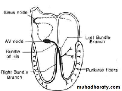
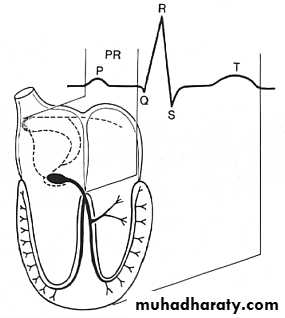
Impulse Conduction & the ECG
Sinoatrial node
AV node
Bundle of His
Bundle Branches
Purkinje fibersPacemakers of the Heart:
SA Node - Dominant pacemaker with an intrinsic rate of 60 - 100 beats/minute.AV Node - Back-up pacemaker with an intrinsic rate of 40 - 60 beats/minute.
Ventricular cells - Back-up pacemaker with an intrinsic rate of 20 - 45 bpm.
The “PQRST“:
P - wave - Atrial depolarization
QRS - Ventricular depolarizationT wave - Ventricular repolarization
The standard 12–lead ECG:The 12-lead ECG is generated from ten physical electrodes that are attached to the skin. One electrode is attached to each limb and six electrodes are attached to the chest
ECG leads:
Leads are electrodes which measure the difference in electrical potential between either:Two different points on the body (bipolar leads)
One point on the body and virtual reference point with zero electrical potential, located in the center of the heart (unipolar)
Three dipole limb leads (I, II,III), three augmented voltage limb leads(avL,avR,avF)
Six unipolar chest leads (V1-V6)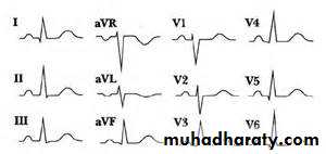
Exercise (stress) ECG :
Exercise electrocardiography is used to detect myocardial ischaemia during physical stress and is helpful in the diagnosis of coronary artery disease. A 12-lead ECG is recorded during exercise on a treadmill or bicycle ergometerAmbulatory ECG (Holter)
Continuous (ambulatory) ECG recordings can be obtained using a portable digital recorder. These devices usually provide limb lead ECG recordings only, and can record for between 1 and 7 days. Ambulatory ECG recording is principally used in the investigation of patients with suspected arrhythmia , such as those with intermittent palpitation, dizziness or syncopy
Chest X-ray
This is useful for determining the size and shape of the heart, and the state of the pulmonary blood vessels and lung fields. Most information is given by a posteroanterior (PA) projection taken in full inspiration
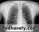
Chest x-ray
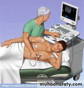
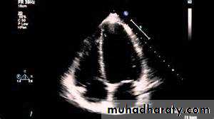
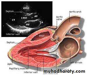
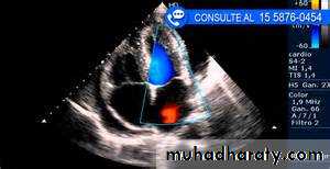
Echocardiography (echo) :Echocardiography, or cardiac ultrasound, is obtained by placing an ultrasound transducer on the chest wall to image the heart structures as a real-time,
two-dimensional ‘slic
Transthoracic echo.
Transoesophageal.
Doppler echocardiography:
This depends on the Doppler principle that sound waves reflected from moving objects, such as intracardiac red blood cells, undergo a frequency shift. The speed and direction of the red cells, and thus of blood, can be detected in the heart chambers and great vessels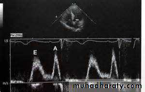
Transoesophageal Echocardiography:
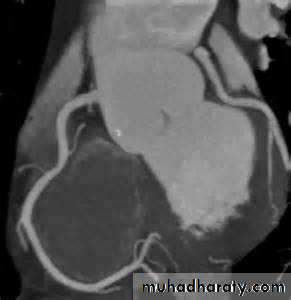
Endoscope -like ultrasound probe which is passed into the oesophagus under light sedation and positioned behind the LA. This produces high-resolution images.Computed Tomographic Imaging :
Useful for imaging the cardiac chambers, great vessels , pericardium, and mediastinal structures and masses
And recently even the coronaries (CT coronary angiography).
Magnetic resonance imaging
Magnetic resonance imaging (MRI) requires no ionising radiation and can be used to generate cross-sectional images of the heart, lungs and mediastinal structures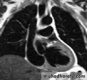
Cardiac catheterization
This involves passage of a reshaped catheter via a vein or artery into the heart under X-ray guidance, which allows the measurement of pressure and oxygen saturation in the cardiac chambers and great vessels, and the performance of angiograms by injecting contrast media into a chamber or blood vessel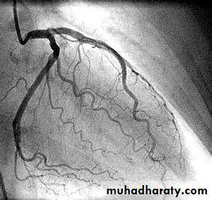
Coronary catheterization
Electrophysiology study
For assessment , diagnosis and treatment of arrhythmiasRadionuclide imaging
Blood pool imagingMyocardial perfusion imaging
Cardiac biomarkers
Brain natriuretic peptide-diagnosis and assess prognosis and response to therapy in patients with heart failure.Cardiac troponins Troponin I and troponin T are structural cardiac muscle proteins that are released during myocyte damage and necrosis, and represent the cornerstone of the diagnosis of acute myocardial infarction.