1
4th stage
يةٌطاب
Lec-7
.د
اسواعيل
18/10/2015
Motility disorders of oesophagus
A. Primary
1. Pharyngeal pouch
This occurs because of incoordination of
swallowing within the pharynx, which leads to
herniation through the cricopharyngeus muscle
and formation of a pouch.
It is rare, and it usually develops in middle life but can arise at any age.
Many patients have no symptoms, but regurgitation, halitosis and
dysphagia can be present. Some notice gurgling in the throat after
swallowing. The investigation of choice is a barium swallow, which
demonstrates the pouch and reveals incoordination of swallowing, often
with pulmonary aspiration. Endoscopy may be hazardous, since the
instrument may enter and perforate the pouch. Surgical myotomy
(‘diverticulotomy’), with or without resection of the pouch, is indicated
in symptomatic patients.
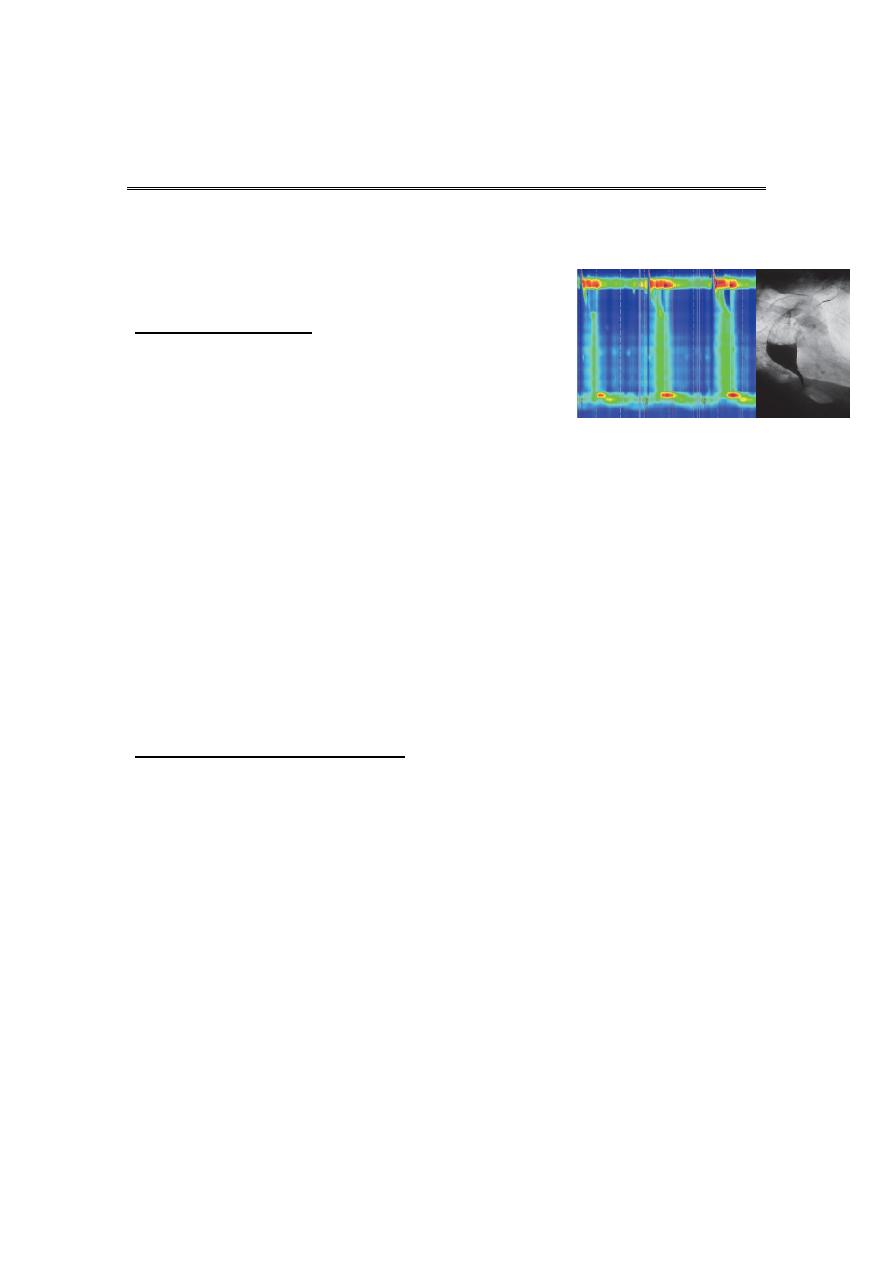
2. Achalasia of the oesophagus
Achalasia is characterised by: a hypertonic lower oesophageal sphincter,
which fails to relax in response to the swallowing wave failure of
propagated oesophageal contraction, leading to progressive dilatation of
the gullet. The cause is unknown. Defective release of nitric oxide by
inhibitory neurons in the lower oesophageal sphincter has been
reported, and there is degeneration of ganglion cells within the
sphincter and the body of the oesophagus. Loss of the dorsal vagal
nuclei within the brainstem can be demonstrated in later stages.
Infection with Trypanosoma cruzi in Chagas’ disease causes a syndrome
that is clinically indistinguishable from achalasia.

2
Clinical features
The presentation is with dysphagia. This develops slowly, is initially
intermittent, and is worse for solids and eased by drinking liquids, and
by standing and moving around after eating. Heartburn does not occur
because the closed oesophageal sphincter prevents gastro-oesophageal
reflux. Some patients experience episodes of chest pain due to
oesophageal spasm. As the disease progresses, dysphagia worsens, the
oesophagus empties poorly and nocturnal pulmonary aspiration
develops. Achalasia predisposes to squamous carcinoma of the
oesophagus.
Investigations:
Endoscopy should always be carried out because carcinoma of the cardia
can mimic the presentation and radiological and manometric features of
achalasia (‘pseudo-achalasia’). A barium swallow shows tapered
narrowing of the lower oesophagus and, in late disease, the oesophageal
body is dilated, aperistaltic and food filled. Manometry confirms the high
pressure, non-relaxing lower oesophageal sphincter with poor
contractility of the oesophageal body
Management
Endoscopic
Forceful pneumatic dilatation using a 30–35-mm diameter
fluoroscopically positioned balloon disrupts the
oesophageal sphincter and improves symptoms in 80%
of patients. Some patients require more than one dilatation
but those needing frequent dilatation are best
treated surgically.
Endoscopically directed injection of botulinum toxin into the lower
oesophageal sphincter induces clinical remission but relapse is common.

3
Surgical
Surgical myotomy (Heller’s operation), performed either
laparoscopically or as an open operation, is effective but
is more invasive than endoscopic dilatation. Both pneumatic
dilatation and myotomy may be complicated by
gastro-oesophageal reflux, and this can lead to severe
oesophagitis because oesophageal clearance is so poor.
For this reason, Heller’s myotomy is accompanied by a
partial fundoplication anti-reflux procedure. PPI therapy
is often necessary after surgery. Recently, a complex
endoscopic technique has been developed in specialist
centres (peroral endoscopic myotomy, POEM).
3. Other oesophageal motility disorders
Diffuse oesophageal spasm presents in late middle age with episodic
chest pain that may mimic angina, but is sometimes accompanied by
transient dysphagia. Some cases occur in response to gastro-
oesophageal reflux. Treatment is based upon the use of PPI drugs when
gastro-oesophageal reflux is present. Oral or sublingual nitrates or
nifedipine may relieve attacks of pain. The results of drug therapy are
often disappointing, as are the alternatives: pneumatic dilatation and
surgical myotomy. ‘Nutcracker’ oesophagus is a condition in which
extremely forceful peristaltic activity leads to episodic chest pain and
dysphagia. Treatment is with nitrates or nifedipine. Some patients
present with oesophageal motility disorders which do not fit into a
specific disease entity. The patients are usually elderly and present with
dysphagia and chest pain. Manometric abnormalities, ranging from poor
peristalsis to spasm, occur. Treatment is with dilatation and/or
vasodilators for chest pain

4
B. Secondary causes of oesophageal dysmotility
In systemic sclerosis or CREST syndrome, the muscle of the oesophagus
is replaced by fibrous tissue, which causes failure of peristalsis leading to
heartburn and dysphagia. Oesophagitis is often severe, and benign
fibrous strictures occur. These patients require long-term therapy with
PPIs. Dermatomyositis, rheumatoid arthritis and myasthenia gravis may
also cause dysphagia.
Common causes of benign oesophageal stricture
Benign oesophageal stricture is usually a consequence of gastro-
oesophageal reflux disease and occurs most often in elderly patients
who have poor oesophageal clearance. Rings, due to submucosal
fibrosis, are found at the oesophago-gastric junction (‘Schatzki ring’) and
cause intermittent dysphagia, often starting in middle age. A post-cricoid
web is a rare complication of iron deficiency anaemia (Paterson–Kelly or
Plummer– Vinson syndrome), and may be complicated by the
development of squamous carcinoma. Benign strictures can be treated
by endoscopic dilatation, in which wireguided bougies or balloons are
used to disrupt the fibrous tissue of the stricture.
Other uncommon causes of oesophageal stricture
• Eosinophilic oesophagitis
• Extrinsic compression from bronchial carcinoma
• Corrosive ingestion
• Post-operative scarring following oesophageal resection
• Post-radiotherapy
• Following long-term nasogastric intubation
• Bisphosphonates
Tumours of the oesophagus

5
Benign tumours
The most common is a leiomyoma. This is usually asymptomatic but may
cause bleeding or dysphagia.
Carcinoma of the oesophagus
Squamous oesophageal cancer is relatively rare in Caucasians but is
more common in Iran, parts of Africa and China. Squamous cancer can
occur in any part of the oesophagus, and almost all tumours in the upper
oesophagus are squamous cancers. Adenocarcinomas typically arise in
the lower third of the oesophagus from Barrett’s oesophagus or from
the cardia of the stomach. The incidence is increasing , this is possibly
because of the high prevalence of gastro-oesophageal reflux and
Barrett’s oesophagus in populations. Despite modern treatment, the
overall 5-year survival of patients presenting with
oesophageal cancer is only 13%.
Squamous carcinoma: aetiological factors (risk factors)
Smoking
• Alcohol excess
• Chewing betel nuts or tobacco
• Achalasia of the oesophagus
• Coeliac disease
• Post-cricoid web
• Post-caustic stricture
• Tylosis (familial hyperkeratosis of palms and soles)

6
Risk factors for adenocarcinoma (lower part) of oesophagus
1. Barrett’s oesophagus.
2. Smoking.
3. Obesity.
Clinical features
Most patients have a history of progressive, painless dysphagia for solid
foods. Others present acutely because of food bolus obstruction. In late
stages, weight loss is often extreme; chest pain or hoarseness suggests
mediastinal invasion. Fistulation between the oesophagus and the
trachea or bronchial tree leads to coughing
after swallowing, pneumonia and pleural effusion. Physical signs may be
absent but, even at initial presentation, cachexia, cervical
lymphadenopathy or other evidence of metastatic spread is common.
Investigations
The investigation of choice is upper gastrointestinal endoscopy with
biopsy. A barium swallow demonstrates the site and length of the
stricture but adds little useful information. Once a diagnosis has been
made, investigations should be performed to stage the tumour and
define operability. Thoracic and abdominal CT, often combined with
positron emission tomography (CT-PET), should be carried out to identify
metastatic spread and local invasion. Invasion of the aorta, major
airways or coeliac axis usually precludes surgery, but patients with
resectable disease on imaging should undergo EUS to determine the
depth of penetration of the tumour into the oesophageal wall and to
detect locoregional lymph node involvement
These investigations will define the TNM stage of the disease

7
Management
The treatment of choice is surgery if the patient presents at a point at
which resection is possible. Patients with tumours that have extended
beyond the wall of the oesophagus (T3) or which have lymph node
involvement (N1) have a 5-year survival of around 10%. However, this
figure improves significantly if the tumour is confined to the
oesophageal wall and there is no spread to lymph nodes. Overall survival
following ‘potentially curative’ surgery (all macroscopic tumour
removed) is about 30% at 5 years, but recent studies have
suggested that this can be improved by neoadjuvant chemotherapy.
Although squamous carcinomas are radiosensitive, radiotherapy alone is
associated with a 5-year survival of only 5%, but combined
chemoradiotherapy for these tumours can achieve 5-year survival rates
of 25–30%. Approximately 70% of patients have extensive disease at
presentation; in these, treatment is palliative and should focus on relief
of dysphagia and pain. Endoscopic laser therapy or self-expanding
metallic stents can be used to improve swallowing. Palliative
radiotherapy may induce shrinkage of both squamous cancers and
adenocarcinomas but symptomatic response may be slow. Quality of life
can be improved by nutritional support and appropriate analgesia.
Perforation of the oesophagus
The most common cause is endoscopic perforation complicating
dilatation or intubation. Malignant, corrosive or post-radiotherapy
strictures are more likely to be perforated than peptic strictures. A
perforated peptic stricture is managed conservatively using broad-
spectrum antibiotics and parenteral nutrition; most cases heal within
days. Malignant, caustic and radiotherapy stricture perforations require
resection or stenting. Spontaneous oesophageal perforation
(‘Boerhaave’s syndrome’) results from forceful vomiting and retching.
Severe chest pain and shock occur as oesophago-gastric contents enter
the mediastinum and thoracic cavity. Subcutaneous emphysema, pleural
effusions and pneumothorax develop. The diagnosis can be made using
a water-soluble contrast swallow but, in difficult cases, both CT and
careful endoscopy (usually in an intubated patient) may be required.

8
Treatment is surgical. Delay in diagnosis is a key factor in the high
mortality associated with this condition.
إى
حظي كدلي
ك
فىق
شىن
ٍثروً
.....
ثن لالىا لحفاة
يىم ريح
ٍاجوعى
..... ٍصعب االهر عليهن للت يا لىهي اتركى
إى
ٍتن تسعدوًا فيك يبر ٍامشا يه
^^
