Dr.khaled MEDICINE
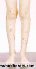
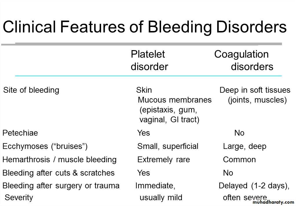
BleedingHistory• Site of bleed• Duration of bleed• Precipitating causes, includingprevious surgery or trauma• Family history• Drug history• Age at presentation• Other medical conditions, e.g.liver diseaseExaminationThere are two main patterns of bleeding:1. Mucosal bleedingReduced number or function of platelets (e.g.bone marrow failure or aspirin) or von Willebrandfactor (e.g.von Willebrand disease)Skin: petechiae, bruisesGum and mucous membrane bleedingFundal haemorrhagePost-surgical bleeding2. Coagulation factor deficiency(e.g. haemophilia or warfarin)Bleeding into joints (haemarthrosis) or musclesBleeding into soft tissuesRetroperitoneal haemorrhageIntracranial haemorrhagePost-surgical bleedingVessel wall abnormalitiescongenital, such as hereditary haemorrhagic telangiectasia• acquired, as in a vasculitis or scurvyHereditary haemorrhagic telangiectasia(HHT)Autosomal dominant .Telangiectasia and small aneurysms are found on the fingertips, face and tongue,and in the nasal passages,lung and gastrointestinal tract.Pulmonary arteriovenous malformations (PAVMs) that cause arterial hypoxaemiadue to a right-to-left shunt. These predispose to paradoxical embolism, resultingin stroke or cerebral abscess. =All patients with HHT should be screened for PAVMs; if these are found, ablationby percutaneous embolisation should be considered.Recurrent bleeds, particularly epistaxis, or with iron deficiency due to occult •gastrointestinal bleeding.
TREATMENT •1-Iron replacement for IDA2-Local cautery or laser therapy may prevent single lesions from bleeding •
Platelet function disorders Primary Hemostasis Platelet Plug formation Dependent on normal platelet number & function Initial manifestation of clot formation1-Thrombocytopenia2-Thrombasthenia
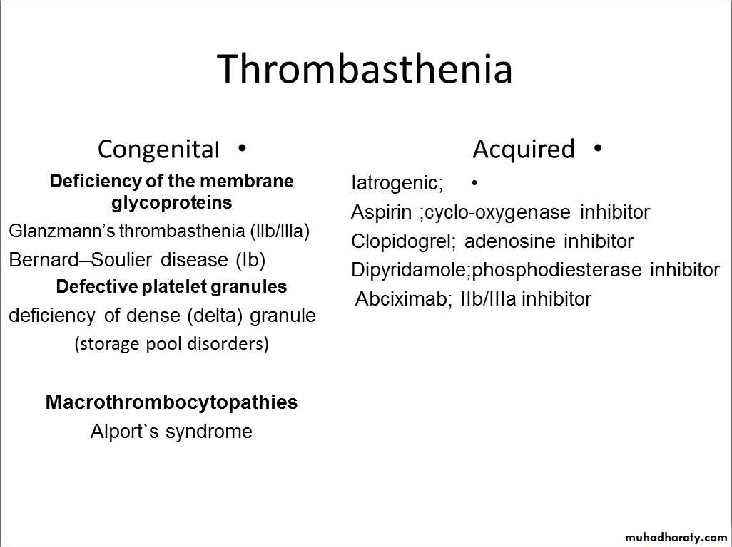
Laboratory Tests for PrimaryHemostasis Function Platelet count Bleeding time Platelet Aggregation Studies clot retraction Flow cytometric studies for Glycoproteins
Glanzmann thrombasthenia Background: Thrombasthenia was first describe in 1918 by Glanzmann when he noted purpuric bleeding in patients with normalplatelet counts Typically, thrombasthenia is diagnosed at an early age Pathophysiology: Autosomal recessive trait The production and assembly of the platelet membraneglycoprotein IIb-IIIa is altered, preventing the aggregationof platelets and subsequent clot formation Treatment 1-local measure.2-antifibrinolytic agent such as tranexamic acid.3-platelet transfusion.4-Recombinant factor VII
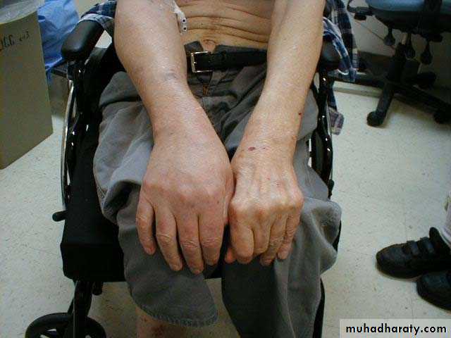
Idiopathic thrombocytopenic purpura Autoantibodies, most often directed against the plateletmembrane glycoprotein IIb/IIIa, which sensitise the platelet,resulting in premature removal from the circulation by cells of thereticulo-endothelial system. 1-Isolated condition.2-Association with connective tissue diseases,HIV infection,B cell malignancies, pregnancy and certain drug therapies.Clinical featuresPurpura andhaematomas
Mucosal bleeding
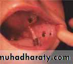
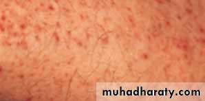
Classification of ITP disease phasesITP phase DefinitionNewly diagnosed Within 3 months of diagnosisPersistent 3 to 12 months from diagnosisChronic > 12 months from diagnosisIn adults, ITP usually has an insidious onset, with no precedingillness.Nearly one-quarter of patients present asymptomatically andreceive a diagnosis of ITP through incidental routine bloodtestsPetechiae or purpura • Unusual or easy bruising (haematoma)•Persistent bleeding symptoms from cuts or other injuries• Mucosalbleeding• Frequent or heavy nose bleeds (epistaxis)• Haemorrhage from any site (usually gingival or menorrhagia inwomennPetechie
Recommended diagnostic approaches for ITP Patient history Family history Physical examination Complete blood count and reticulocyte count Peripheral blood smear Quantitative immunoglobulin level measurement* Bone marrow examination (in selected patients) Blood group (rhesus) Direct antiglobulin test Helicobacter pylori Human immunodeficiency virus (HIV) Hepatitis C virus (HCV)Bone marrow aspiration is indicated in older patients (particularlythose over 60 years of age to excludemyelodysplastic syndrome), in those with an atypical presentation(e.g. abnormalities observed onperipheral blood smear suggestive of other haematological disorders), in those with a poor response to first-linetherapy and in those being considered forsplenectomy..
Treatmentwhen to treat?
If a patient has two relapses,or primary refractory disease, splenectomy is considered.Splenectomy produces complete remission in about 70% of patients and improvement ina further 20–25%, so that,following splenectomy, only 5–10% of patients require further medical therapy.Second-line therapy with the thrombopoietin analogue romiplostim or the thrombopoietin receptor agonist eltrombopagRituximab, ciclosporin and tacrolimus should be consideredin cases where the approaches above are ineffective.
Coagulation disorders
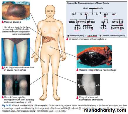
Haemophilia A the "ROYAL DISEASE"
Epidemiology:more than 400.000persons affectedAbout 1 in 10.000 people is born with heamphilia A
Clinical features
A prolonged aPTT: a normal aPTT does not exclude mild hemophilia A because the aPTT may not be sufficiently sensitive to detect slightly reduced levels of FVIII-C in the approximate 20-30% rangeNormal PT
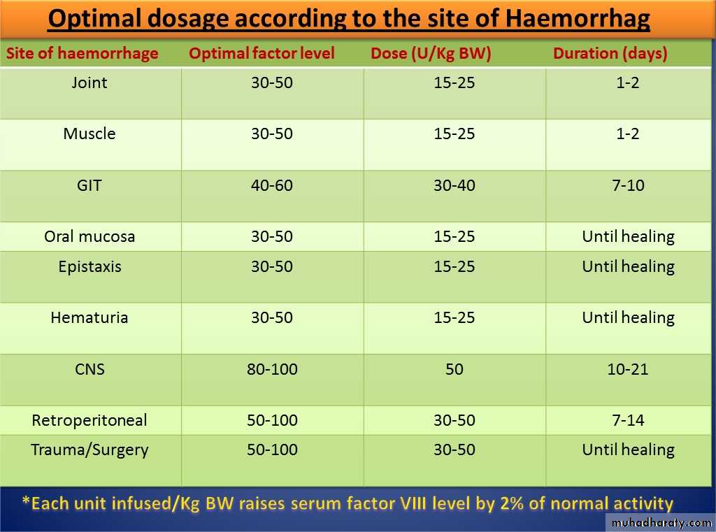
Treatment

Haemophilia B (Christmas disease)• Due to deficiency of factor IX .• X-linked• Clinical manifestation indistinguishable fromhaemophilia A• Treatment ; factor IX concentrate , indication anddosing same as to haemophilia A.• Complication of therapy similar to haemophilia Aregarding transmission of infection BUT theincidence of inhibitor is < 1%.Von Willebrand disease von Willebrand factor– Synthesis in endothelium and megakaryocytes– Forms large multimer– Carrier of factor VIII– Anchors platelets to subendothelium– Bridge between platelets
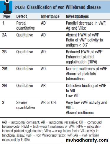
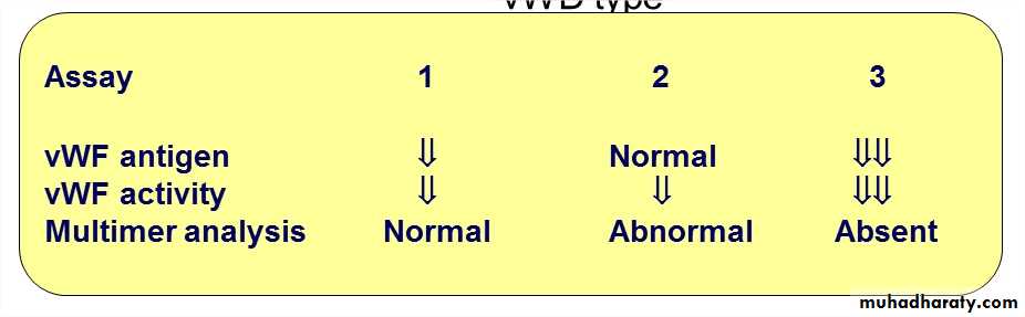
VWD VWD is the most common inherited bleeding disorder Males and females are affected equallyDeficiency of VWF results in defective platelet adhesion andcauses a secondary deficiency in factor VIII The result is that VWF deficiency can cause bleeding thatappears similar to that caused by platelet dysfunction orhaemophilia The most common symptoms include nosebleeds, skin bruises. Prolonged bleeding from trivial wounds, oral cavity bleeding,and excessive menstrual bleeding are common.Lab Studies:Screening tests typically include prothrombin time (PT) activated partial thromboplastin time (aPTT), FVIII level ristocetin cofactor (RCoF) activity vWF antigen (vWF:Ag).Laboratory evaluation of von Willebrand disease Classification– Type 1 Partial quantitative deficiency– Type 2 Qualitative deficiency– Type 3 Total quantitative deficiency*Bleeding time ↑*aPTT ↑vWD type
Treatment of von Willebrand Disease Cryoprecipitate– Source of fibrinogen, factor VIII and VWF– Only plasma fraction that consistently contains VWF multimers DDAVP (des -demino-8-arginine vasopressin)– plasma VWF levels by stimulating secretion from endothelium– Duration of response is variable– Not generally used in type 2 disease– Dosage 0.3 µg/kg q 12 hr IV Factor VIII concentrate (Intermediate purity)– Virally inactivated product– The treatment of choice for patients with vWD type IIIis virus-inactivated, vWF-containing FVIII concentrates
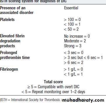
Disseminated intravascular coagulation (DIC)DIC is a clinicopathologic syndrome in whichwidespread intravascular coagulation is inducedby procoagulant that are introduce or produce incirculation and overcome the natural anticoagulantmechanisms.DIC may cause tissue ischemia from occlusivemicrothrombi as well as bleeding from bothconsumption of platelet and coagulation factor andanticoagulation effect of product of secondaryfibrinolysis
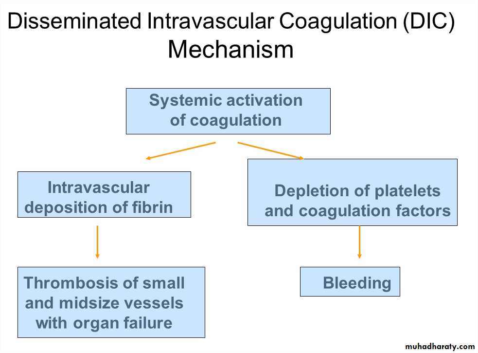
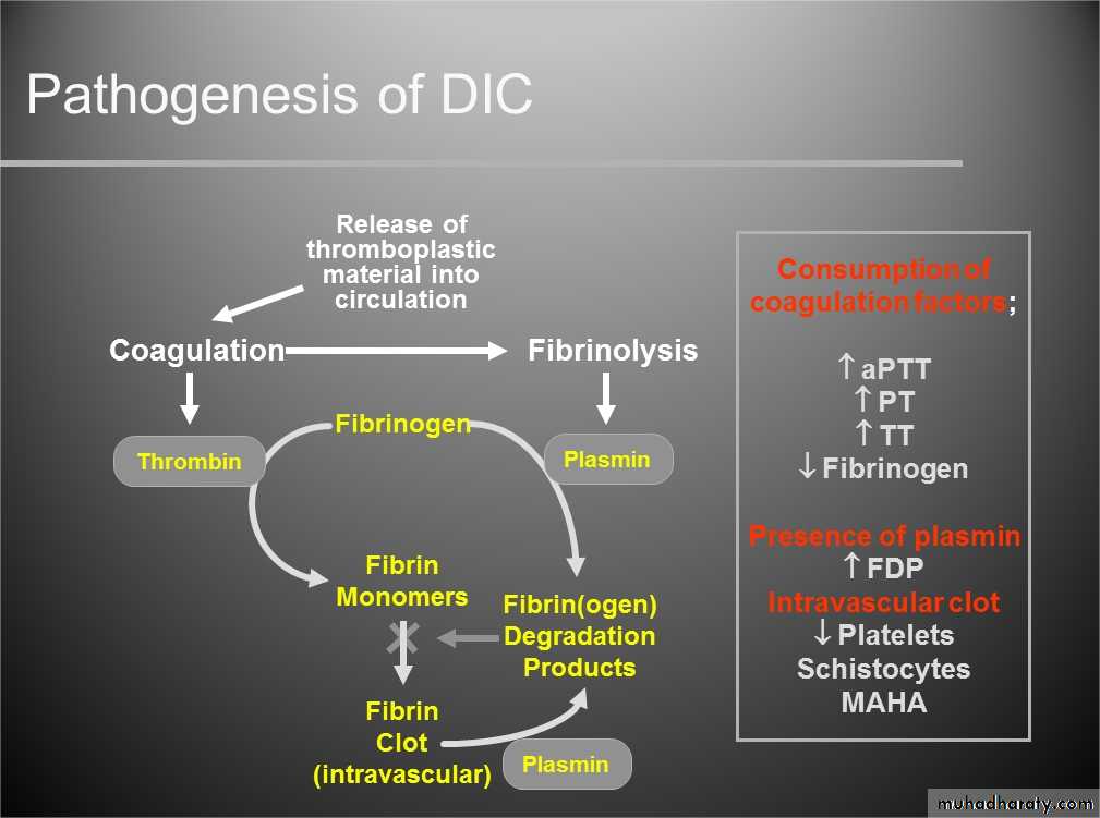
DIC
Treatment Treatment of underlying disorder Platelet transfusion (6-10 U plat (ideally rise to morethan 50000-100000 Fresh frozen plasma;1-2 unit For coagulation factor depletion Hypofibrinogenaemia; 8-10 U cryopercipitate Anticoagulation with heparin; unless there is a clearcontraindication Coagulation inhibitor concentrate (ATIII) Patients with DIC should not be treated with antifibrinolytictherapy, e.g.tranexamic acid.
Thrombotic thrombocytopenic purpura(TTP)thrombosis is accompanied by paradoxicalthrombocytopenia, TTP is characterised by a pentad of findings,although few patients have all five components:• thrombocytopenia• microangiopathic haemolytic anaemia(MAHA)• neurological sequelae• fever• renal impairmentTTP-Cont. It is an acute autoimmune disorder mediated by antibodiesagainst ADAMTS-13 (a disintegrin and metalloproteinase witha thrombospondin type-1 motif).It is a rare disorder (1 in 750 000 per annum), which may occuralone or in association with drugs (ticlopidine, ciclosporin), HIV,shiga toxins and malignancy.It should be treated by emergency plasma exchange.Corticosteroids, aspirin and rituximab also have a role inmanagement Untreated mortality rates are 90% in the first 10 days, andeven with appropriate therapy, the mortality rate is 20–30% at6 months
THROMBOTIC DISORDERSVirchow’s Triad Pathogenesis of a ThrombusEndothelial injuryAbnormal blood flowHypercoagulability Genetic acquired
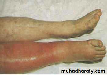
Signs & Symptoms DVT: 50% with no clinical signs ?Edematous extremity Plethoric,Warm,Painful extremity PE: Cough, SOB, Hemoptysis Tachycardia
Thrombophilia Physiologic Inhibitors ofcoagulation Antithrombin Activated Protein C + protein S InactivatesVa andVIIIa (viaproteolysis) Thrombomodulin Binds to thrombin activate Protein C Hereditary Thrombophilias Protein C pathway Factor V Leiden Protein C deficiency Protein S deficiency Prothrombin G20210Amutation Antithrombin deficiency Hyperhomocystinemia C677T MTHFR mutation
Hereditary Thrombophilias None of them is strongly associated with arterial thrombosis. • All are associated with a slightly increased incidence ofadverse outcome of pregnancy,including recurrent early fetalloss, but there are no data to indicate that any specificintervention changes that outcome. • Apart from in antithrombin deficiency and homozygous factorV Leiden, most carriers of these genes will never have anepisode of VTE; if they do, it will be associated with thepresence of an additional temporary risk factor. • There is little evidence that detection of these abnormalitiespredicts recurrence of VTE. • None of these conditions per se requires treatment withanticoagulants
Antiphospholipid Antibody Syndrome Autoimmune Acquired Prothrombotic Disorder Very High Risk for recurrent thromboembolic disease both venous and arterial Indefinite duration anticoagulation recommended +/-immunosuppression Strict Diagnostic Criteria
Clinical criteria (≥1 must be present):1. Vascular thrombosis:- ≥ 1clinical episode of, objectively confirmed, arterial, venous, or smallvessel thrombosis2. Pregnancy morbidity:- ≥ 1 unexplained fetal death @ ≥ 10 weeks EGA- ≥ 1 premature birth (≤ 34th week of gestation) due to eclampsia, severepre-eclampsia, or placental insufficiency- ≥ 3 unexplained consecutive spontaneous abortions @ <10 weeks EGA
Laboratory criteria (≥1 must be present): Lupus anticoagulant {LA} (+) ≥ 2 occasions, at least 12 weeksapart, according to ISTH guidelines: prolonged aPTT, lack of correction with 1:1 mix, and correction with Anticardiolipine antibody(ACLA) and/or anti-β2 glycoprotein-Iantibody: medium or high IgG and/or IgM isotype titer ≥ 2 occasions, at least 12weeks apart Standardized ELISA assays