
GIT slides
1
2
3
Description :-
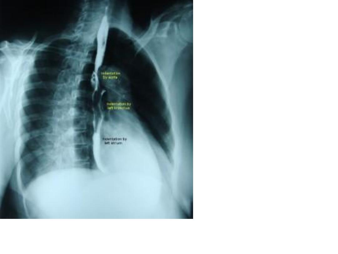
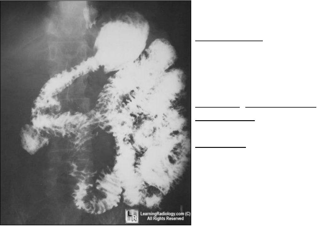
Ba swallow
visualizes the constrictions
areas “normallly”in
esophagus :-
1.at the level of the body of
cervical vertebrae (C2-C3)
2.arch of aorta
3.left atrium
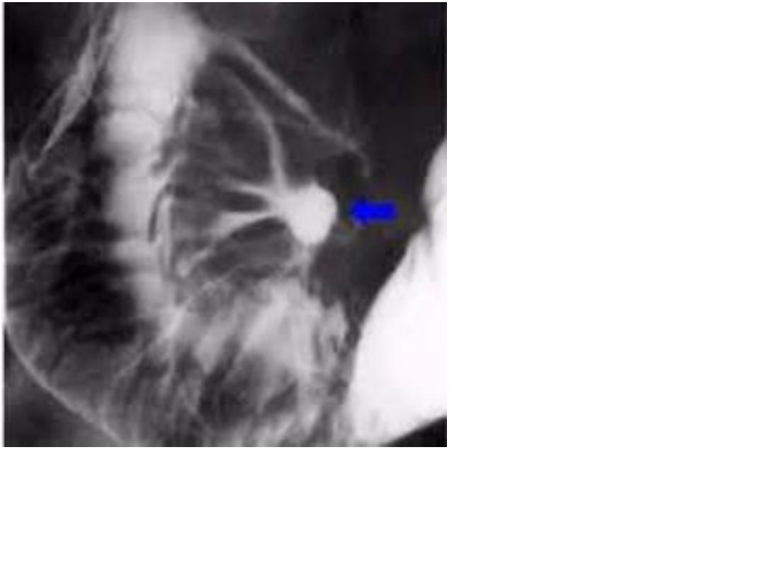
4.diaphramatic hiatus.
Note :-Esophagus
length=25cm
.

4
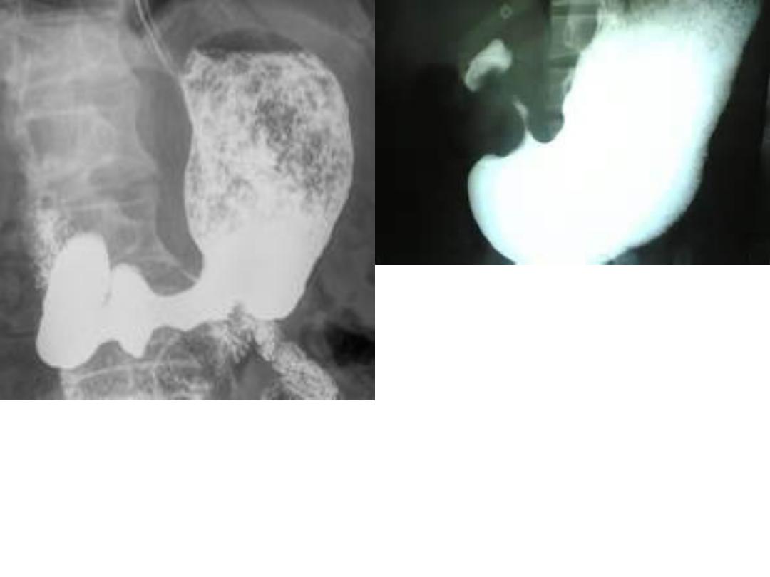
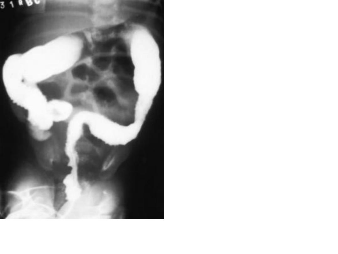
Description:-Ba meal (normal stomach
configuration)
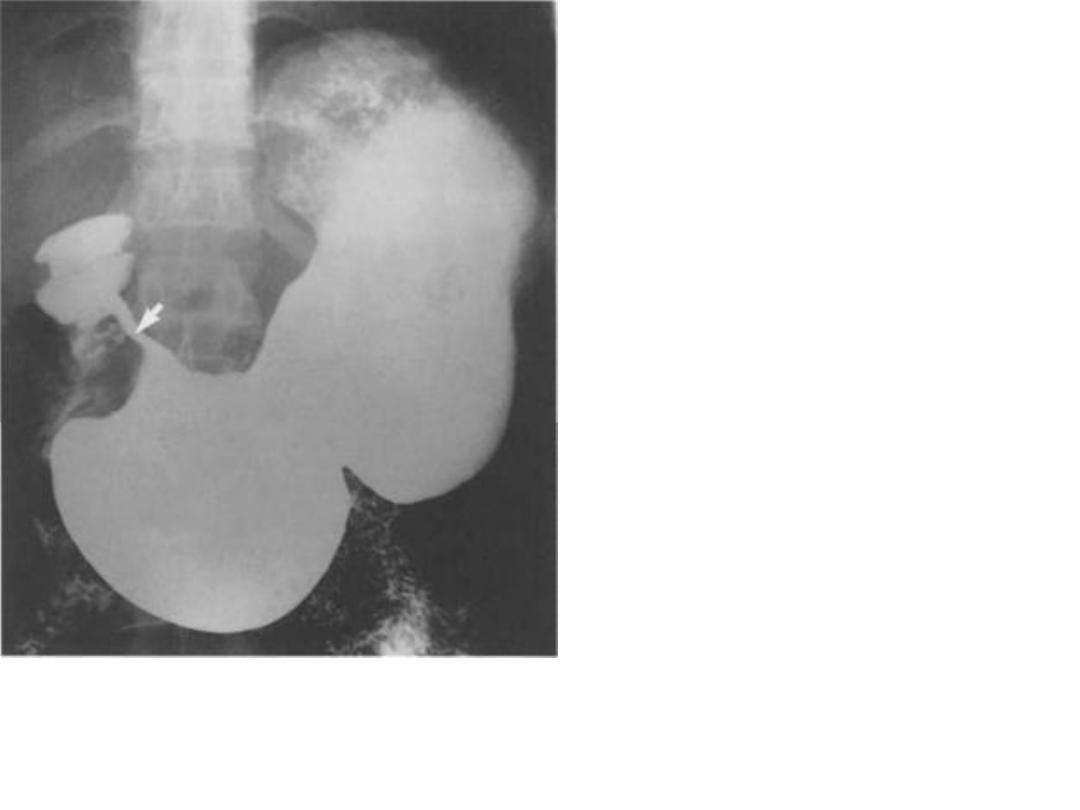
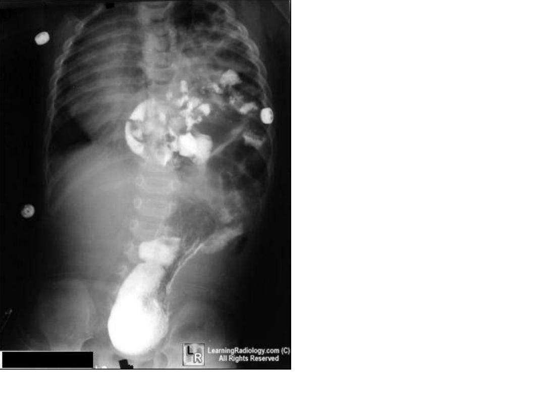
5
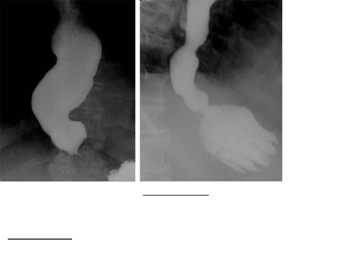
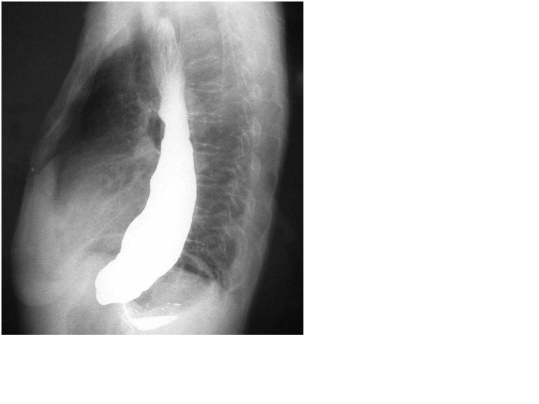
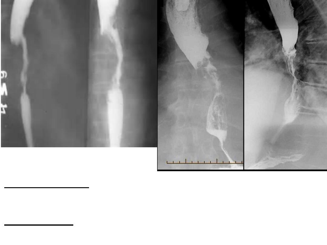
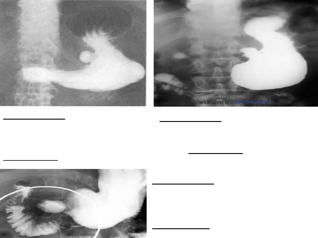
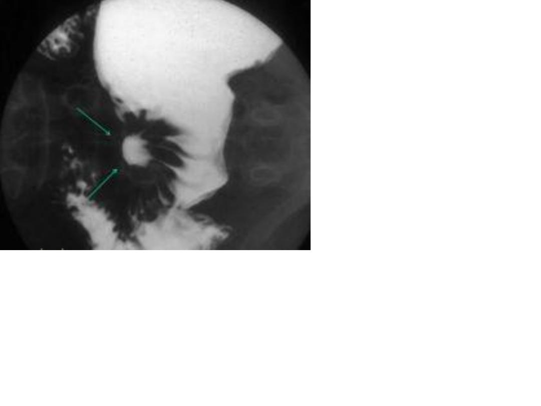
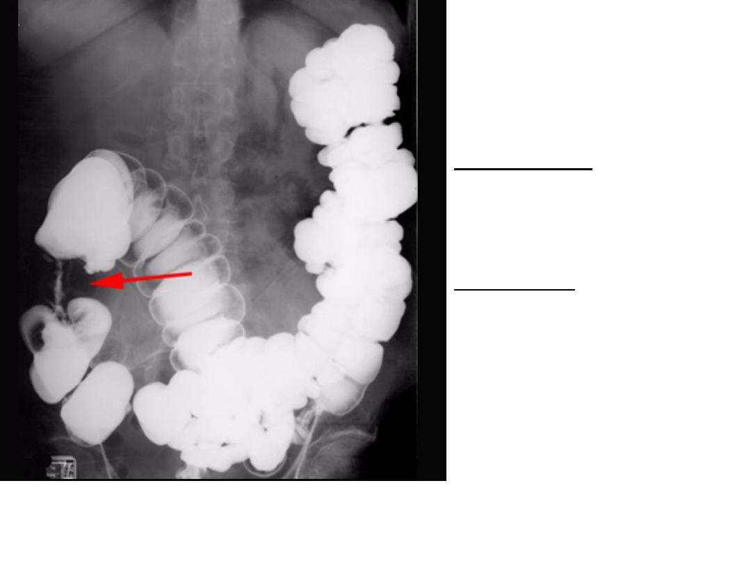
Description:-
Ba swallow shows prominent sac like dilatation of esophagus along with
reactionary peristalsis(3ary contraction).
Diagnosis :-
Achalasia cardia.
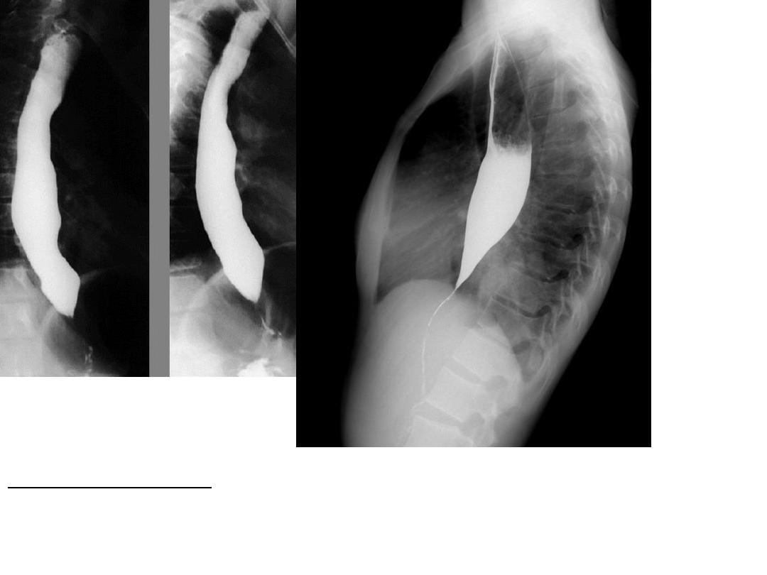
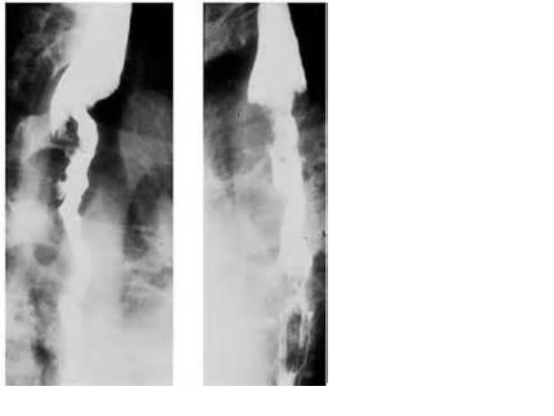
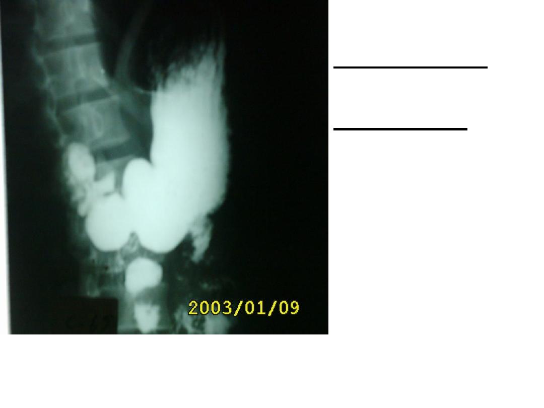
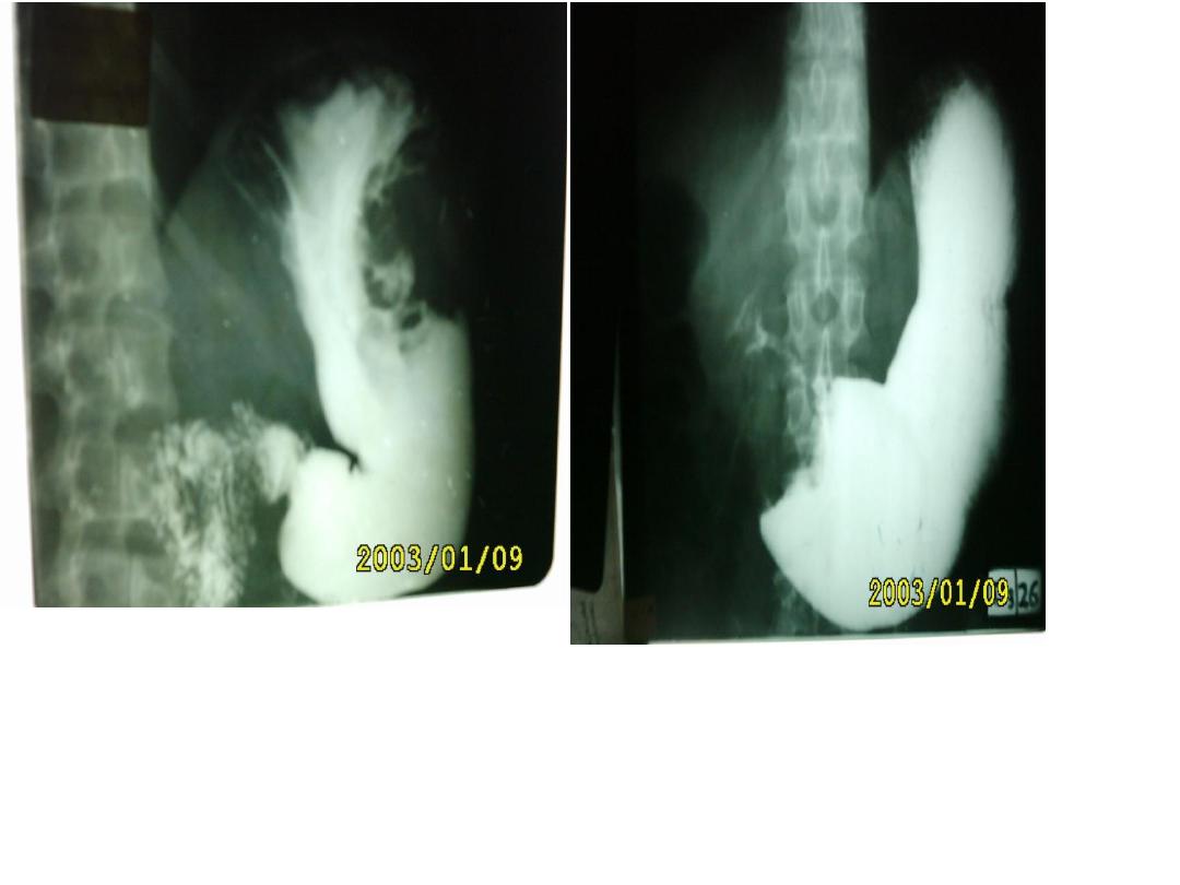
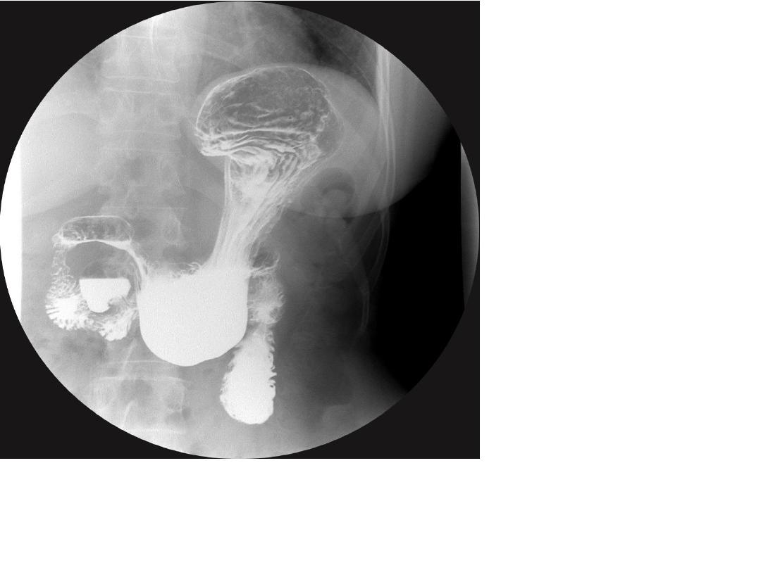
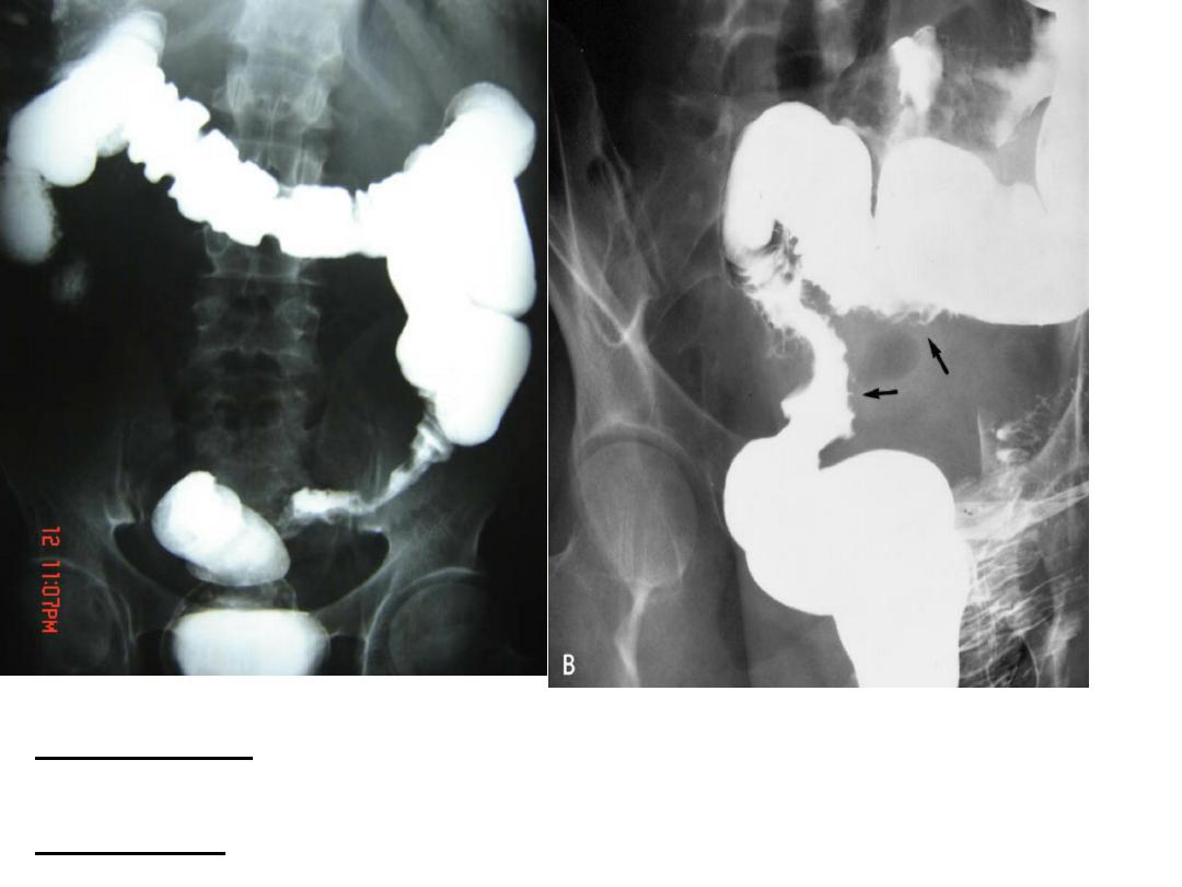
6
Barium swallow show dilatation of the esophagus with rat
tail narrowing of distal end .
Dx. Achalasia cardia
NB; differentiate it from rectum by presence of ribs .
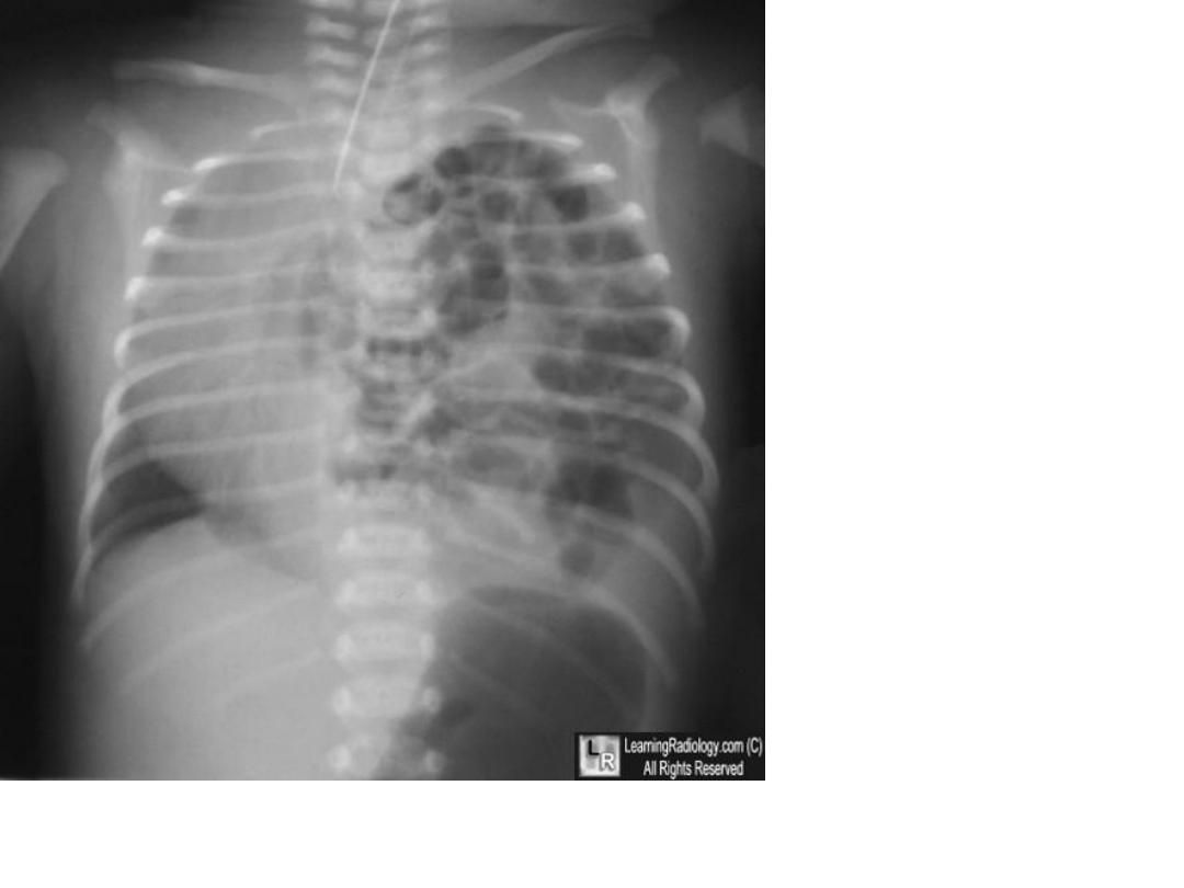
7
8
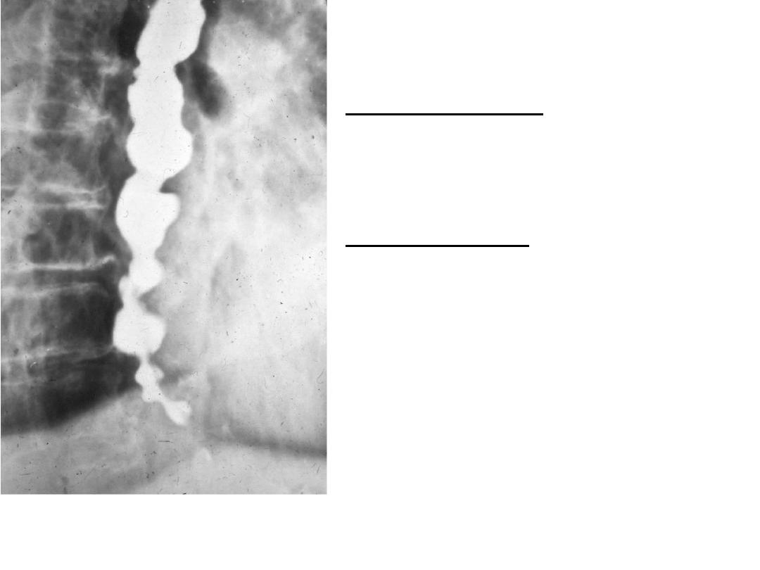
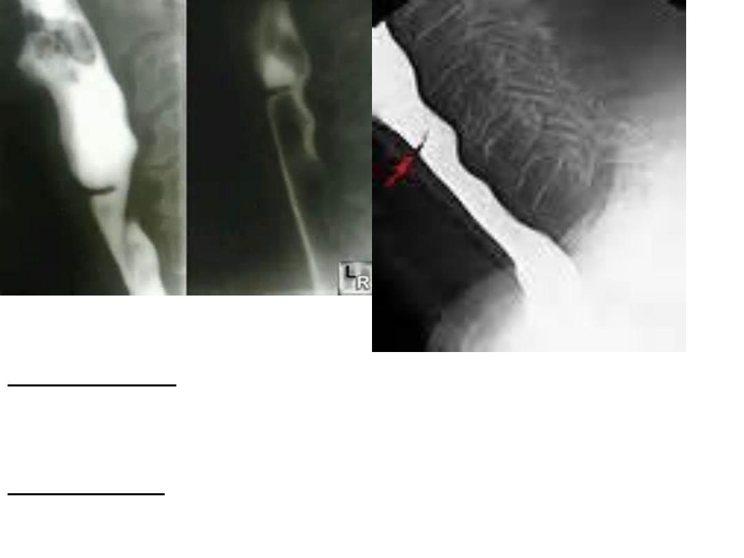
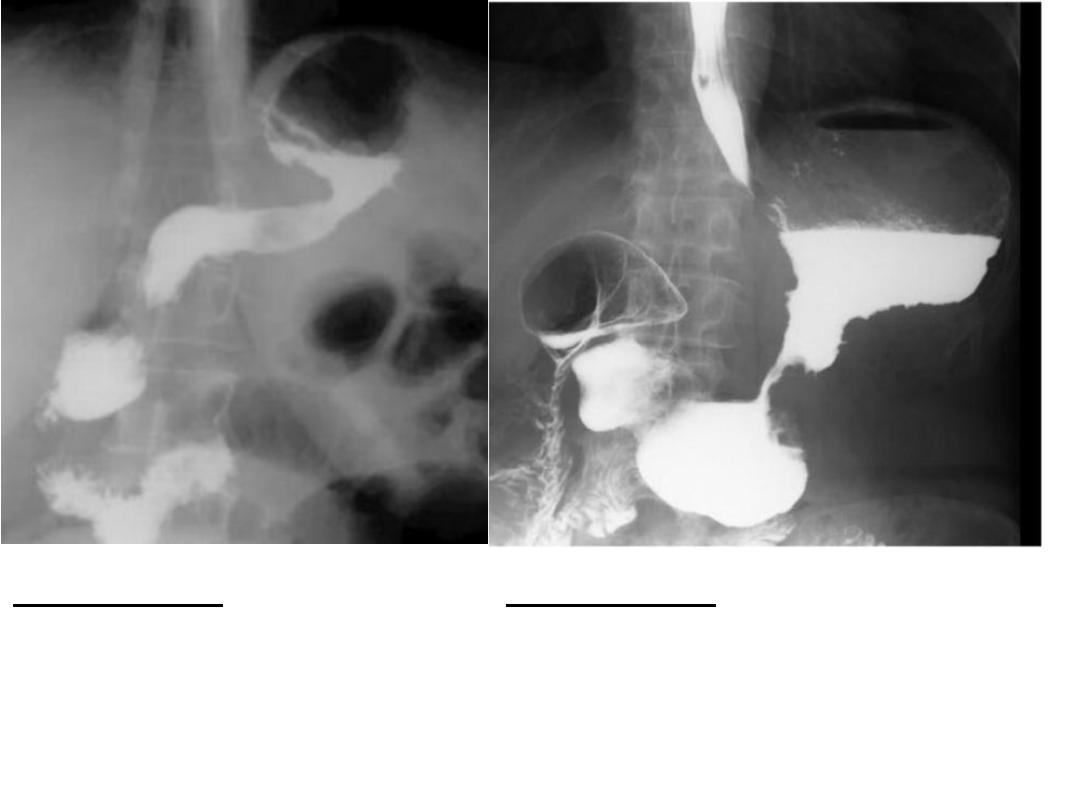
Description:-
Ba swallow
showing diffuse esophageal
spasm (tertiary peristalsis ) .
Diagnosis :-
cork screw
esophagus.
Note :- it is benign condition,pt
usually present with
dyspahgia.
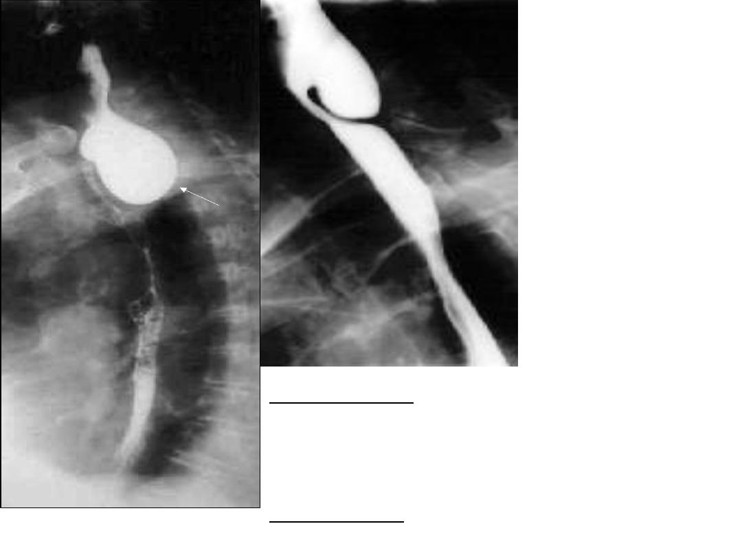
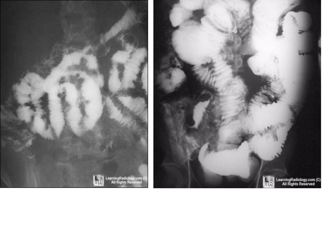
9
Description:-
Ba swallow
& meal ,showing regular
,smooth,well defined&long
segement of esophageal
stricture .
Diagnosis:-
corrosive
ingestion.
10
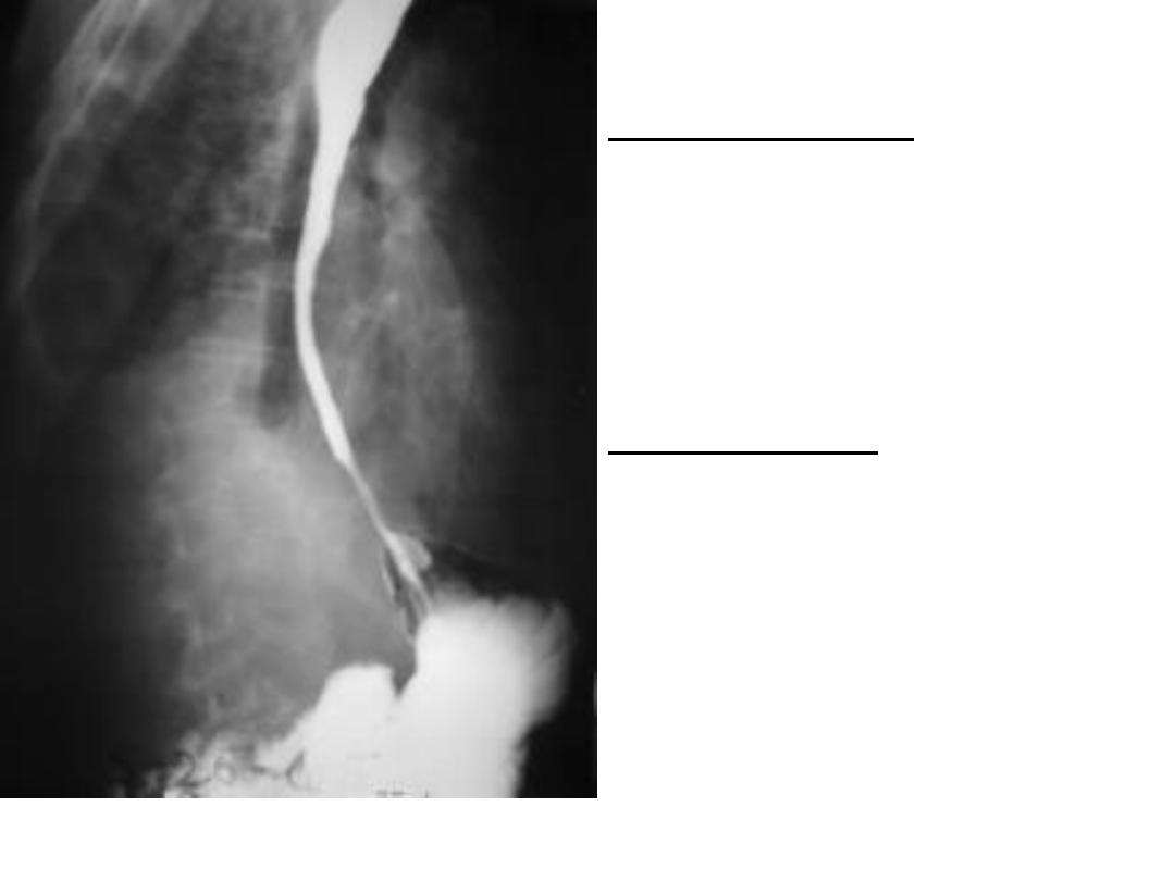
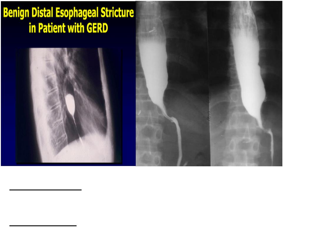
Description:-
Ba swallow showing short area of narrowing
of lower esophagus.
Diagnosis :-
GERD.
11
Description:-
Ba swallow showing : abrupt irregular
narrowing area of esophagus (shoulder sign)
Diagnosis :-
esophageal carcinoma(malignant stricture).
12
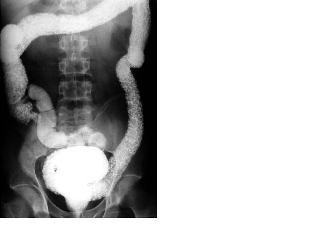
Same as
previuosly
13
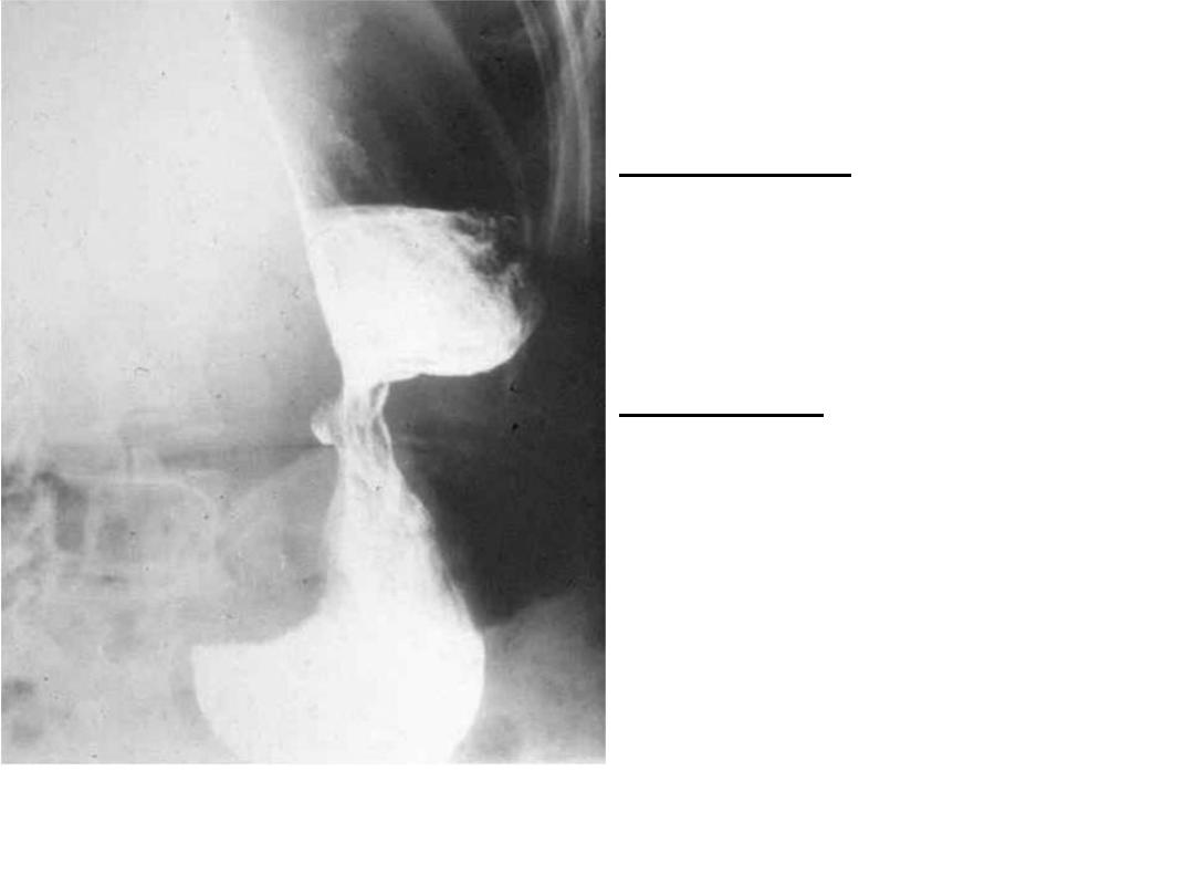
Description:-
Ba swallow showing abnormal mucosal
thickening ,shelf like fold in the cervical esophagus project from
anterior wall.
Diagnosis :-
esophageal web.
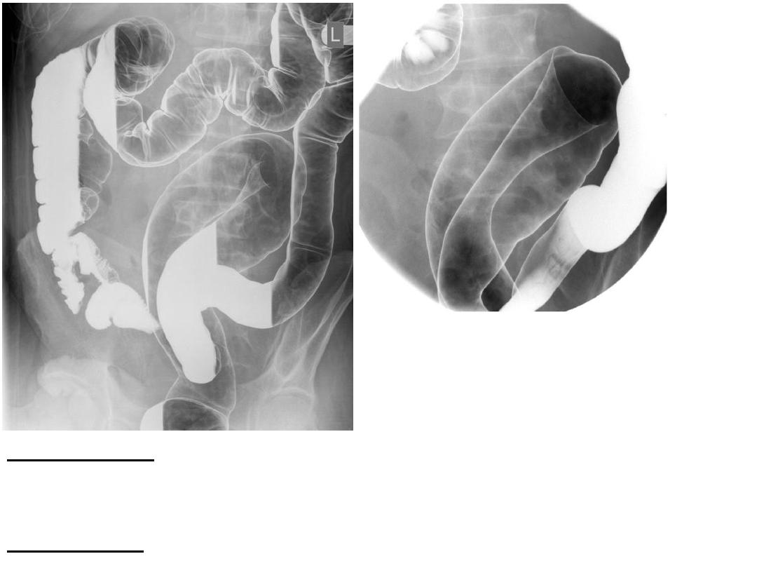
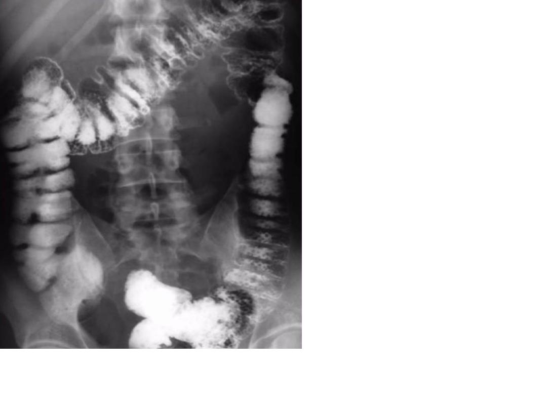
Note :-always esophageal web occurs in cervical esophagus as
incomplete shelf like projection that NOT occludes the lumen
.
14
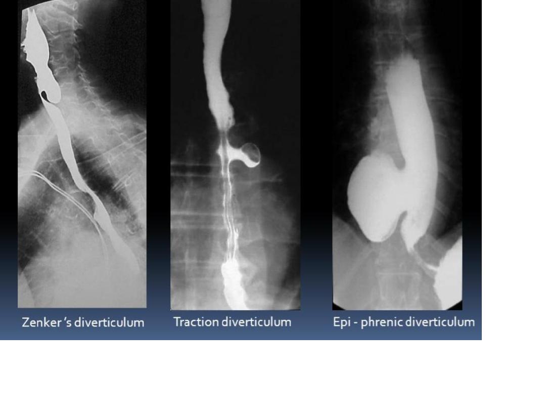
Description:-
Ba swallow showing
abnormal dilatation & outpouching of
cervical esophagus located posteriorly (just
anterior to spine).
Diagnosis :-
Zenker diverticulum.
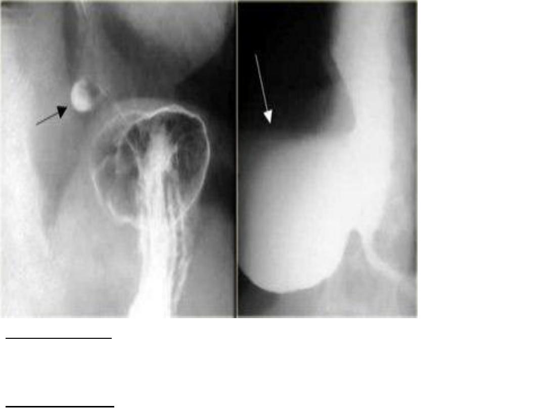
15
Description:-Ba swallow & meal showing abnormal
mucosal bulging & dilatation above the stomach.
Diagnosis :-
Epiphrenic diverticulum.
16
Same as previously
17
18
19
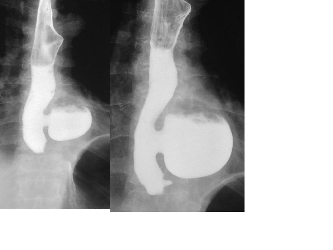
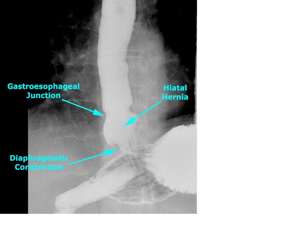
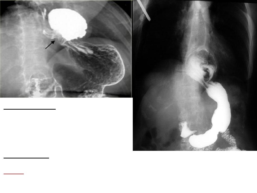
Description :-
Ba swallow & meal
,showing GEJ above the diaphragm
with pooling or accumulation of Barium
along with bulding of part of stomach
above the
diaphragm.
Diagnosis:-
. sliding hernia
Note :-sliding hernia is more common
than paraesophageal hernia.
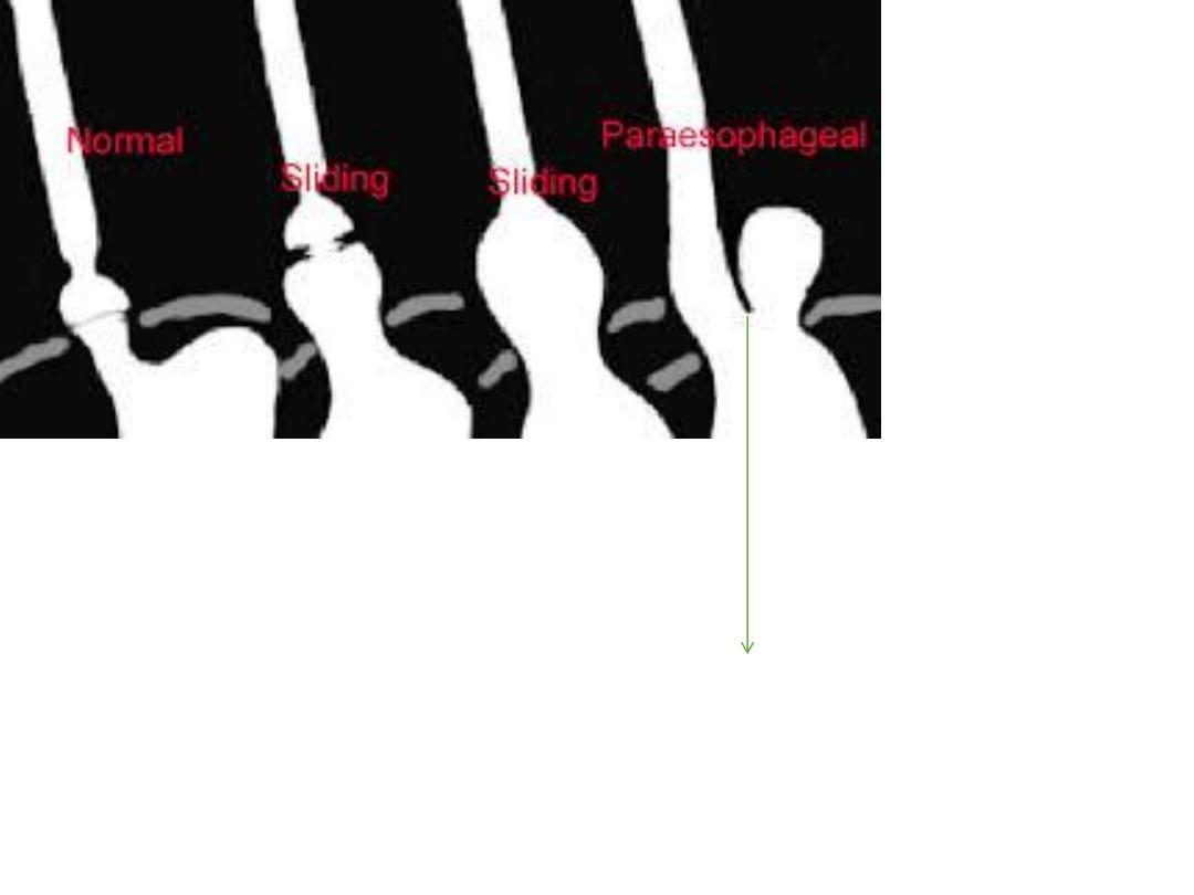
20
GEJ still below the diaphragm
21
Description:-
outpouching from
outside wall of stomach from lesser
curvature filled with Barium(profile
view)
Diagnosis:-
peptic ulcer
Description:-
Ba meal (profile
view),showing an outside pouching
of the lesser curvature of stomach
filled with barium.
Diagnosis:-
peptic ulcer.
Description:-
Ba meal showing area
of ulcer nitch or ulcer crater in pyloric
area of stomach.
Diagnosis :-
peptic ulcer.
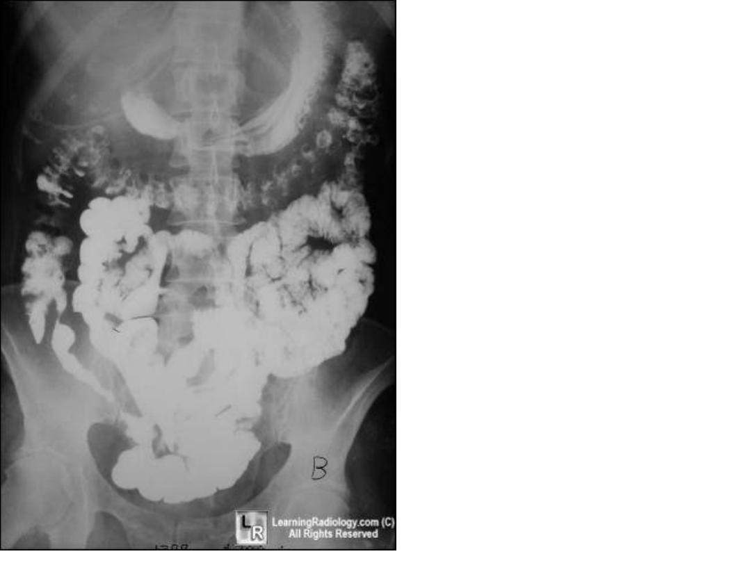
22
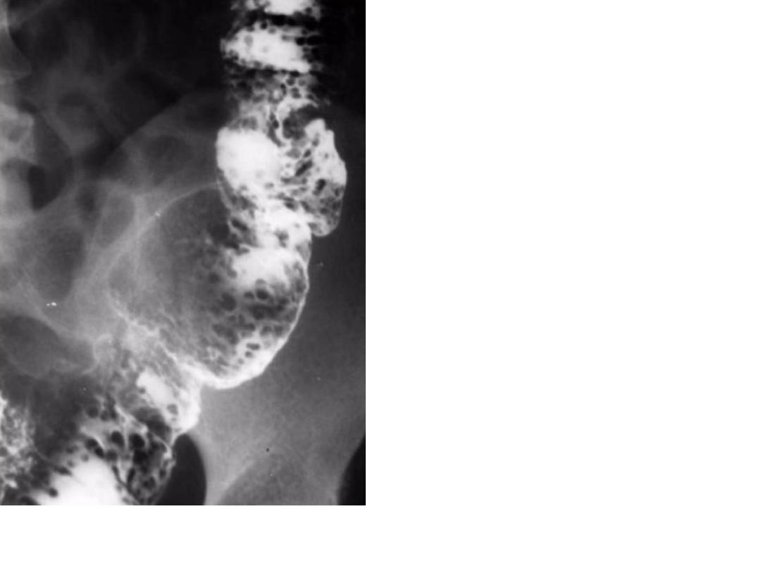
Description:-Ba meal showing
outpouching from outside
wall of stomach filled with
Barium , with constriction of
body of stomach.
Diagnosis:-peptic ulcer.
23
24
25
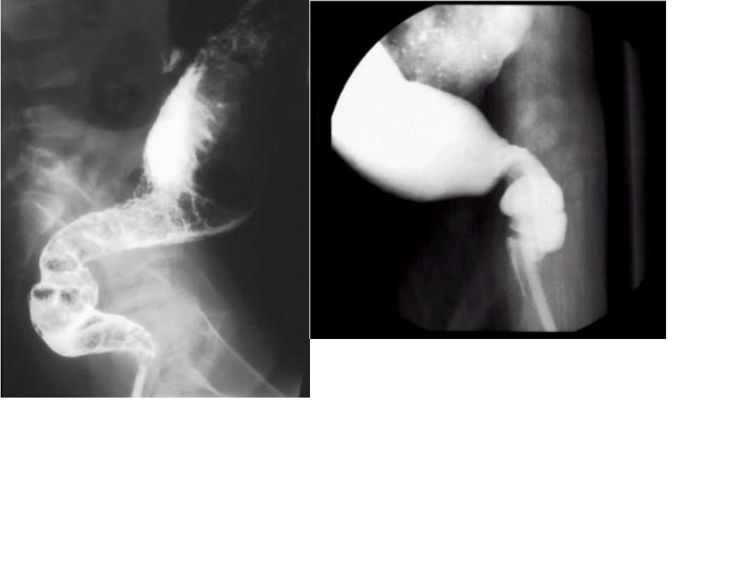
Description:-
tri foil deformity.
Diagnosis:-
chronic duodenal
ulcer.
26
Description:-
Ba meal show abnormal
configuration of stomach
Dx. Infiltrative carcinoma
(generalized )
Description:- Ba meal show
filling defect in the body of
the stomach.
dx. Infiltrative carcinoma (
localized )
•Ba meal show
narrowing of the distal
pyloric antrum with
shouldering sign .
•Dx . Infiltrative ca of
the stomach
27
28
29
30
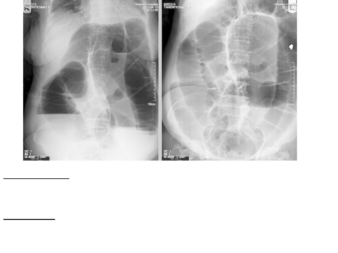
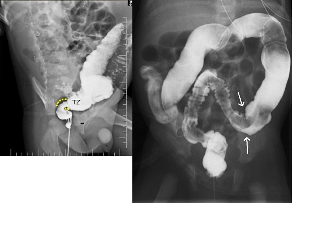
31
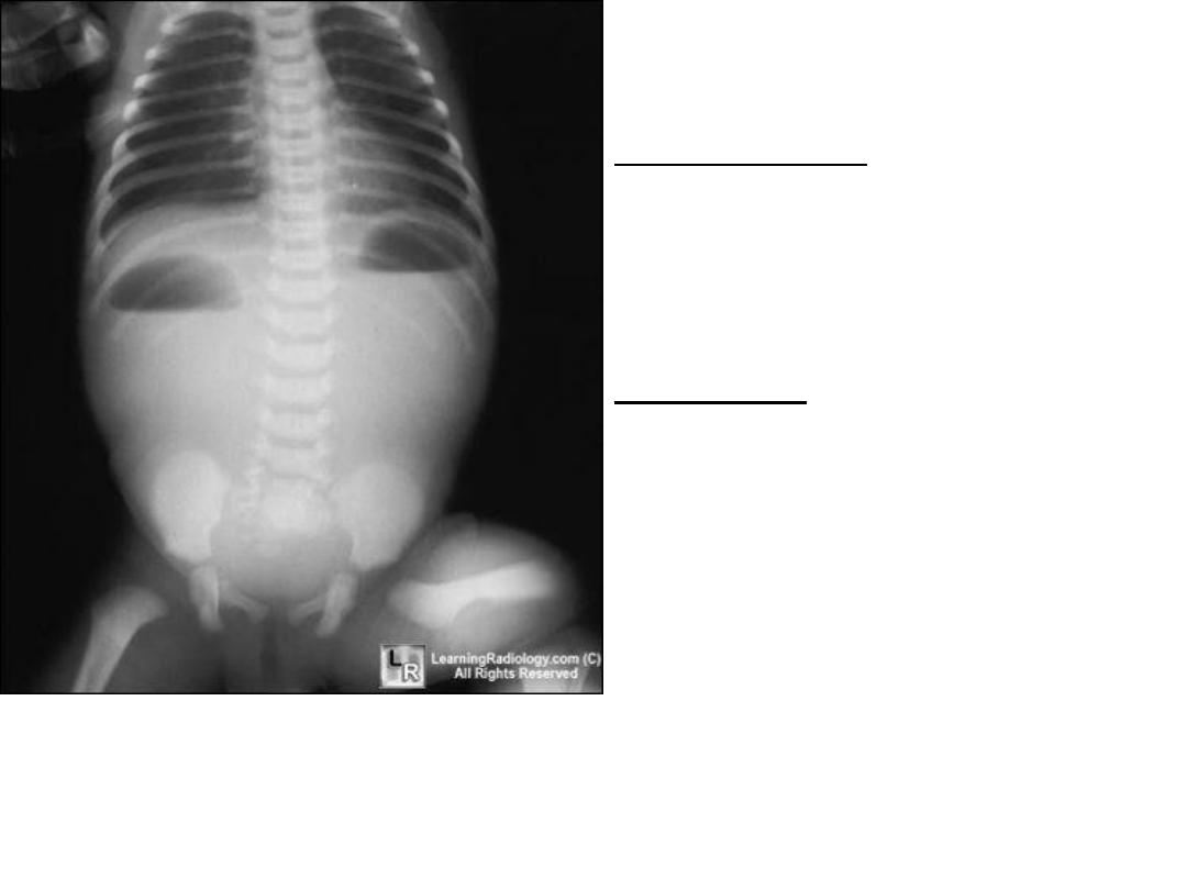
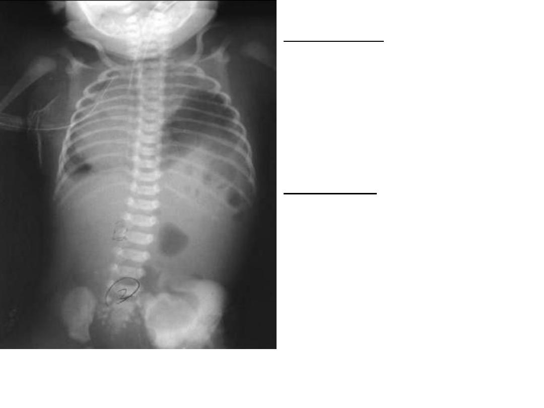
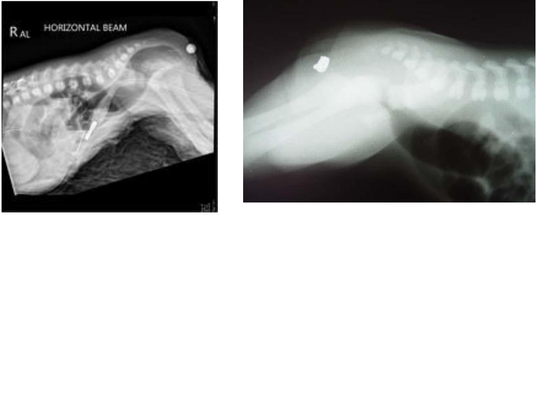
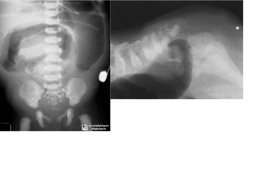
Description :- area of gas
collection in stomach &
duodenum full with gas
,double bubble sign .
Diagnosis :-duodenal atresia

The double bubble sign is seen in infants and
represents dilatation of the proximal
.It is seen in both
radiographs and ultrasound, and can be
identified antenatally
32
33
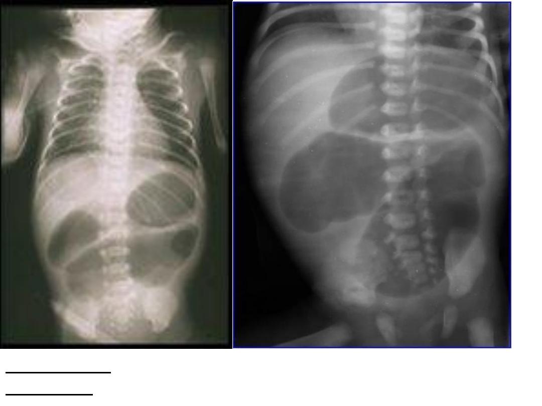
Description:-KUB study showing triple bubble sign .
Diagnosis :-jejunal atresia.
34
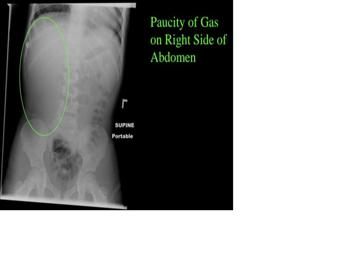
Description :-
abnormal gaseous
distribution throught the
hemi thorax with absent
normal configuration of
diaphragm.
Diagnosis :-
Bockdalek hernia
( diaphragmatic hernia )
35
Description :- gaseous distension in the bowel
(bowel in the chest).
Diagnosis :-diaphragmatic hernia
36
Same as previously
37
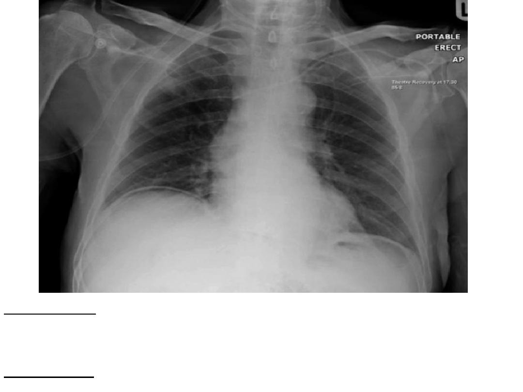
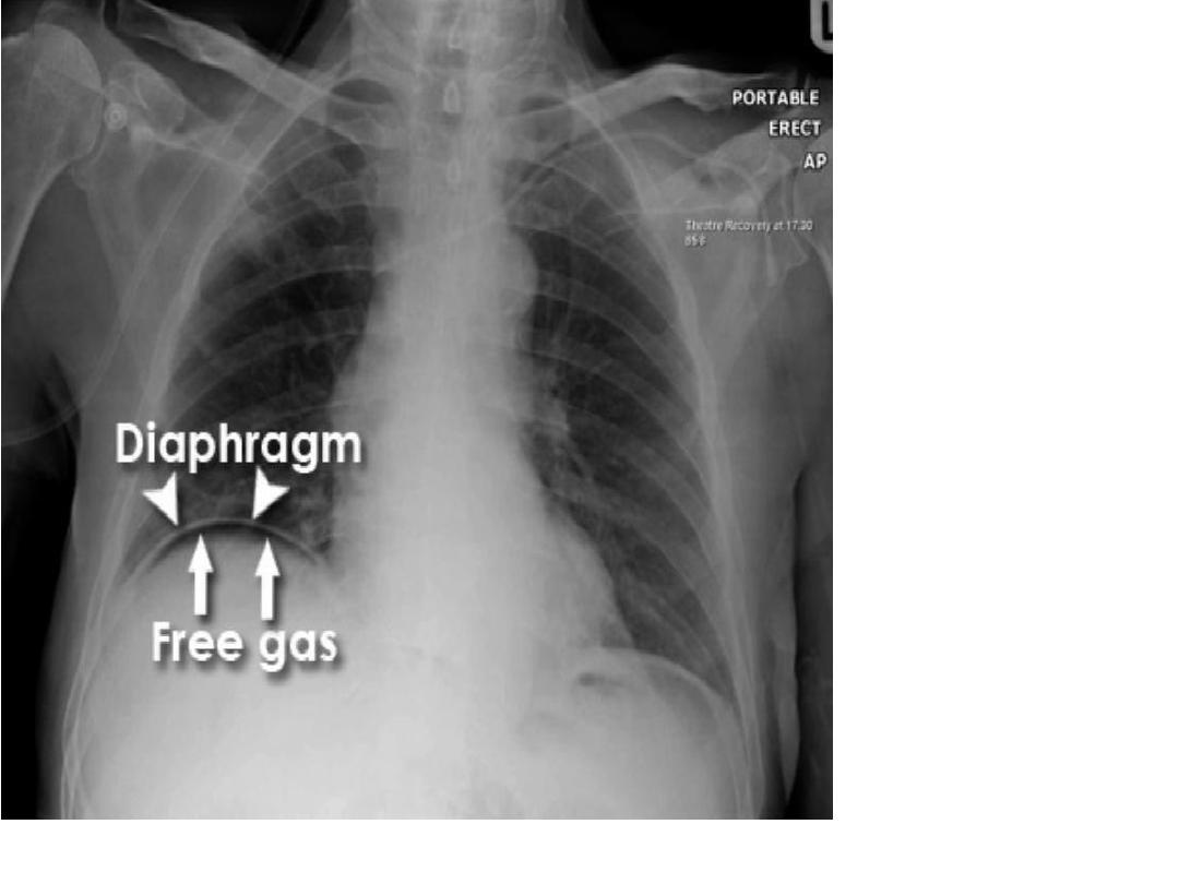
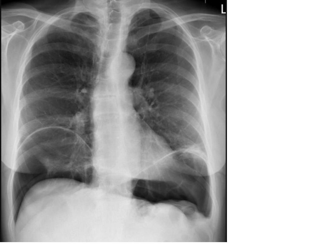
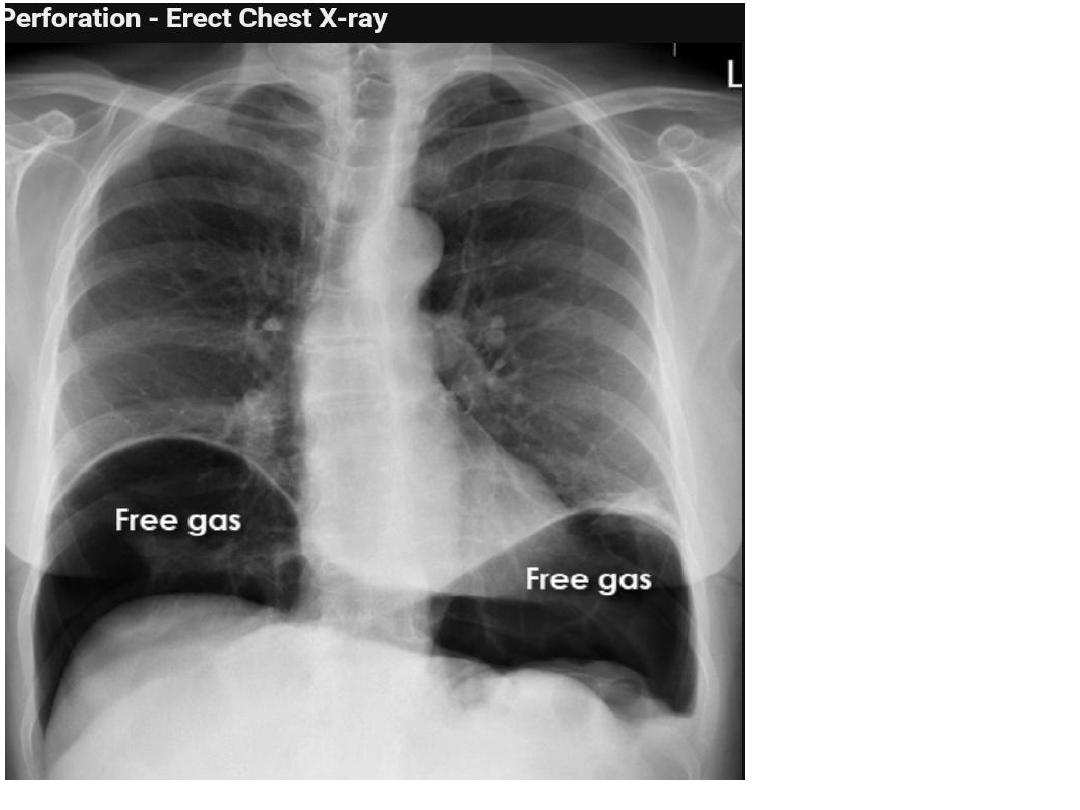
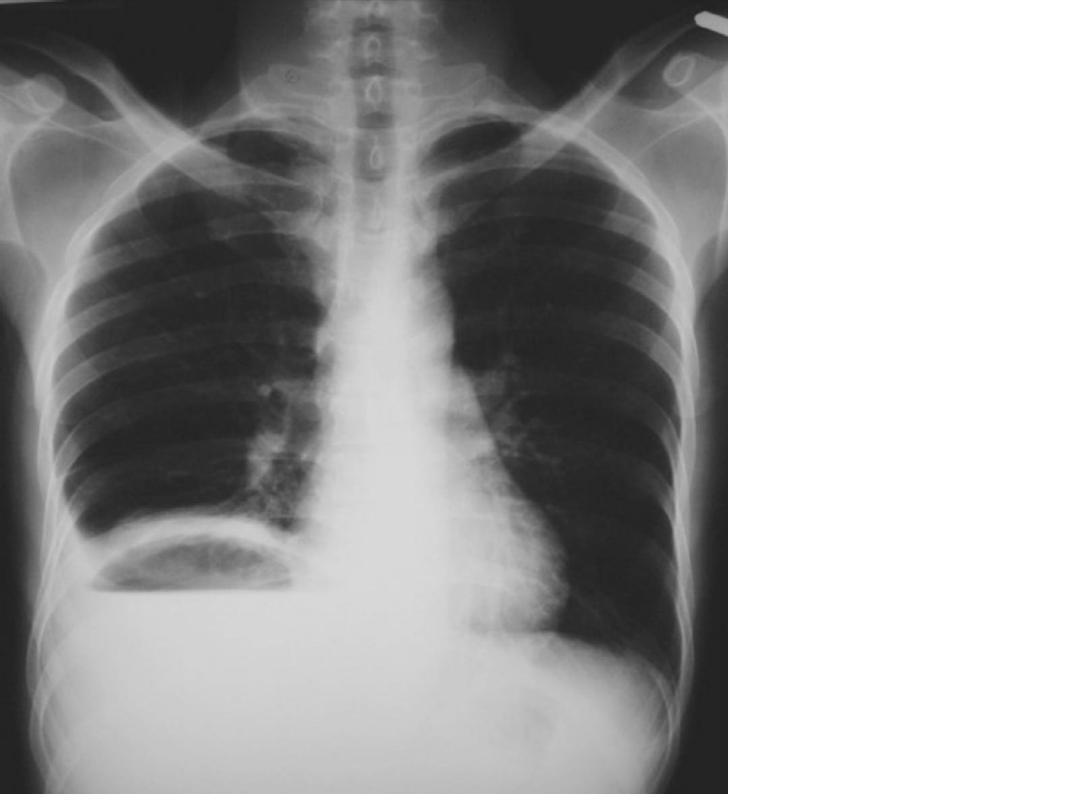
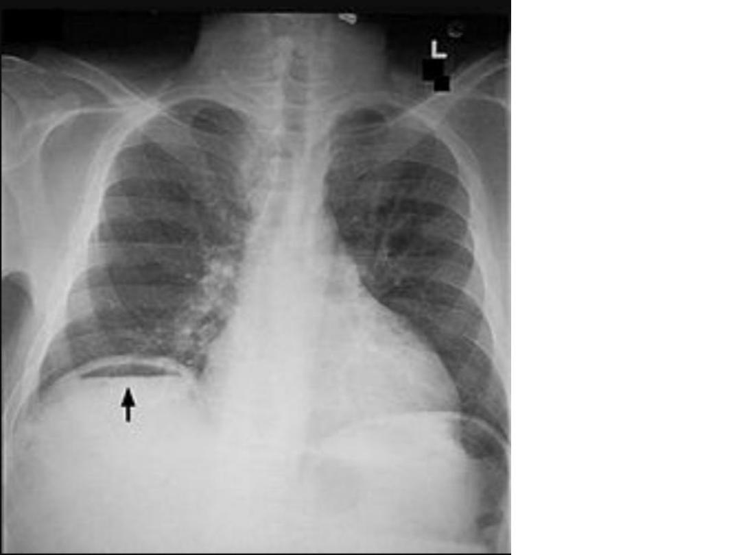
38
Description:-
chest x-ray showing free gas (area of crescent shape
lucency)under right side the diaphragm .
Diagnosis:- pneumo peritoneum ( perforated viscus )
39
40
41
CXR:
showing
dome shape
lucency
below the
diaphragem
Dx.
subphrenic
abscess
42
43
44
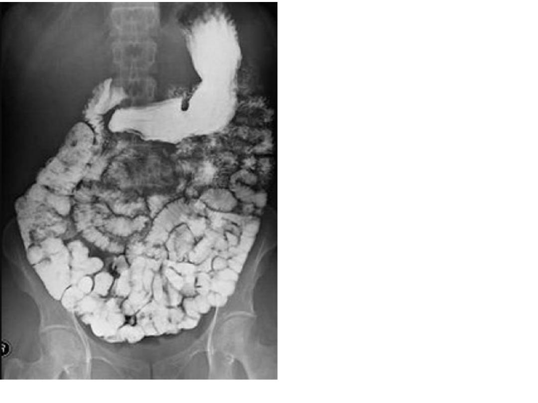
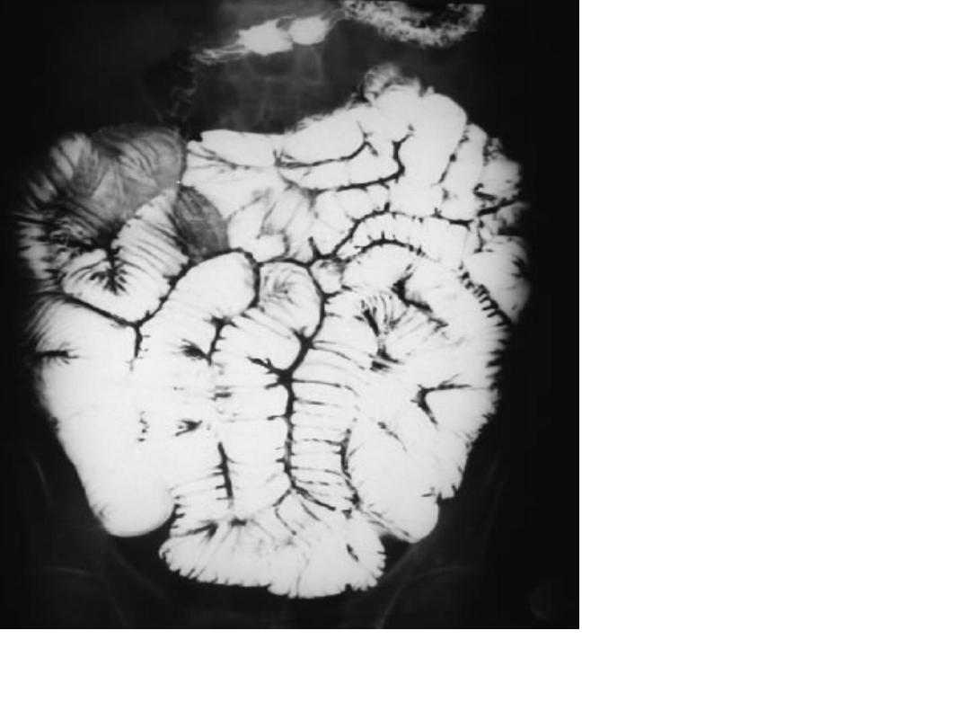
Ba follow through
showing Normal
feathery
appearance of
small bowel
45
46
47
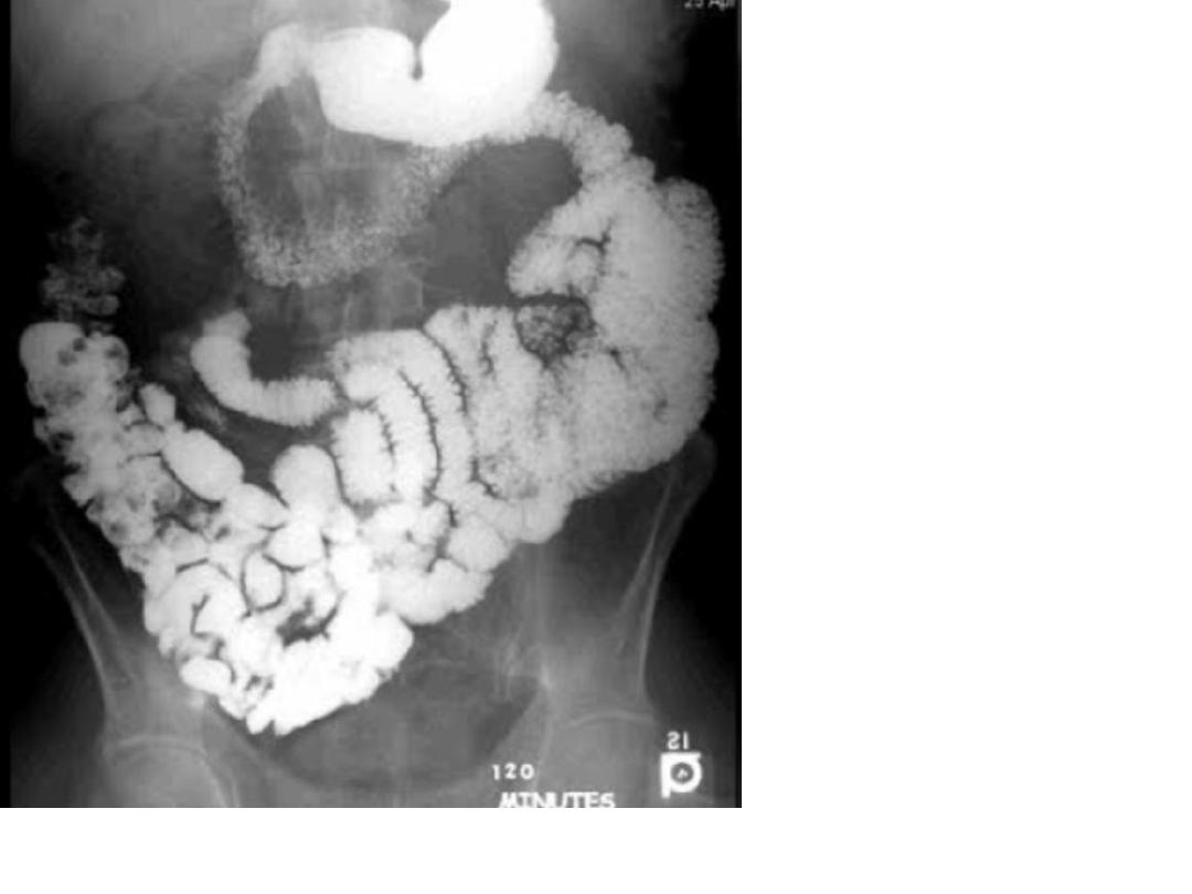
Ba follow through showing loss of normal bowel feathery
appearance flocculation and segmentation of barium and
dilation of intestinal loops.
Dx . Malabsorption syndrome
48
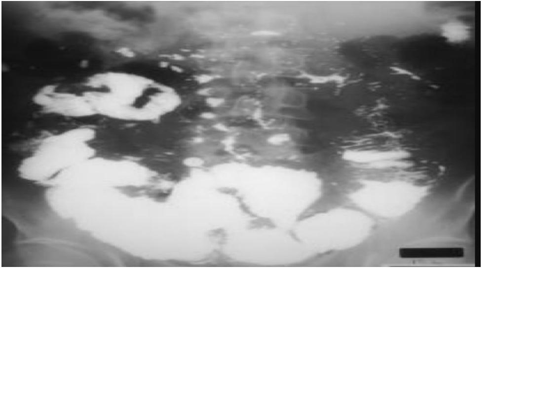
Description:-:Ba follow
through showing loss of
normal configuration of
small bowel (feathery
appearance) with
splaying ,segmentation &
flocculation of bowel
loops.
Diagnosis:- lymphoma
note :-in lymphoma
,there will be thick
valvulae conviennentes
,nodular shaped .
49
50
51
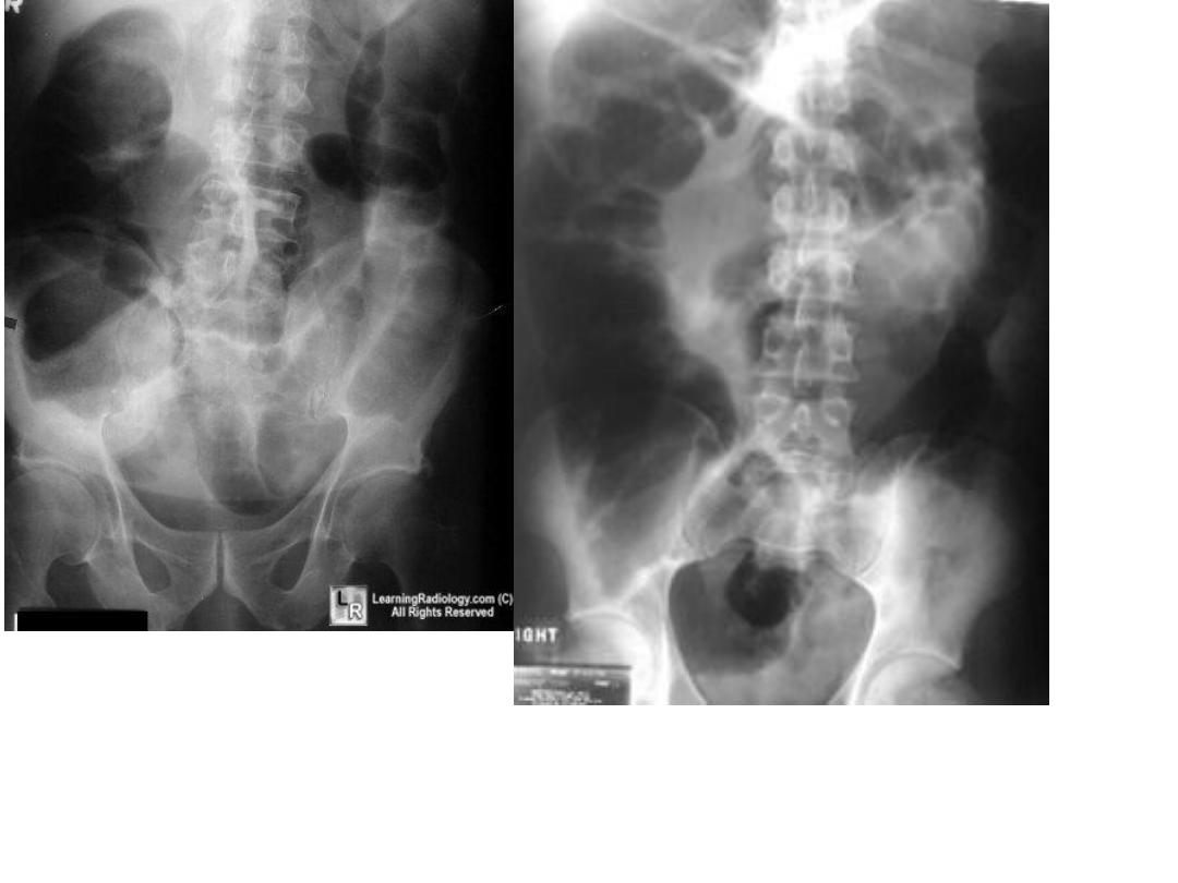
52
Description:-Ba enema shows loss of normal haustral
markings in large bowel (lead pipe colon) in rectosigmoid
area.
Diagnosis :-chronic UC
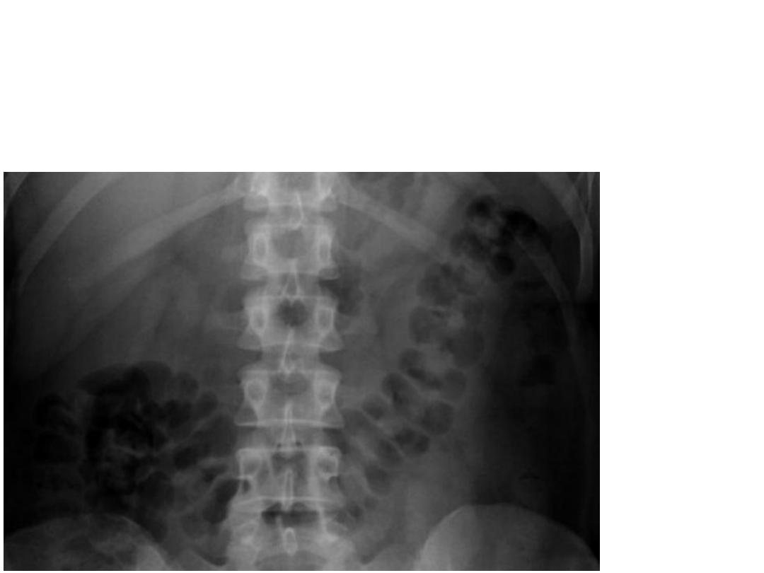
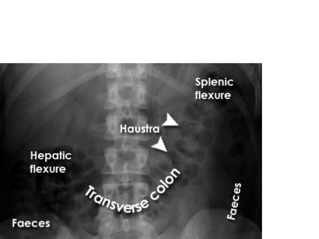
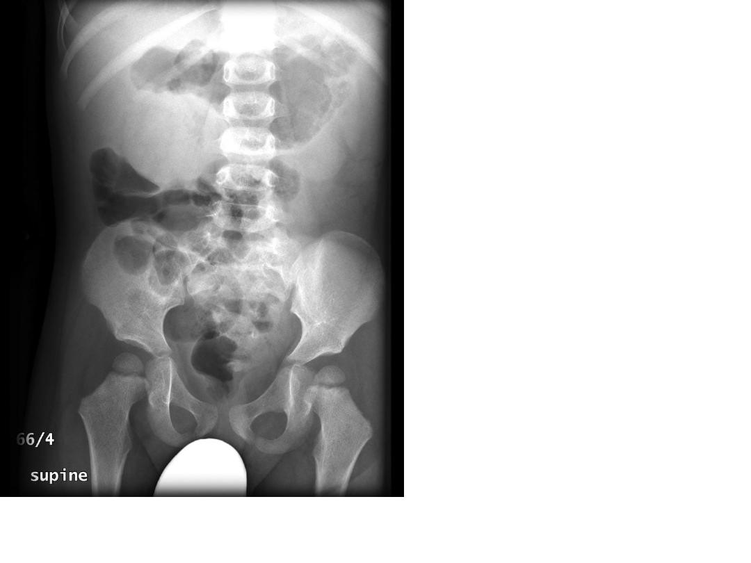
•KUB showing normal gas shadow of transverse
colon
53
54
55
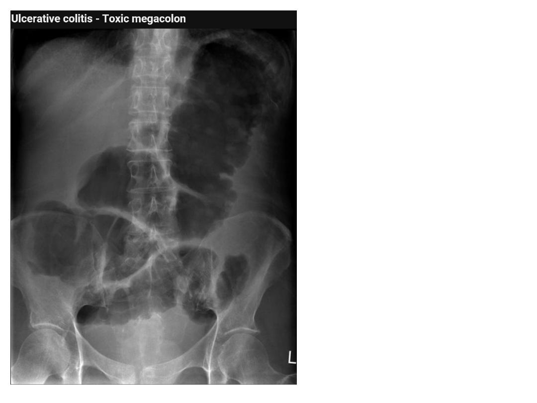
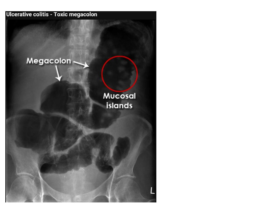
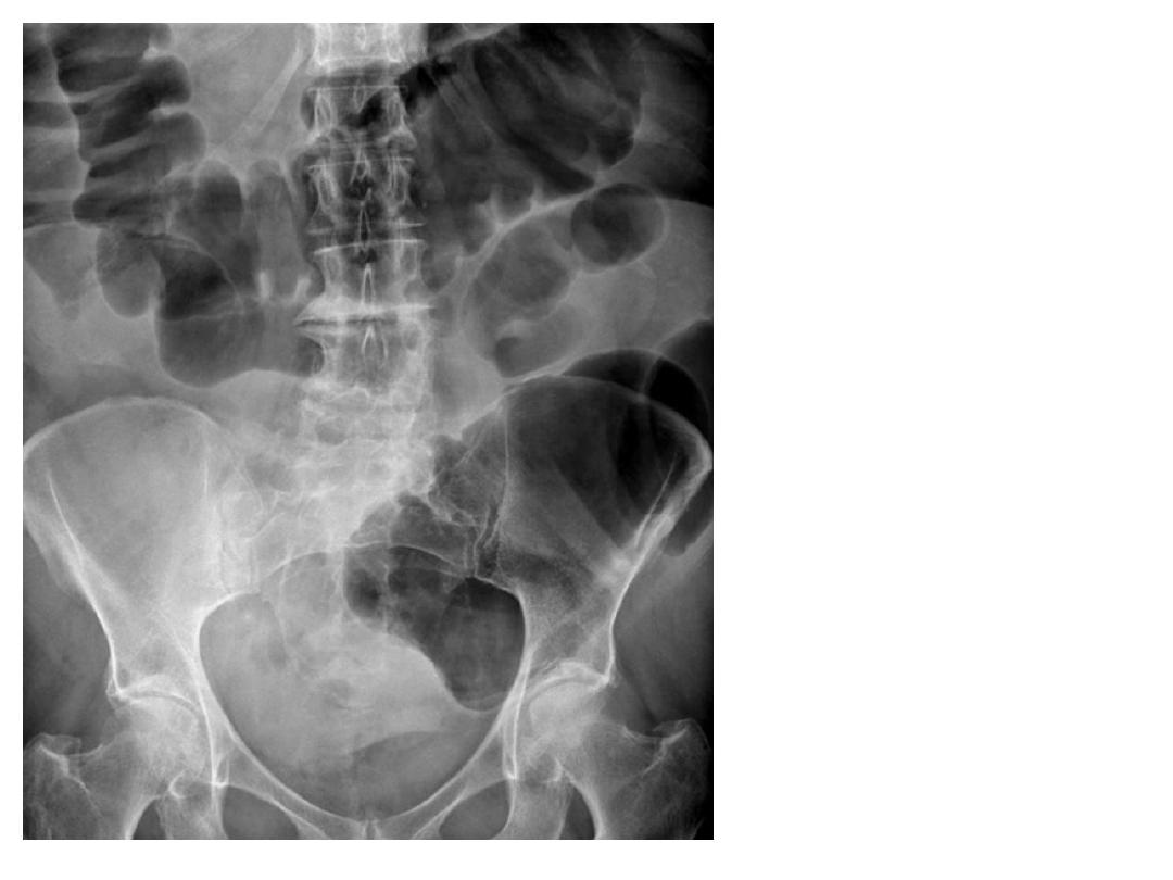
Description:-
KUB film showing
massive dilatation
of the colon ,
haustral marking
are apparent.
Diagnosis:-
Toxic megacolon
56
57
58
59
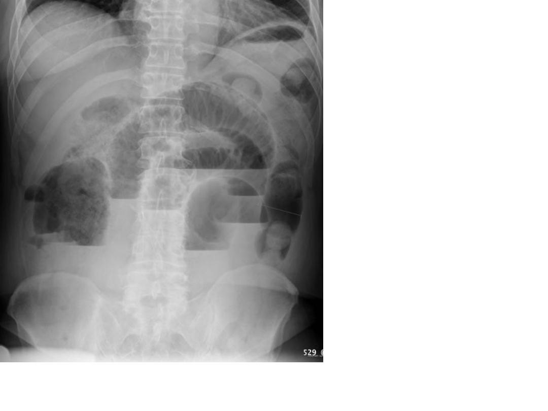
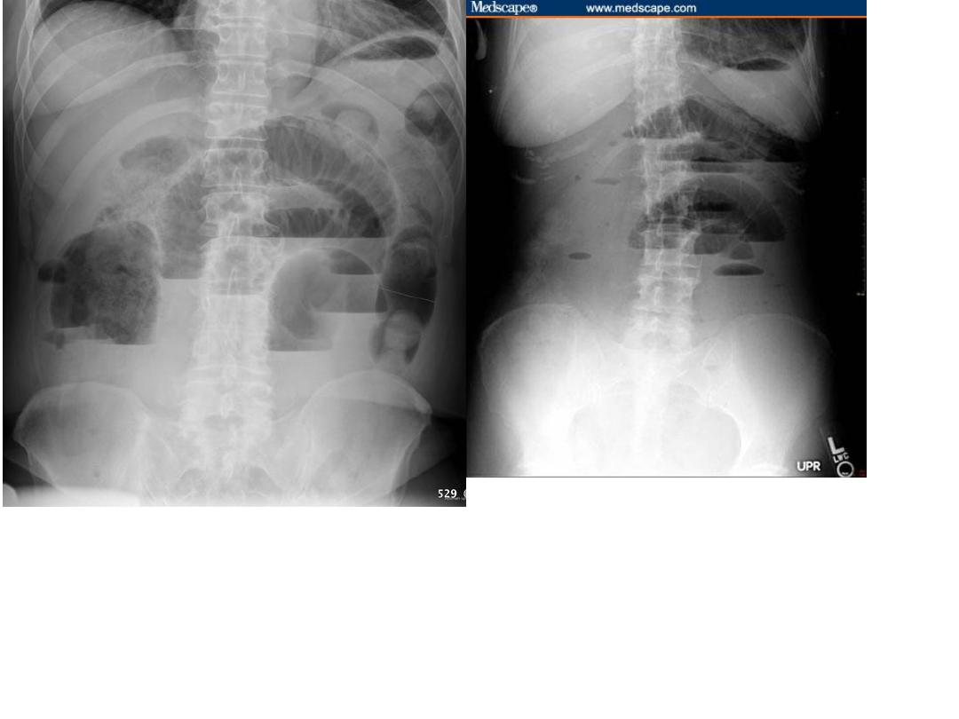
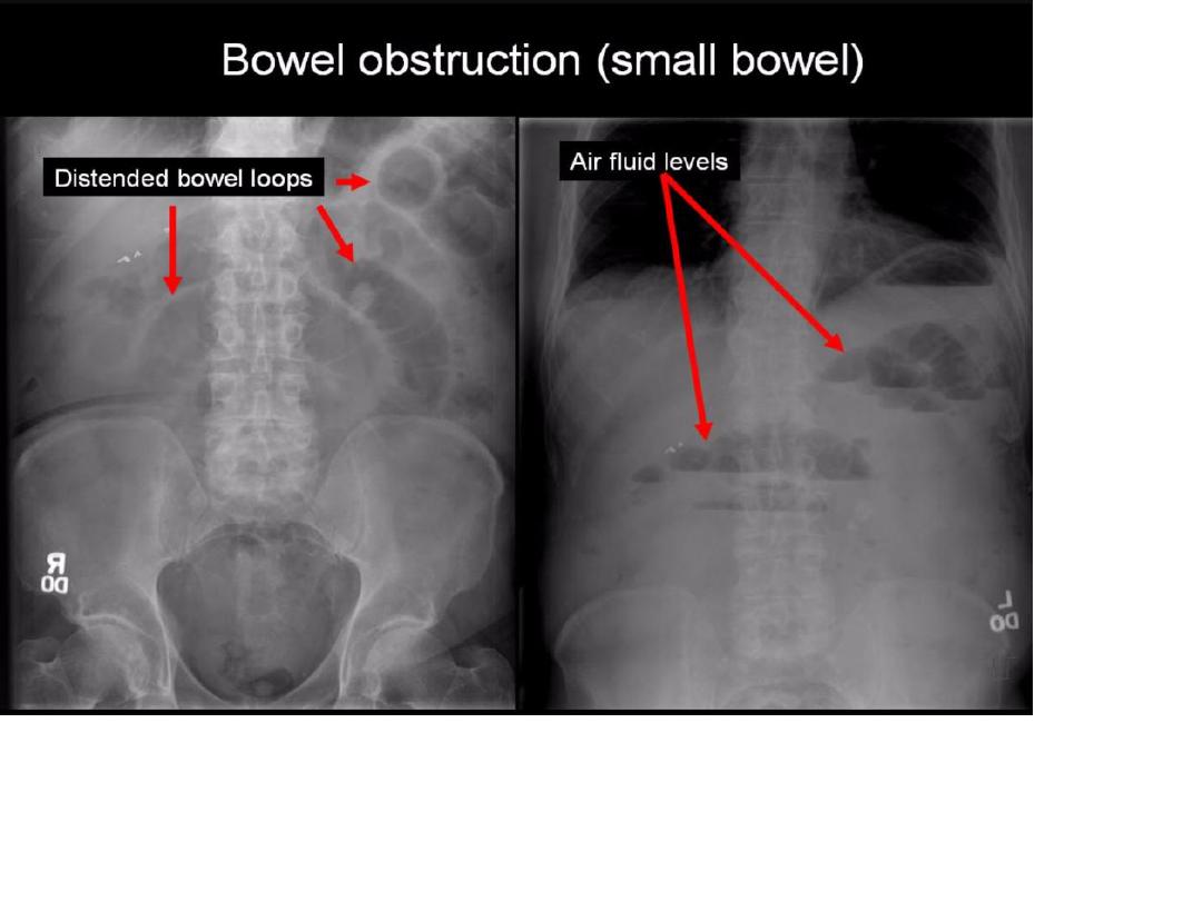
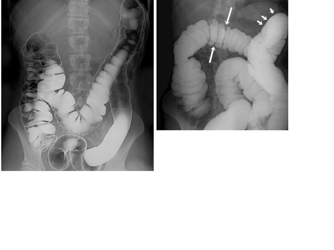
Plain radiograph show
Centrally located
multiple dilated loops of
gas filled small bowels
with multiple air fluid
levels & visible Valvulae
conniventes .
Dx. :
Small bowel obstruction
60
61
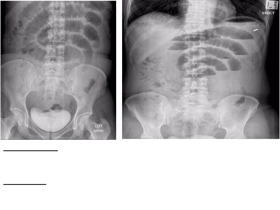
Description:-KUB study shows stepladder
configuration,multiple fluid levels,centrally located in
erect position.
Diagnosis :-SBO.
62
63
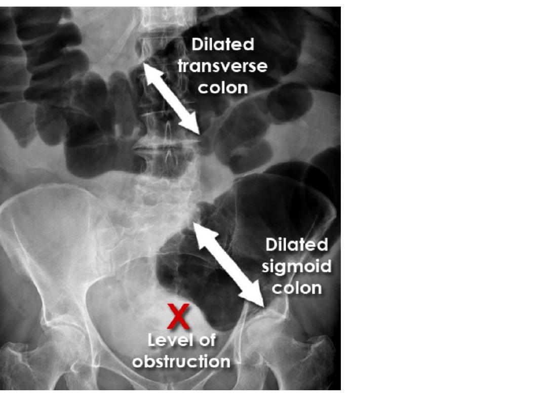
64
Description:- KUB study shows dilated colon more thn 6 cm
with effacement of haustra peripherally located and
multiple air fluid level
Diagnosis :-Large Bowel Obstruction .
65
66
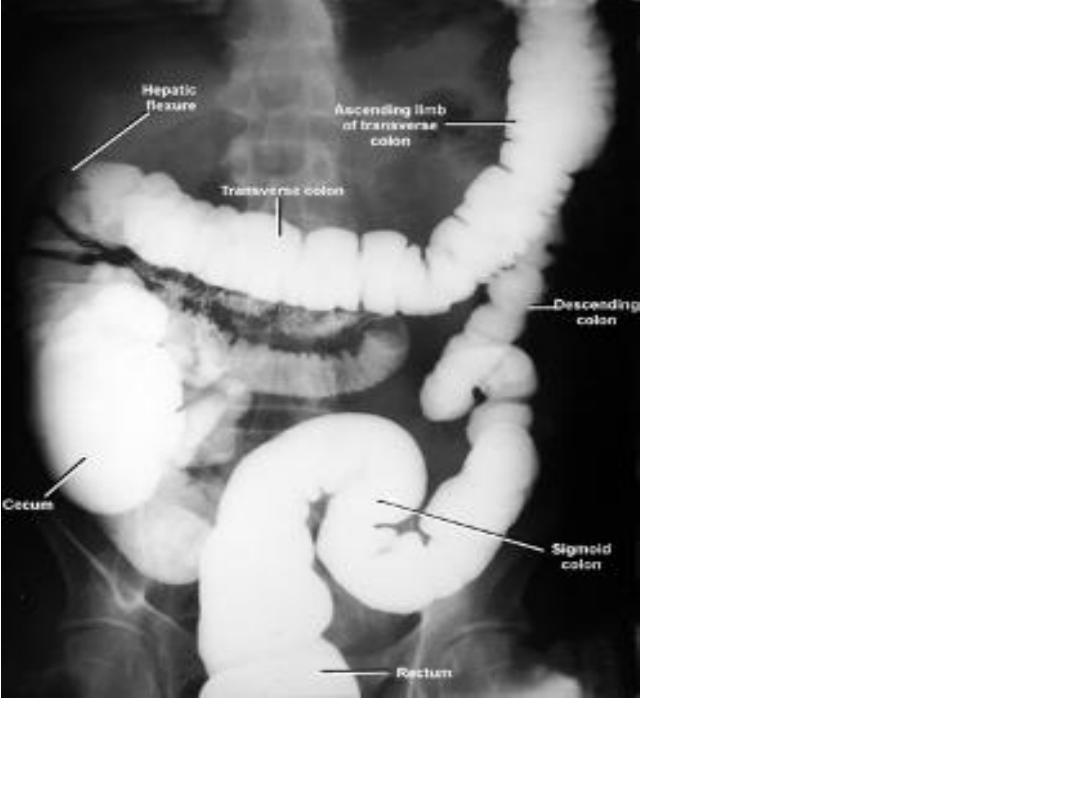
Ba enema showing
normal appearance
of colon and
rectum .
67
Ba enema showing
normal appearance
of colon and rectum .
68
Description :-Ba enema showing meniscus or apple
core sign of colon .
Diagnosis :-Ca colon
69
Description :-Ba enema
showing meniscus or
apple core sign of
colon .
Diagnosis :-Ca colon
70
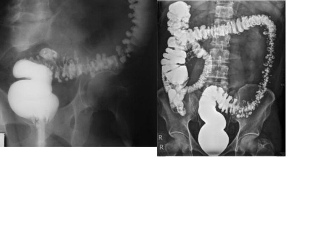
KUB showing an
elongated soft tissue
mass (meniscus sign) in
the right upper quadrant
with bowel obstruction
proximal to it.
dx. Intussuception
71
72
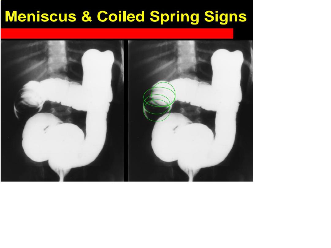
Ba enema showing the meniscus and coiled spring sign.
Dx. Intussusception
73
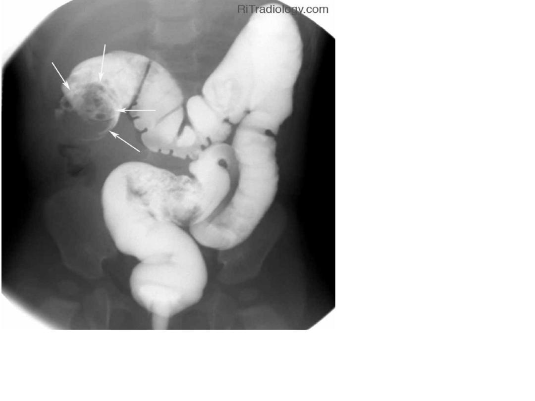
Ba enema showing
the meniscus and
coiled spring sign in
the iliocolic region .
Dx.
Intussusception
74
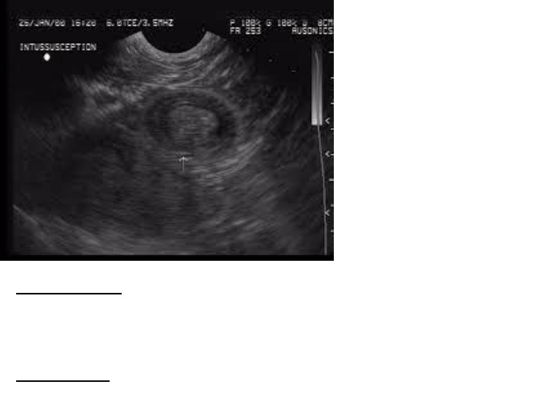
Description:-ultrasound of large bowel showing target
sign (doughnut sign) as hypochoic area surrounding
hyperechoic area.
Diagnosis:-intussusception.
75
Ba enema (DC) showing multiple Ba filled out pouchings of mucosa .
dx. Colonic diverticulosis
76
Ba enema examination
(DC) showing multiple
filling defect within the
lumen of the bowel .
Dx. FAP
77
Ba enema examination
(DC) showing multiple
filling defect within the
lumen of the bowel .
Dx. FAP
78
79
80
81
82

1 .
GIT slide presentation
2 .Normal Ba-swallow of esophagus
3 . Normal Ba-meal of stomach
4 . Normal Ba-meal of stomach
5 . Achalasia cardia
6 . Achalasia cardia
7. Achalasia cardia
8 . Tertiary contraction of the esophagus ( crock screw esophagus )
9 . Benign stricture of the esophagus
10 . Benign stricture of the esophagus
11 . Ca esophagus
12 . Ca esophagus
13 .osophageal web
14 .osophageal diverticulum ( pulsion type )
15 .epi phrenic diveticulum
16 .epi phrenic diveticulum
17 .zenkers D. … traction D. …. Epi phrenic D .
18 .hiatus hernia ( sliding hernia )
19 .sliding HH
20 .HH types .
21 .
Ba - meal gastric ulcers profile view
22. Ba - meal gastric ulcers
23. Ba - meal gastric ulcers En phase view
24 . Ba - meal gastric ulcers En phase view
83

25. Ba-meal chronic DU ( trifoil deformity ) .
26.Ba-meal infiltrative CA
Generalized left side
Localized Right side
27. Infiltrative CA of the stomach (distal pyloric antrum )
28 . Ba-meal Ca stomach
29 . Ba-meal Ca stomach
30 . Ba-meal duedenal diverticulum
31. Duodenal atresia ( double bubble sign )
32 . Duodenal atresia ( double bubble sign )
33 . Juejenal atresia ( triple bubble sign )
34 .Bockdalek hernia ( diaphragmatic hernia )
35 . diaphragmatic hernia
36 . diaphragmatic hernia
37 . diaphragmatic hernia
38 . pneumo peritoneum
39 . pneumo peritoneum
40 . pneumo peritoneum
41 . pneumo peritoneum
42 . Sub phrenic abscess
43 . Sub phrenic abscess
84

44.Normal Ba-follow through
45.Normal Ba-follow through
46.Normal Ba-follow through
47.Malabsorption syndrome
48.Lymphoma
49.Lymphoma
50.Cronhns disease
51.Ulcerative colitis acute phase
52. Ulcerative colitis chronic phase
53.Normal gas shadow of the KUB ( transverse colon )
54.Normal gas shadow of the KUB ( transverse colon )
55.Toxic megacolon
56.Toxic megacolon
57. Toxic megacolon
58.Toxic megacolon
59.Toxic megacolon
60.Small bowel obstruction
61.SBO
62.SBO
63.SBO
64.LBO
65.LBO
85

66.Normal Ba-enema
67.Normal Ba-enema
68.Ca colon
69.Ca colon
70.Intussuceptions
71.Intussuceptions
72. Intussusceptions
73.Intussuceptions Ba-enema
74. Intussusceptions US of abdomen
75.Ba-enema diverticulosis
76.Ba-enema FAP
77.Ba-enema FAP
78.Hirshprungs disease
79.Hirshprungs disease
80.Hirshprungs disease
81.Imperforated anus
82.Imperforated anus
86