1
Fifth stage
Surgery-Ortho
Lec-3
د. يقضان
29/11/2015
Introduction to fractures and trauma – 3
Complications of the fractures :
A- general complications .
B- local complications .
A – general complications .
1- blood loss and shock .
2- cardiopulmonary failure .
3- fat embolism .
4- DVT .
5- tetanus .
6- gas gangrene .
7- crush syndrome .
Fat embolism :
Fat globules larger than 10 micro meter in diameter can enter the circulation after closed
fractures of the long bones . It's source is from the bone marrow , it can be deposited in
any site in the body mainly in the lung or even in the brain , the condition is more common
in patients with multiple fractures .
Clinically :
The patient usually with lower limb fracture , early signs (within 72 hours of the injury ) has
slight rise in temperature and pulse rate , then the patient develop breathlessness and mild
mental confusion or restlessness ; petechiae can be looked for on the chest , back , axilla
and conjunctival folds .
In sever cases there will be marked respiratory distress and coma due to hypoxia or brain
emboli .
Treatment : there is no infallible test for embolism ; monitoring of the patient is
mandatory , oxygen supply and even in sever cases ventilator can be used , but in sever
cases recovery is unpredictable and the mortality rate is high … early fixation of the
fractures help in decrease its possibility .

2
Crush syndrome :
the crushed limb is deprived from the blood flow , also in case of interruption of blood
supply for the limb for any cause ; tissues begin to die and toxic metabolite accumulate and
when reach to the circulation it causing a lot of problems :
The resultant hyperkalemia , hypocalcaemia and metabolic acidosis can arrest the heart ;
the large molecules of myoglobin which released from dead muscles may lead to acute
renal failure .
Treatment :
The most important measure is prevention .
High urine flow must be ensured by giving large volume of intravenous crystalloid ; manitol
alkaline diuretic can be given .
Extensive wound excision (remove all the dead muscle if there is wound) is mandatory .
Some time amputation is the resolution to save the life .
Gas gangrene :
This condition is caused by clostridial infection , mainly clos. Welchii ; which are anaerobic
organism which can live and multiply in tissues with low oxygen tension ; so the dirt wound
with dead muscle that has been closed without adequate debridement is the most suitable
media for the growth of this micro organism .
The toxin which produced by this m.o. destroy the cell wall and rapidly lead to tissue
necrosis and spread of the infection .
Clinically :
Clinical features appear within 24 hours of the injury .
The patient has sever pain and swelling around the wound and brownish discharge may be
seen , little or no pyrexia , high pulse rate , gas in the tissue can be detected by x – ray but it
is not very marked ; characteristic smell become evident .
Rapidly the patient become toxemic and may pass into coma and death .
Prevention :
Any deep wound in the muscle should be explored , all dead tissue should be totally
removed , if there is doubt about tissue validity , then the wound should be left opened .

3
Treatment :
Early diagnosis is the key for success of the treatment which include :
I.V fluid replacement , intravenous antibiotic ( benzyl penicillin in high doze ,
metronidazole ) .
Hyperbaric oxygen has been used to decrease spread of infection ;
Decompression of the wound and remove the dead tissue .
In advance cases amputation may be essential .
B- local complication of the fractures :
a- early complications .
b- late complications .
a- early complications :
1- vascular injuries and compartment syndrome .
2- nerves injuries .
3- tendons injuries .
4- visceral injuries .
5- haemoarthrosis .
6- infection .
Compartment syndrome :
Fractures of upper and lower limbs can give rise to sever ischemia even if there is no
damage to a major vessels .
Bleeding , edema or inflammation may increase the pressure within one of the osteofacial
compartment ; there is reduced capillary flow which result in muscle ischemia , further
edema , still greater pressure and yet more profound ischemia .
Vicious circle that end after 12 hours or less , in necrosis of nerve and muscle within the
compartment .
Nerve is capable of regeneration but muscles once infarcted , can never recover and it
replaced by inelastic fibrous tissue (Volkmann's ischemic contracture ) .
A similar events may be caused by swelling of a limb inside a tight plaster cast .

4
Late complications of the fractures :
1- delayed union and non union .
2- malunion .
3- avascular necrosis .
4- growth disturbance of the bone .
5- myositis ossificans .
6- tendons ruptures .
7- nerves compression .
8- bed sore .
9- joint stiffness .
10- joints instability .
11- sudeck dystrophy .
12- osteoarthritis .
1- delayed union and non union :
non union :it is failure of the fracture to unite after double of the
expected time of healing which is determined by perkin`s
table .
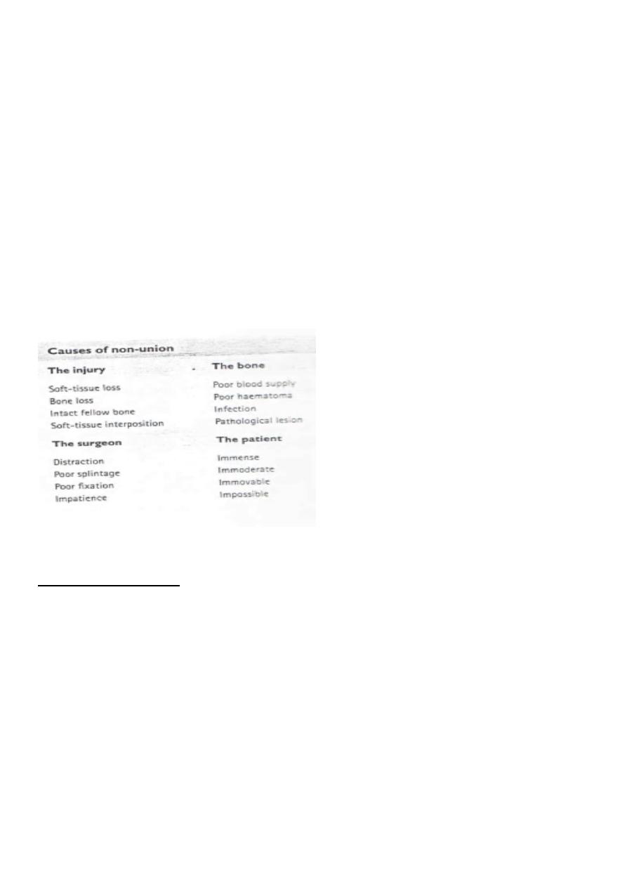
Causes of non union :
A- causes related to the type of injury :
1- extensive soft tissue damage and loss i.e in compound fractures .
2- bone loss in compound fractures .
3- soft tissue inter position .
4- intact fellow bone .
5- infection .
B- causes related to the bone itself .
1- poor blood supply .
2- poor hematoma formation .
3- flimsy periosteum .
4- diseased bone ( pathological fractures )
C- causes related to the surgeon : these includes technical faults like:
1- over traction .

5
2- poor splintage .
3- poor fixation .
4- impatience .
D- cause related to the patient :
1- immense .
2- immoderate .
3- immovable .
4- impossible .
Types of non union :
1- atrophic non union .
2- hypertrophic non union .
Treatment of non union :
A- conservative treatment :
1- functional brace .
2- pulse electromagnetic fields .
3- low frequency pulsed ultra sound .
B- operative treatment :
1- refreshment of the fracture site .
2- perfect reduction .
3- rigid fixation with compression .
4- bone grafting .

6
2- malunion :
When the fracture is in unsatisfactory position e.g unacceptable angulation , rotation or
shortening and unite in this position , so the deformity will persist and it is called malunion.
It is caused by , either failure of reduction or failure of holding of the fracture .
3- avascular necrosis :
In certain regions , when fracture occur it may complicated with interruption of blood
supply to certain parts of the bone lead to avascular necrosis . e.g of these parts of bones :
1- femoral head necrosis after fracture neck of femur .
2- proximal part of the scaphoid after fracture waist of the scaphoid .
3- body of the talus after fracture neck of talus .
4- growth disturbance :
This complication occur when there is damage to the growth plate of the bone by the
fracture .
5- bed sore :
It is pressure sore or ulceration occur in bed ridden patient at the areas which sustained
pressure mainly lower back , buttock .
It is occur very rapidly and healed very slowly .
The prevention is better than treatment .
6- myositi`s ossificans :
Deposition of calcium in the muscles lead to stiffness of the near joint .
Indomethacin is helpful in treatment .
7-Tendons lesions :
a- tendonitis e.g tibialis posterior tendonitis in fracture medial malioli .
b- rupture of tendon e.g rupture of extensor pollicis longus tendon in
Cole's fracture .
8- sudeck dystrophy :
It is post traumatic localized reflex sympathetic over activity also known as algodystrophy .

7
The patient has continuous pain , swelling , redness , the skin look shiny , pinkish ,
decrease in hair distribution warmth , localized tenderness and the near by joints are stiff .
X- ray show localized osteoporosis .
Treatment :
Removal of splintage of the fracture (pop) and start active physiotherapy , analgesic anti
inflammatory drugs , sympatholytic drugs e.g guanithidin i. v or even sympathetic block .
9- muscles contracture :
Usually follow arterial injuries with the fractures or compartment syndrome . e.g for it is
Volkmann's ischemic contracture in the forearm .
10- nerve compression . By the mal united bone or by the callus .
11- joint instability .
12- joint stiffness . It is occur due to immobilization of the limb during
the period of healing of the fracture . It can reduced by avoid prolonging the period of
holding of the fracture , using functional orthosis , or by using internal or external fixators
which they permit movement of the joints at the time of healing of the fractures .
13- osteoarthritis :
This occur when the fracture involve the articular surface of the joint .
It can be minimized by perfect reduction of the articular surface and internal fixation .
Stress fractures :
Stress or fatigue fracture is one occurring in normal bones of healthy patient but it is not
caused by a specific traumatic incident , but by repetitive minor trauma or stress .
It affect many bones but the common examples are Marsch fracture
(fracture metatarsal bones , mainly the second one ) in the military people ; and fractures in
the lower third of the fibula (runner's people) .
Pathological fracture :
It is fracture that occur in abnormal bone or diseased bone , and it suspected when the
force which cause the fracture is less than the ordinary force needed to break the normal
bone .

8
Any condition lead to weaken the bone can lead to pathological fracture e.g osteomyelitis ,
bone tumors , metabolic bone diseases …..etc.
Dislocation and subluxation of the joints :
Dislocation : it mean the joint surfaces are completely displaced and
no longer in contact (complete separation of the two
articular surfaces ) .
Subluxation : it represent a lesser degree of displacement . Such that
articular surfaces are still partly apposed .
Injuries of the physis :
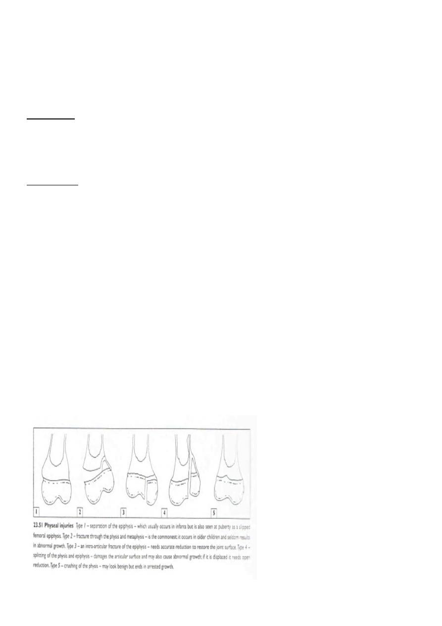
Classification : Salter Harris classification :
Type one : transverse fracture through the growth plate .
Type two : it is similar to type one but it contain triangular piece from the metaphysis .
Type three : the fracture split the epiphysis vertically .
Type four : splitting the epiphysis vertically and extend to the metaphysis .
Type five : compression of the growth plate (crushing) and it result in
growth disturbance of the bone ( baddest type ) .
