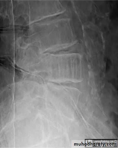
What it could be
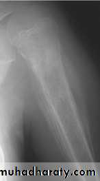
The sign & findingsWide zone of transition is being
Benign or malignant ????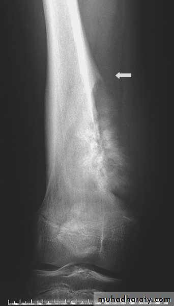
Codman's triangle
Elevation of the periosteum at the side of the tumor margin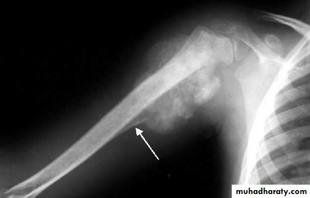
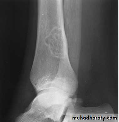
lytic, expansile lesion, Sub articular in location give the soap bubble appearance
Giant cell tumor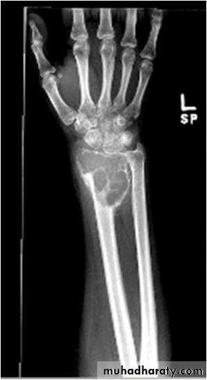
onion peal periosteal reaction
Characteristic ofEwings Sarcoma
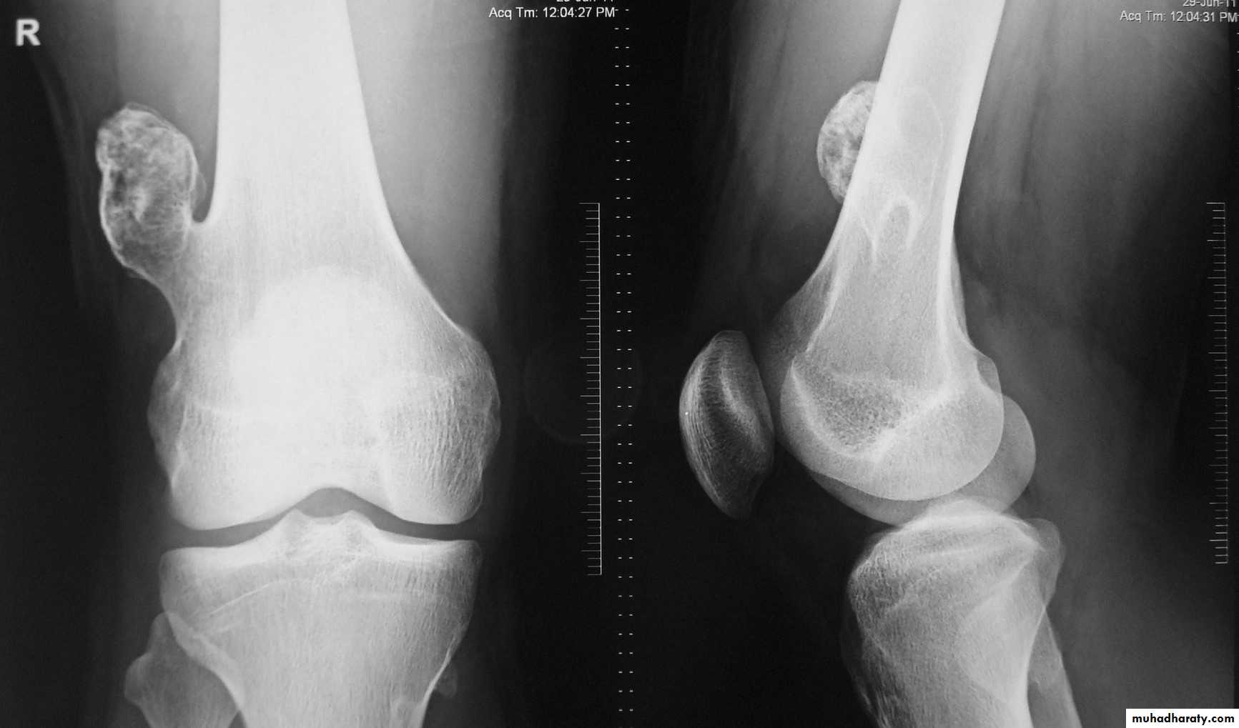
Tumor have slender pedicle directed from growth plate ,,, what it could be ???
Is the direction of pedicle toward joint or away ?????What is being have at upper aspect ???
What is their significance ???
Answer .Osteochondroma Most common benign bone lesion which have their own growth plate)
Pedunculated: slender pedicle directed away from growth plate .The thickness of the cartilage cap above the bony projection is very important
if the thickness > 1 cm of cartilaginous cap by CT, > 2 cm by MRI give high possibility of Malignant transformation
Also Dispersed calcifications in the cap
*
(Ollier's disease) means multiple enchondromatosis
Diaphyseal acalsia means multiple exostosisMultiple ivory osteoma associated with Gardner's syndrum
*shepherd crook deformity seen in fibrous dysplasia
*Codmans triangle seen in malignant bone tumor (OS)*Soap bubble appearance (giant cell tumor )
*Fallen fragment sign (simple bone cyst with # )
*moth eaten appearance (rarifaction ) acute osteomylitis
*sequestrum dead bone
*involucrum new bone formation
*cloaca The cloaca is an opening
Gibbous deformity wedging of the vertebra (anterior loss of vertebral height)
Seen in TB infection of vertebra
*spina ventosa means TB Dactylitis
(* Rain drop appearance ( multiple lytic lesion in skull MM
* Rackety rosary of the ribs mean widening of the anterior end of the rib seen in (rickets)* codfish appearance Vertebral collapse resulting in biconcave vertebra with widened disc (osteomalasia )
*Triradiate pelvis in which pelvic side walls bend inward (sever cases of osteomalasia )
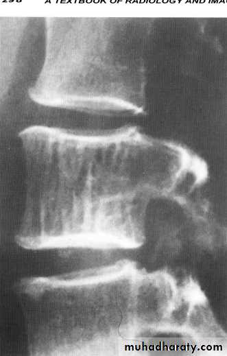
* Looser's zones these are thin short lucent lines with sclerotic margins running across the cortex at right angle, best seen in the scapula , femoral neck , pubic rami (osteomalacia )
Subperiosteal bone resorption seen particularly in the radial aspect of the middle phalange (hyper parathyriodism)
*Salt and pepper sign multiple tiny hyper lucent areas in the skull vault (hyper parathyriodism )
*rugger jersey spine band of increased density within the vertebral body & across the metaphysis of long bones in renal osteodystrophy .
*hair on end appearance Increase diploic space thickness with vertical striation I seen in haemolytic anaemia (thalacimia) .
*Bullets vertebrae anterior vertebral beaking ( achondroplasia )
*Ballooning of the sella ( acromegaly )
*Bamboo spine (ankylosing spondylitis )
*square shape vertebra (ankylosing spondylits )
*punched out peri articuler erosion of the bone (Gout )
*charcot joint (nueropathic arthritis )
*Legg-Calve-Perthes disease osteochodrosis of the femur head
*Mushroom deformity of femoral Neck (perthes diseade )• *Scheuermann's disease : (adolecent Kyphosis) osteochodrosis of the vertebral end plates
• *Osgood-Schlatter osteochodrosis of the tibial tubercle
• *Blount's disease tibial epiphysis
• *Kohler's Navicular bone
• *Kienbock's lunate bone
• *Osteochondritis dissecans separation of the affected part in to the joint space resulting in intra-articular loose body seen in large joint
• * Psueodoarthrosis of the femoral head with iliac bone (old neglected CDH)
Bamboo spine … Ankylosing spondylits

Mushroom Deformity of the femoral neck in perthes disease
being flattened femoral head with a contiguous broad neck, accompanied by awidened articular space .
Fallen fragment sign
shepherd crook deformity seen in fibrous dysplasia
A shepherd crook deformity refers to a coxa varus angulation of the proximal femur, classically seen in femoral involvement by fibrous dysplasia .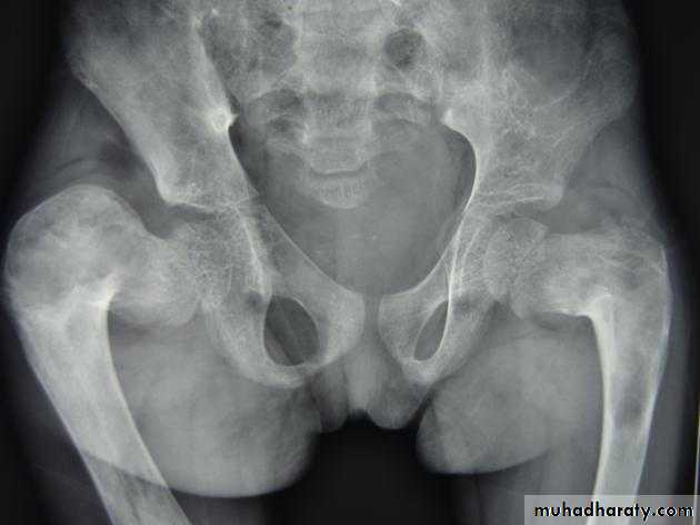
The whole vertebral body marked by vertical striation … being Haemangioma
Osteoid osteoma:
lucent area surrounded by calcification nidus surrounded by sclerotic rim with or without periosteal reaction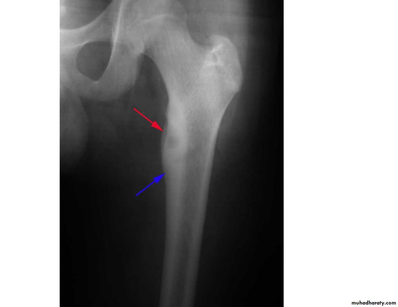
Both secondary metastasis & MM being cause multiple lytic lesion within the skeletal system who can you differentiated secondary from MM if each one of them affecting vertebra ????
* secondary M primarily affecting & have predilection to pedicle
* MM spares vertebral pedicle.
causes of reduced bone density:
Metastatic carcinoma.Multiple myeloma.
Hyperparathyroidism.
Osteomalacia.
Osteoporosis .
Causes of generalized increase in the density of the bone
1.Sclerotic metastases.
2.Osteopetrosis (Marble bone disease)
3.renal osteodystrophy
4.Fluorosis
5.Myelosclerosis
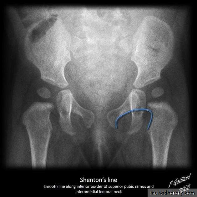
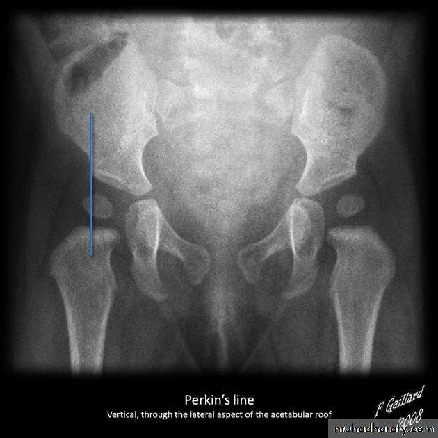
Who can you do Von Rosen view
At 3-6 months :abduction of the thigh 45 degree and internal rotation
Define the following
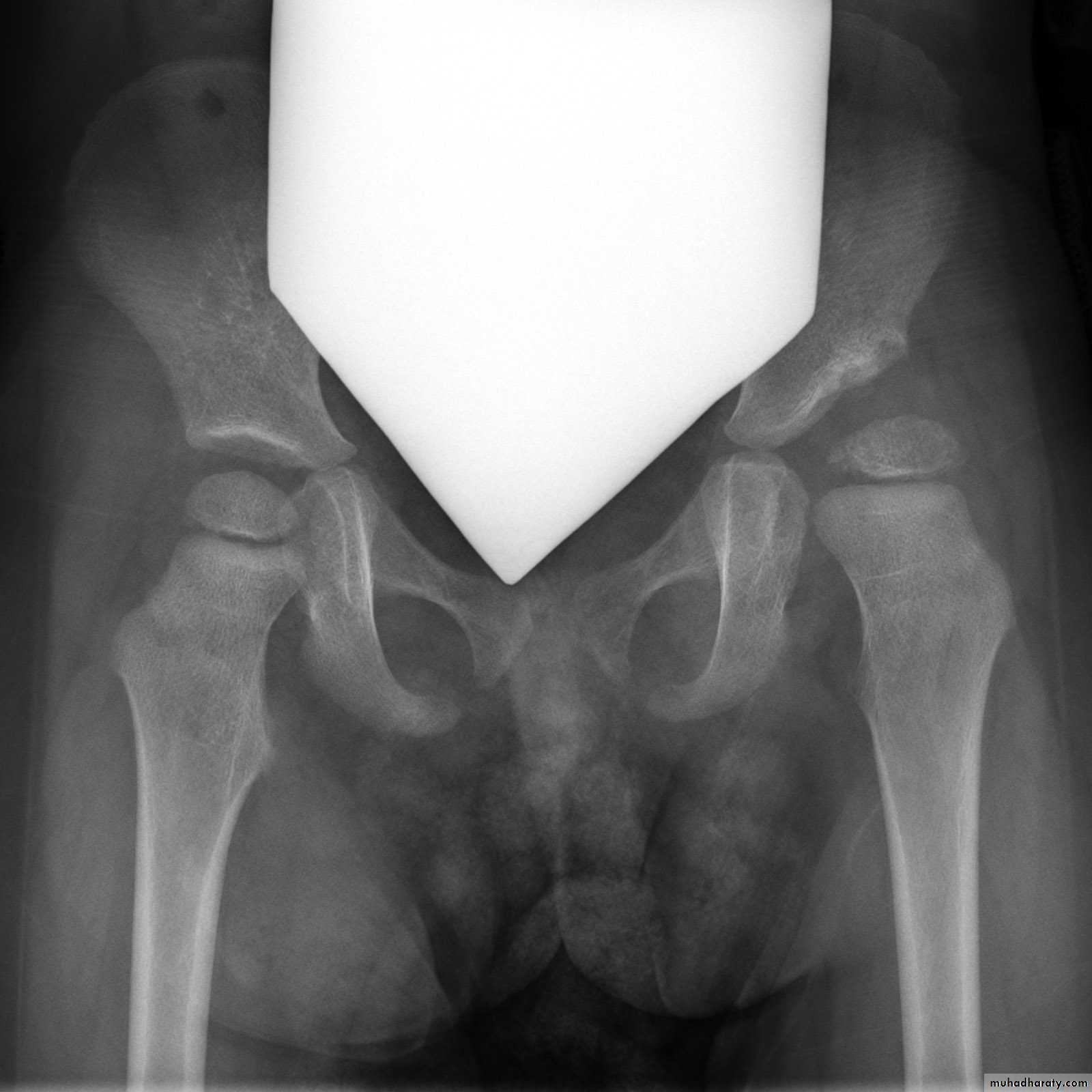
Shenton line is drawn along the inferior border of the superior pubic ramus and should continue laterally along the infero medial aspect of the proximal femur as a smooth line. If there is supero lateral migration of the proximal femur due to DDH then this line will be discontinuousPerkin line is drawn intersecting the lateral most aspect of the acetabuler roof & iliac creast
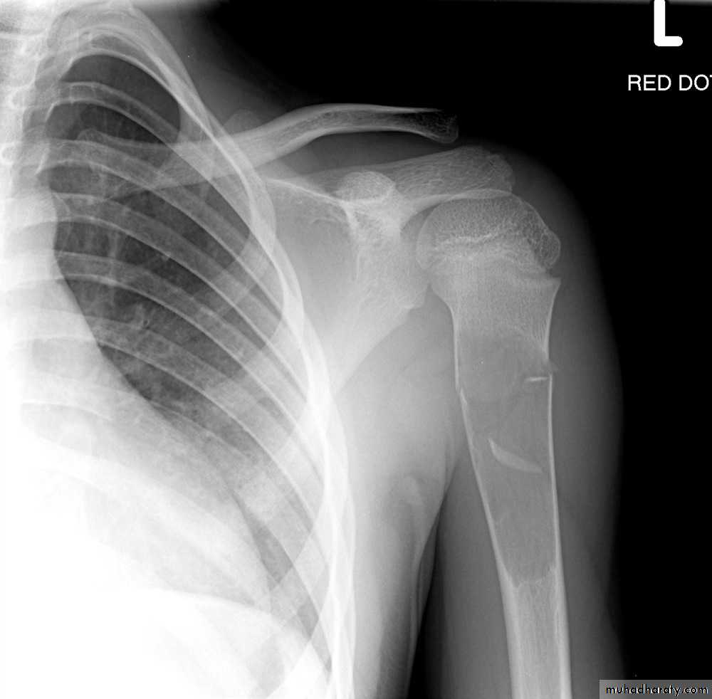
Periosteal reaction is a non specific radiographic finding that occurs with periosteal irritation.
Codman's triangle is a type of periosteal reaction seen with aggressive bone lesions could be singlelayer and mulitlayered periosteal reaction), only the edge of the raised periosteum will ossify.
Brodies abscess is an intraosseous abscess infavouer of subacute pyogenic osteomyelitis
Gary's osteomylitis is a specific type of chronic osteomyelitis. It mainly affects children and young adults . It typically affectes the mandible
Potts disease also known as tuberculous spondylitis, refers to vertebral body and intervertebral disc involvement with tuberculosis (TB)
Gibbus deformity is a short-segment thoracolumbar kyphosis causing sharp angulation.
A sequestrum is usually a complication of osteomyelitis and represents devascularisation of a portion of bone with necrosis and resorption of surrounding bone leaving a 'floating' piece
An involucrum is a complication of osteomyelitis and represents a thick sheath of periosteal new bone surrounding a sequestrum
A cloaca is an opening in a involucrum which allows drainage of purulent and necrotic material out of the dead bone
Osteomalacia (OM) is bone softening due to insufficient mineralisation of the osteoid secondary to any process results in vitamin D deficiency
Looser zone are wide transverse lucencies traversing part way through a bone, usually at right angles to the involved cortex and are associated most frequently with osteomalacia and rickets
Code fish vertebra fish vertebra (or codfish vertebra) describes the biconcave appearance of vertebrae (espaically lumbar vertebrae).
Rachitic rosary refers to expansion of the anterior rib ends at the costochondral junctions and is most frequently seen in rickets as nodularity at the costochondral junctions
Define
Brown tumor: more common in the primary type seen as lytic expansile lesion in any bone particularly the mandible & pelvis.Rugger-jersey spine describes the prominent subendplate densities at multiple contiguous levels to produce an alternating dense-lucent-dense appearance.
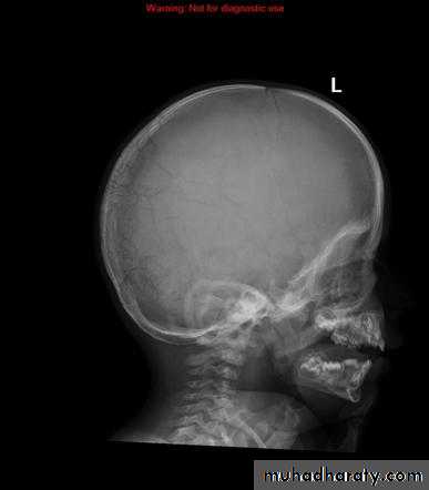
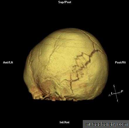
Wormian bone are a subset of the small intra sutural bones that lie between the cranial sutures mainly seen around the lambdiod sutures
Prognathismwidening of angle between the body and ramus of the mandible.
A shepherd crook deformity refers to a coxa varus angulation of the proximal femur, classically seen in femoral involvement by fibrous dysplasia
Osteo petrosis is metabolic skeletal disease defined as a reduction of bone mineral density (BMD) below a defined lower limit of normal.
Acromegaly & prognathism
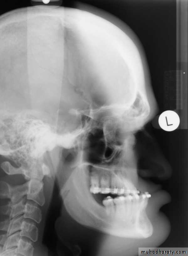
Wormian bone
What is it being ??? Wormian bone are a subset of the small intra sutural bones that lie between the cranial sutures mainly seen around the lambdiod suturesCauses ?????
osteogenesis imperfecta
rickets
cleidocranial dysostosis
Hypothyroidism
Down syndrum
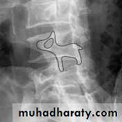
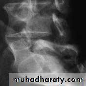
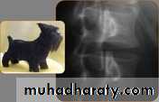
Spondylolysis and spondylolisthesis
Spondylolisthesis is a term denoting fore ward or backward movement of a vertebra relative to the vertebral segment below, typically due to spondylolysis (pars interarticularis defects) .Spondylolysis - “Spondylolysis is a defect in the pars interarticularis of the neural arch, the portion of the neural arch that connects the superior and inferior articular facet in which cause defect in the neck of scotty dog .
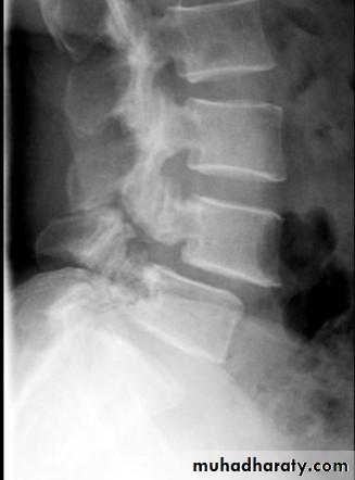
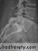
scotty dog sign refers to the normal appearance of the lumbar spine when seen on oblique radiographic projection. On oblique views, the posterior elements of vertebra form the figure of a Scotty dog with:
the transverse process being the nose
the pedicle forming the eye
the inferior articular facet being the front leg
the superior articular facet representing the ear
the pars interarticularis (the portion of the lamina that lies between the facets) equivalent to the neck of the dog.
sacroiliitis & facet joint ankylosis
Q..Being charct. Of which one of the following ???*Ankylosing spondylitis
*DISH
Ans…. Ankylosing spondylitis ( is the true answer )
Q..What do you mean the symbol ( DISH ) ????
Ans….DIFFUSE IDIOPATHIC SKELETAL HYPEROSTOSIS*Erosive changes of DIP and PIP (osteoarthritis )
*Peri articuler erosive changes at 2nd and 3rd MCP joint( Rheumatoid arthritis )*punched out peri articuler lytic lesion affecting 1st MTP joint (of the big toe ( Gout
Sub periosteal resorption at the radial aspect of the middle phalanges (hyper parathyriodism )
Types of metastasis
Osteolytic (most common causes) : neuroblastoma (in children), breast (adult female), bronchus (adult male), thyroid, kidney, colon The vertebral pedicles are often involved.Osteoblastic : prostate, breast , carcinoid , TCC of bladder,, neuroblastoma.
Mixed: breast, prostate, lymphoma.
Solitary expansile bubbly metastases with soft tissue involvement: thyroid, kidney.
Bone metastases with sun burst periosteal reactions: prostate, retinoblastoma and neuroblastomaVery important note
Myeloma resembles metastases in everything except :*it's more well defined,
*cause bone expansion if solitory called plasmacytoma
*spares vertebral pedicle
Skull manifestation of the acromegaly
1.Thickening of the skull vault &diplioc space2.Thickening of the posterior occipital tubercle
3.Prognathism
4.Froehead bossing with enlargement of the frontal sinuses also with enlargement of the mastiod air cell .
5.Balloning with Double floor appearance of the sella tursica .
Osteophytes,
Subchondral sclerosisUneven loss of articular space
These signs being of which one of the following ???
1.Metabolic arthritis
2.Degenerative arthritis
3.Inflammtory arthritis
True answer is Degenerative arthritis
Q….What are the main 5 radiological sign of degenerative arthritis ?? ???
Q….What sign in spine being pathgnomonic for degenerative spondylosis of the spine ???
Vacuum phenomenon: gas (N2),is pathognomonic of the degenerative processQ…what are the types of inflammatory arthritis
Q… what are he early changes seen in Rheumatoid arthritis ??
Q…CDH occurs most commonly in ????(70%) in the left hip . Bilateral involvement is seen in 5%
Q…By simple diagram draw pelvis with lines (shenton’s & Perkins line)
Very important notes
* Well defined clear cut with narrow zone of transition indicates benign or slowly growing lesion.* Ill-defined wide zone of transition indicates aggressive rapidly growing lesion as osteo myelitis and malignant tumors.
* Metastases & myeloma lie in the middle of the spectrum (well defined lytic lesion with no sclerotic margin).
* commonest malignant tumors affecting the
bone ??? secondary metastasis*the commonest primary malignant bone tumor in young adults ?? Osteosarcoma
Q….lytic, expansile lesion, Sub articular in location, Not clearly defined margin with thinning of the cortex ???
Giant cell tumor
What is the most conmen causative organism affecting the bone ???
staphylococcus aurous : 80-90% of all infections
vertebral metastases (94%) causing primerly affection of the ????
Pedicle