Forth stage
MedicineLec-5
د.جاسم محمد
9/11/2015
Aortic Stenosis
Overview:
Normal Aortic Valve Area: 3-4 cm2Symptoms: Occur when valve area is 1/4th of normal area.
Types:
Supravalvular
Subvalvular
Valvular
Etiology of Aortic Stenosis
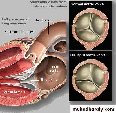
Congenital (Bi-cusped).
Rheumatic.
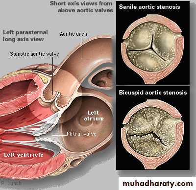
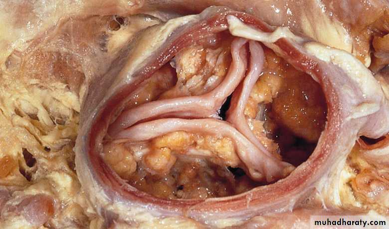
Degenerative/Calcific.
Patients under 70: >50% have a congenital cause
Patients over 70: 50% due to degenerative
Evaluation of AS:

Cardiac catheterization: Should only be done for a direct measurement if symptom severity and echo severity don’t match OR prior to replacement when replacement is planned.
Pathophysiology of Aortic Stenosis
A pressure gradient develops between the left ventricle and the aorta. (Increased afterload)LV function initially maintained by compensatory pressure hypertrophy
to maintain the cardiac output
When compensatory mechanisms exhausted, LV function declines and pulm.edema supervenes.
Presentation of Aortic Stenosis:
Asymptomatic mild/moderate
Syncope: (exertional)
Angina: (increased myocardial oxygen demand; demand/supply mismatch)
Dyspnea: on exertion due to heart failure (systolic and diastolic)
Sudden death
Physical Findings in Aortic Stenosis
Slow rising carotid pulse (pulsus tardus)Thrusting apex beat (LV pressure overload)
Narrow pulse pressure
Heart sounds- soft and split second heart sound, S4 gallop due to LVH.
Systolic ejection murmur- cresendo-decrescendo character. This peaks later as the severity of the stenosis increases.
Loudness does NOT tell you anything about severity
Investigations
ECG Left ventricular hypertrophy ,LBBBChest X-ray May be normal; sometimes enlarged LV and dilated ascending aorta on PA view, calcified valve on lateral view.
Echo Calcified valve with restricted opening, hypertrophied LV) Doppler Measurement of severity of stenosis Detection of associated aortic regurgitation.
Cardiac catheterization Mainly to identify associated coronary artery disease May be used to measure gradient between LV and aorta.
Management of AS
General- IE prophylaxis in dental procedures with a prosthetic AV or history of endocarditis.
Medical - limited role since AS is a mechanical problem. Vasodilators are relatively contraindicated in severe AS
Aortic Balloon Valvotomy- shows little benefit.
Surgical Replacement: Definitive treatment
Simplified Indications for Surgery in Aortic Stenosis
Any SYMPTOMATIC patient with severe AS (includes symptoms with exercise)Any patient with decreasing EF
Any patient undergoing CABG with moderate or severe AS
Aortic Regurgitation
Overview:
Definition: Leakage of blood into LV during diastole due to ineffective coaptation of the aortic cusps.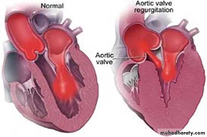
Etiology of Acute AR
Endocarditis
Aortic Dissection
Physical Findings:
Wide pulse pressure
Diastolic murmur
Florid pulmonary edema
Treatment of Acute AR
True Surgical Emergency:Positive inotrope: (e.g., dopamine, dobutamine)
Vasodilators: (e.g., nitroprusside)
Avoid beta-blockers
Do not even consider a balloon pump
Etiology of Chronic AR
Cusps defects
Congenital -Bicuspid aortic valve
Rheumatic
Infective endocarditis
Aortic root dilatation Marfan.
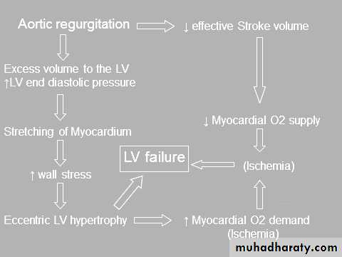
Pathophysiology of AR
Combined pressure AND volume overload
Compensatory Mechanisms: LV dilation, LVH. Progressive dilation leads to heart failure.
Symptoms
Asymptomatic until 4th or 5th decadeProgressive Symptoms include:
Dyspnea: exertional, orthopnea, and paroxysmal nocturnal dyspnea
Nocturnal angina: due to slowing of heart rate and reduction of diastolic blood pressure.
Palpitations: due to increased force of contraction.
Signs
Peripheral signs
Pulses Large volume or ‘collapsing’ pulse.
Corrigan pulse
Increased pulse pressure
Bounding peripheral pulses
Capillary pulsation in nail beds: Quincke’s sign Femoral bruit (‘pistol shot’)
Duroziez’s sign
Head nodding with pulse: de Musset’s sign.
Hill’s sign
JVP may be normal or elevated
Central Signs
Apex: Hyperdynamic and displaced apical impulse.
Diastolic thrill.
Auscultation
High pitched, blowing, decrescendo diastolic murmur at LSB, best heard at end-expiration & leaning forward.
Austin-Flint murmur indicates severity (mid to late diastolic murmur)
Systolic murmur related to high flow state
Investigations
ECG Initially normal, later left ventricular hypertrophy and T-wave inversion
Chest X-ray Cardiac dilatation, maybe aortic dilatation Features of left heart failure
Echo Dilated LV Hyperdynamic, LV Doppler detects reflux , Fluttering anterior mitral leaflet
Cardiac catheterization (may not be required) , Dilated LV , Aortic regurgitation , Dilated aortic root.
Management of AR
General: IE prophylaxis in dental procedures with a prosthetic AV or history of endocarditis.Medical: Vasodilators (ACEI’s), Nifedipine improve stroke volume and reduce regurgitation only if pt. symptomatic or HTN.
Serial Echocardiograms: to monitor progression.
Surgical Treatment: Definitive Rx
Simplified Indications for Surgical Treatment of AR
ANY Symptoms at rest or exerciseAsymptomatic treatment if:
EF drops below 50% or LV becomes dilated.
Tricuspid stenosis
Almost always rheumaticThe low cardiac output state causes fatigue; abdominal discomfort may occur due to hepatomegaly and ascites
The diastolic murmur of tricuspid stenosis is augmented by inspiration.
Medical management includes salt restriction and diuretics.
Surgical treatment in patients with a valve area <2.0cm and a mean pressure gradient >5mmHg.
Tricuspid regurgitation
Most common cause is annular dilatation due to RV failure of any cause
Symptoms and signs result from a reduced cardiac output, ascites, painful congestive hepatomegaly and oedema.The pansystolic murmur of TR is usually loudest at the left sternal edge and augmented by deep inspiration.
Severe functional TR may be treated by annuloplasty or valve replacement. Severe TR due to intrinsic tricuspid valve disease requires valve replacement.
Pulmonary stenosis
Most commonly due to congenital malformationSurvival into adulthood is the rule, infective endocarditis is a risk and right ventricular failure is the most common cause of death.
Carcinoid plaques may lead to constriction of the pulmonary valve ring.