


Biliary Atresia
Dr.Shatha A.Alkawaz
Lecturer ,College of Medicine
Consultant Paediatric Surgeon

Biliary atresia
Biliary atresia is a relatively rare obstructive condition of the bile ducts
causing neonatal jaundice.
There is a variable incidence around the world, in Europe it is 1:18000 live
births, in Japan it is 1:9640, highest in French Polynesia 1:3124.
There is a slight female predominance.
Etiology and pathogenesis:
Despite the intensive investigation, the cause of B.A remains unknown,
various etiologic mechanisms have been postulated including;
1. Intrauterine or perinatal viral infection ; reovirus type 3 ,rotavirus
CMV, papilloma virus and Epstein- Barr virus all have been proposed
as possible etiologic agent , but there is no conclusive evidence.
2. Genetic mutation that result in defective morphogenesis may be
important in syndromic B.A
3. Other causes; including vascular or metabolic insult to the developing
biliary tree, immunologically mediated inflammation.
Two distinct forms are described:
1. Syndromic B.A (embryonic) type account for 10-20% of all cases is
associated with other congenital anomalies including interrupted IVC,
preduodenal portal vein, intestinal malrotation, situs inversus,
cardiac defects and polysplenia.
Is likely to be due to developmental insult occurring during
differentiation of the hepatic diverticulum from foregut of the
embryo
2. Non –syndromic (perinatal) may have its origin later in gestational
age.
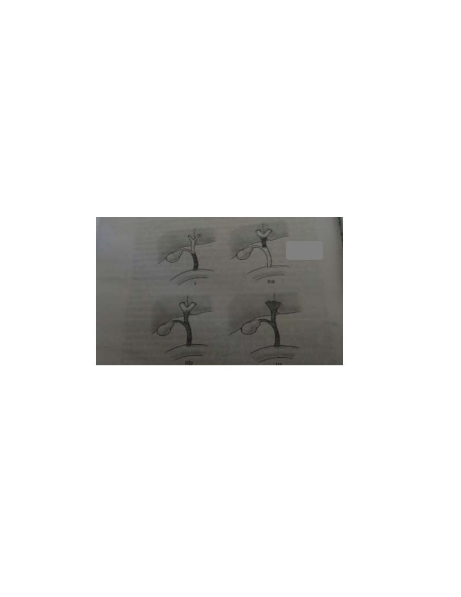
Classification
B.A can be classified by using macroscopic appearance and
cholangiography finding into three main categories:
The most common one is type III.
Pathology:
Although the term B.A implies a static process with complete
obstruction or absence of bile ducts, it is more a dynamic process of
progressive bile duct obliteration and sclerosis.
Early in the course of B.A the liver is enlarged, firm and green. The
gall bladder is small and filled with white mucus, or it may be
completely atretic.

Microscopically:
The biliary tracts contain inflammatory and fibrous cells
surrounding miniscule ducts that are probably remnants of the
original duct system.
Liver parenchyma is fibrotic and shows signs of cholestasis.
Proliferation of biliary neoductules.
This process develops into end state cirrhosis if good drainage
cannot be achieved.
These early changes are not specific to B.A and may be
confused with neonatal hepatitis and metabolic disease.
Physical findings:
The cardinal sign and symptoms of B.A are jaundice, clay -
colored stools, and hepatomegaly. In the neonatal period feces
are yellow or in more than half of patients.
The newborn pass dark brown urine.
The neonate with B.A is typically active, full term and may
manifest normal growth and weight in the first few months of
life. Anemia, malnutrition and growth retardation develop
gradually because of malabsorption of fat-soluble vitamins.
Jaundice that persists beyond 2 weeks of life should no longer
be considered as physiological, particularly if the elevation in
bilirubin is mainly in the direct fraction.
Diagnosis:
Routine examination
C
olor of stool
C
onsistency of the liver
C
onventional liver function test:
Serum bilirubin (total and direct) conjugated
hyperbilirubinemia ,defined as any level exceeding either
0.2mg/dL or 20%of total bilirubin, infants with B.A typically

show moderate elevation in total bilirubin, which is
commonly 6-12mg/dL with the direct(conjugated) fraction
composing 50-60% of total serum bilirubin.
Alkaline phosphatase (AP), 5’nucleotidase, gamma-glutamyl
transpeptidase (GGTP), serum aminotransferase, serum bile
acid.
C
oagulation time (PT, PTT)
Special examination
Special biochemical studies
Hepatitis A, B, C serologic studies
TORCH titer
Serum alpha 1-antitrypsin; alpha 1-antitrypsin deficiency is the most
common inherited liver disease that present with neonatal jaundice.
Sweat chloride (CL). Biliary tract involvement is well recognized
complication of Cystic fibrosis, meconium ileus in neonate and
cholestasis
Confirmation of patency of patency of extrahepatic bile duct.
Duodenal fluid aspiration.
Ultrasonography should be performed on all jaundice patients, to
exclude other surgical causes of jaundice such as choledochal cyst
and inspisssated bile syndrome.
In B.A the gall bladder is small, shrunken, and not contractile; the presence
of other congenital anomalies like polysplenia syndrome is pathognomonic
for B.A.
The triangular cord sign at porta hepatis represent fibrotic ductal remnant.
Hepatobiliary scintigraphy with technetium-labeled agents.
Endoscopic retrograde cholangiopancreatography(ERCP) .
Needle biopsy of the liver for histopathologic studies

Laparoscopy-assisted cholangiography .
Surgical cholangiography.
Treatment:
Once B.A is suspected, surgical intervention is the only mechanism available
for a definitive diagnosis (intraoperative cholangiogram) and therapy (Kasai
portoenterostomy).
Pre-operative management:
VK daily
Oral antibiotics
Bowel preparation glycerin enema
Oral feeding is discontinued for 24-72 hours before operation.
Surgical technique
Hepatic Portoenterostomy
Complications:
1. cholangitis
2. cessation of bile flow
3. portal hypertension
4. hepatic malignancy