
Lecture 4
النسائية
د. أحمد جاسم
Cervical Cancer
Page 1 of 10
Cervical Cancer
o Cervical cancer is cancer that starts in the cervix, the lower part of the
uterus that opens at the top of the vagina.
o It is the most common form of cancer in women in developing countries
(because of lack screening programs), and the second most common
form of cancer in the world as a whole.
Incidence
o Cervical cancer accounts for 6% of all malignancies in women.
o It affect women of middle age or older mainly 45-55 years, but it may be
diagnosed in any reproductive-aged woman.
Causes
Human papillomavirus (HPV) infection with high-risk types has been
shown to be a necessary factor in the development of cervical cancer.
HPV DNA may be detected in virtually all cases of cervical cancer.
Not
all of the causes of cervical cancer are known. Several other contributing
factors have been implicated.
risk factors for cervical cancer:
1. human papillomavirus (HPV) infection,
2. smoking,
3. HIV infection,
4. chlamydia infection,
5. stress and stress-related disorders,
6. dietary factors,
7. hormonal contraception,
8. multiple pregnancies,
9. exposure to the hormonal drug diethylstilbestrol (DES) and
:العدد
5
2/3/2014

Lecture 4
النسائية
د. أحمد جاسم
Cervical Cancer
Page 2 of 10
10. a family history of cervical cancer.
11. There is a possible genetic risk associated with HLA-B7.
Pathology
o Squamous cell carcinoma from squamo-columnar junction comprise
approximately (80–85%) of cervical cancers.
o Adenocarcinomas from the columnar cells inside the cervical canal
o Cancer may appear as a fungating, cauliflower – like growth which may
completely fill the vagina or more commonly as an ulcer on the cervix.
Cancer may expand cervix into barrel shaped.
Cervical cancers can spread by:
1. Direct Spread may be to cervical stroma, corpus, vagina, bladder and
parametrium.
2. Lymphatic spread to pelvic and then para-aortic lymph nodes
3. Hematogenous spread particularly to lungs, liver, and bone.
Clinical presentations
o The early stages of cervical cancer may be completely asymptomatic.
Vaginal bleeding, contact bleeding or (rarely) a vaginal mass may
indicate the presence of malignancy. Also, moderate pain during sexual
intercourse and vaginal discharge are symptoms of cervical cancer. In
advanced disease, metastases may be present in the abdomen, lungs or
elsewhere.
o Symptoms of advanced cervical cancer may include: loss of appetite,
weight loss, fatigue, pelvic pain, back pain, leg pain, single swollen leg,
heavy bleeding from the vagina, leaking of urine or feces from the
vagina,
and bone fractures.

Lecture 4
النسائية
د. أحمد جاسم
Cervical Cancer
Page 3 of 10
1.Abnormal vaginal bleeding:
Post-coital bleeding.
Inter-menstrual bleeding.
Menorrhagia. (Some times)
Post menopausal bleeding.
Vaginal bleeding in pregnancy.
2. offensive vaginal discharge which may be blood stained.
3. Pain. indicates extension of the growth beyond the limits of
the cervix.
4. Leg swelling.
5. Urinary frequency.
6. Incontinence of urine and some times of faeces may occur.
7. bowel changes
8. malaise and weight loss.
o In early-stage cervical cancer, physical examination findings can be
relatively normal.
o As the disease progresses, the cervix may become abnormal in
appearance, with nodule, ulcer, or mass. Enlarged cervix hard and barrel
shaped.
o There is free bleeding on examination and offensive watery discharge.
o Cervix feels hard and bleeds on touch.
o Mobility of cervix varies and eventually become fixed.
o Bimanual examination findings often reveal pelvic metastasis.
o Rectal examination which is essential to determine the extent of
involvement.
o Pyometra occurs occasionally, causing uterine enlargement.
o There may be enlarged inguinal or supra-clavicular lymph nodes,
oedema of legs, ascites, pleural effusion, or hepatomegally.

Lecture 4
النسائية
د. أحمد جاسم
Cervical Cancer
Page 4 of 10
Differential diagnosis
1. Cervicitis.
2. Cervical ectropion.
3. Endometrial carcinoma.
4. Pelvic inflammatory disease (PID).
5. Vaginal cancer
6. Metastatic cancer to cervix (rare).
7. Tuberculosis
8. Syphilitic chancre
9. Choriocarcinoma.
Investigations
o Diagnosis should be based on histology and appropriate biopsies.
o After the diagnosis is established, investigation which needed are:
1. Complete blood cell count
2. Renal functions test
3. Hepatic functions test
4. Imaging Studies: for staging purposes
5. Chest radiograph should be obtained to help rule out pulmonary
metastasis.
6. CT scan of the abdomen and pelvis is performed to look for
metastasis in the liver, lymph nodes, or other organs and to help rule
out hydronephrosis/ hydroureter.
7. Barium enema (sometimes).
8. Intravenous urogram.
Staging
Clinical Staged Disease
1. Physical Exam
2. Blood Work
3. Cystoscopy
4. Proctoscopy

Lecture 4
النسائية
د. أحمد جاسم
Cervical Cancer
Page 5 of 10
5. IVP
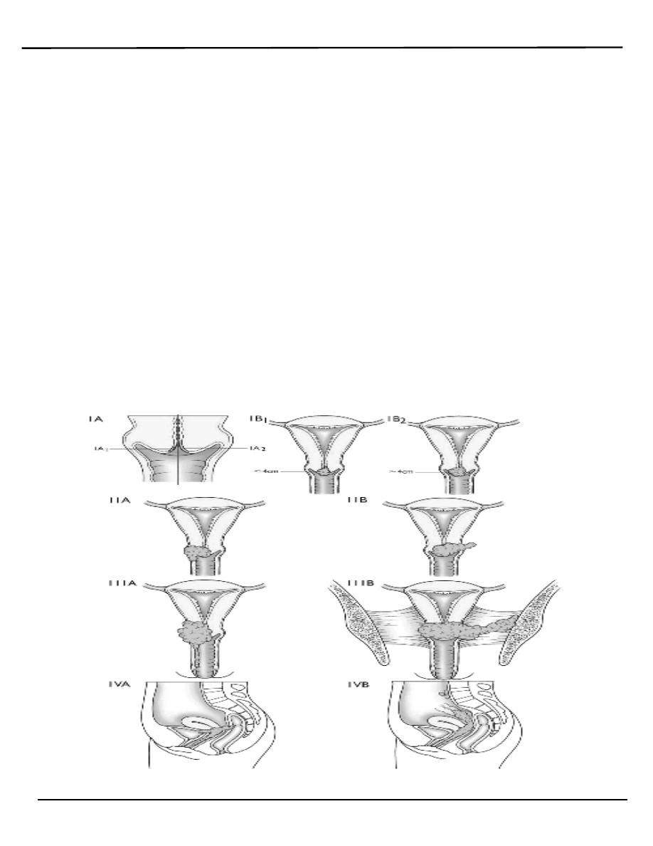
Stage 0 - full-thickness involvement of the epithelium without
invasion into the stroma (carcinoma in situ)
Stage I - limited to the cervix
IA - diagnosed only by microscopy; no visible lesions
IA1 - stromal invasion less than 3 mm in depth and 7 mm
or less in horizontal spread
IA2 - stromal invasion between 3 and 5 mm with
horizontal spread of 7 mm or less
IB - visible lesion or a microscopic lesion with more than 5 mm
of depth or horizontal spread of more than 7 mm
IB1 - visible lesion 4 cm or less in greatest dimension
IB2 - visible lesion more than 4 cm
Stage II - invades beyond cervix
IIA - without parametrial invasion, but involve upper 2/3 of
vagina
IIB - with parametrial invasion
Stage III - extends to pelvic wall or lower third of the vagina
IIIA - involves lower third of vagina
IIIB - extends to pelvic wall and/or causes hydronephrosis or
non-functioning kidney
IVA - invades mucosa of bladder or rectum and/or extends beyond true
pelvis
IVB - distant metastasis
Stage 0 carcinoma-in-situ
Stage I the tumor is confined to the cervix
IA microinvasive disease, with the lesion not grossly visible: no
deeper than 5 mm and no wider than 7 mm
IA1 invasion <3 mm and no wider than 7 mm
IA2 invasion >3 mm but <5 mm and no wider than 7 mm
IB larger tumor than in IA or grossly visible, confined to cervix
Lecture 4
النسائية
د. أحمد جاسم
Cervical Cancer
Page 6 of 10
IB1 clinical lesion no greater than 4 cm
IB2 clinical lesion greater than 4 cm
Stage II extends beyond the cervix, but does not involve the pelvic side
wall or lowest third of the vagina
IIA involvement of the upper 2/3 of vagina, without lateral extension
into the parametrium
IIB lateral extension into parametrial tissue
Stage III involves the lowest third of the vagina or pelvic side wall, or
causes hydronephrosis
IIIA involvement of the lowest third of the vagina
IIIB involvement of pelvic side wall or hydronephrosis
Stage IV extensive local infiltration or has spread to a distant site
IVA involvement of bladder or rectal mucosa
IVB distant metastases

Lecture 4
النسائية
د. أحمد جاسم
Cervical Cancer
Page 7 of 10
Treatment of Early Disease
o Conization or simple hysterectomy (removal of the uterus) -
microinvasive cancer
o Radical hysterectomy - removal of the uterus with its associated
connective tissues, the upper vagina, and pelvic lymph nodes. Ovarian
preservation is possible.
o Chemoradiation therapy
Factors that influence the mode of treatment include:
1. Stage and type of lesion.
2. Age of patient.
3. Health status.
The treatment of cervical cancer frequently requires a multidisciplinary
approach.
should only be considered an option for early disease (stage 1 and
stage 11a).
The standard treatment of cervical cancer may involve:
1. surgery
or
2. radiotherapy
Or
3. a combination of both.
Early cervical cancers (stage I and IIA) may be treated by either procedure.
Radiotherapy is the treatment of choice once the disease has spread beyond
the confines of the cervix and vaginal fornices, when surgery is not
effective.
Stage Ib2-IVa
Lecture 4
النسائية
د. أحمد جاسم
Cervical Cancer
Page 8 of 10
o The standered surgical procedure of cervical carcinoma is a Wertheim's
radical abdominal hysterectomy which involves removal of the uterus,
paracervical tissue, and upper vagina and pelvic lymph nodes.
o Early microinvasive disease can be treated by cone biopsy or excisional
treatment alone.
Complications of radical hysterectomy:
The most frequent complication of radical hysterectomy is:
1. Urinary dysfunction
2. Hemorrhage
3. Infection.
4. Bowel obstruction.
5. Bladder and rectovaginal fistulas.
o Can be used for all stages. Once the disease has spread outside cervix,
radiotherapy is the mainstay of treatment.
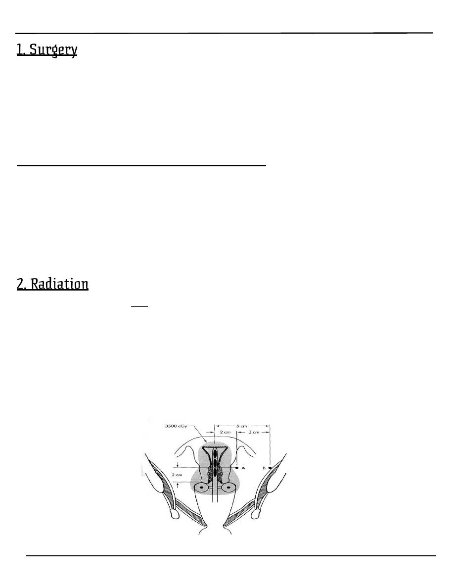
o Radiotherapy of cervical cancer may often involve a combination of:
A. external radiotherapy (for whole pelvis radiation)
B. transvaginal intracavitary irradiation (to the central part of the
disease)
o Palliative radiation often is used individually to control bleeding, pelvic
pain, or urinary or partial large bowel obstructions from pelvic disease.

Lecture 4
النسائية
د. أحمد جاسم
Cervical Cancer
Page 9 of 10
Complications from radiation
1. Acute adverse effect:
A. gastrointestinal effects include diarrhea, abdominal cramping,
rectal discomfort, or bleeding.
B. Cystourethritis can occur, which leads to dysuria, frequency,
and nocturia.
2. Late sequelae of radiation usually appear 1-4 years after treatment.
The major sequelae include rectal or vaginal stenosis, small bowel
obstruction, malabsorption, and chronic cystitis.
Symptoms of Recurrence
1. Weight loss, fatigue and anorexia
2. Abnormal vaginal bleeding
3. Pelvic pain
4. Unilateral leg swelling or pain
5. Foul discharge
6. Signs of distant metastases
NOTE: must distinguish radiation side effects from recurrent cancer
Management of Recurrence
o Chemoradiation may be curative or palliative, especially in women who
have not received prior radiation therapy.
o Isolated soft tissue recurrence may occasionally be treated by resection
with long-term survival.
CERVICAL CANCER DURING PREGNANCY
Prior to 24 weeks: the treatment recommended is the same as for women
who are not pregnant.
After 24 weeks:
o When cancer is detected at the time of fetal viability, radical Caesarean
o hysterectomy can be offered or the fetus can be delivered and therapy

Lecture 4
النسائية
د. أحمد جاسم
Cervical Cancer
Page 10 of 10
instituted thereafter.
o The route of delivery has traditionally been Caesarean section, though
this is more
o related to the possibility of increased bleeding, rather than the older
concept of spread of disease if the vaginal route is chosen.
Prognosis
FIVE YEAR SURVIVAL RATES FOR CERVICAL CANCER
Stage I 80%
Stage II 65%
Stage III 30%
Stage IV 15%
By: Mu’taz Fathi