


2015-2016
Baghdad medical college
Refractive Errors (Ammetropia)
Introduction:
- Axial diameter of the eyeball: is the distance from tip of cornea to the center of
macula, which is normally 23-25 mm (average 24 mm).
- The most important two refractive surfaces of the eyeball are cornea and lens.
- The power of cornea and the lens depends on the curvature of their surfaces,
mostly the anterior surface of each one.
The cornea is a part of a sphere and normally it has a radius of curvature for its
anterior surface which is about 7.8 mm, while the radius of curvature of anterior
surface (capsule) of the lens is 10 mm. so, the cornea is steeper. There is a reverse
relationship between the radius of curvature and steepness of any sphere or a part
of sphere structure e.g. the lens and cornea respectively. If the radius of curvature
of the cornea or lens is decreased, there will be increase in the curvature (more
steepness). The increase in curvature of any of those 2 ocular structures is
associated with increase in refractive power of them and vice versa.
7.8 mm
10 mm
More curvature = less radius and vice versa
cornea
lens
Dr. Najah
Lectures: 2 & 3
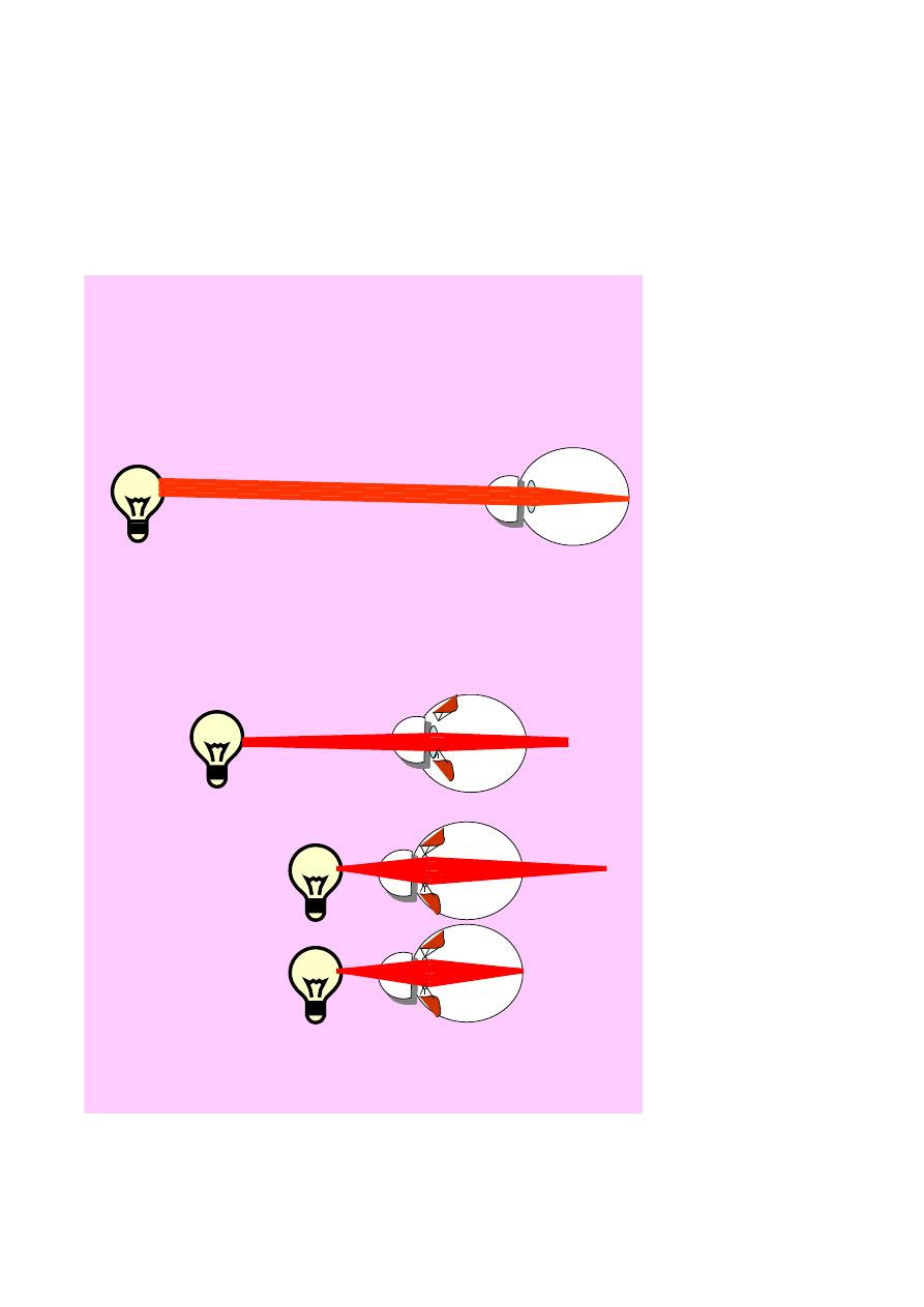
- For any rays of light to come in parallel pattern to the eye, their source must be
at distance of six meters and more from it, and if the source of light is at any
distance less than 6 meters, then the rays will come divergent. The more close
source to the eye, the more divergent rays coming from it.
Infinity ( 6 meter & more )
Less than 6 meter
More near = more divergent
Accommodation
- the total refractive power (converging power) of normal eye (emmetropic eye) is
60 diopters (Diopters) divided between the cornea and the lens. Definitely, the

cornea sharing in highest portion (43D) of this refracting power while the lens
having power of residual 17 D only. So, the cornea is more important than the lens
regarding focusing of light on the retina for the following 2 reasons:
1- the anterior surface of the cornea is steeper than that of the lens.
2- the light is transfer to the cornea passing through the air which having the least
refractive index (1.00) or (least density of any other material) while the light
transfer to lens passing through the aqueous humor (water) which having
refractive index higher than that of the air (1.33). The refractive index for the
cornea is 1.37 while the lens is 1.38.
The deviation of light (refraction) is more when it is passing between 2 media
having more deference in their refractive indices (density). So, the light is deviated
more (more convergence) when passing between air and cornea than when passing
between aqueous and the lens.
The difference in refractive index between cornea and air is: 1.37-1.0= 0.37
While between lens and aqueous is : 1.38-1.33= 0.05.
0.05 is much less than 0.37.
Emmetropic eye (eye with normal refraction):
It is an eye in which parallel rays of light tend to a focus directly on the retina
when the eye is at rest {i.e. without accommodation (the eye is using its normal
power which is 60 D)}.
- Accommodation: contraction of Ciliary muscle in order to increase curvature
of lens (and so increase its refractive power more than 17D) to visualize objects
closer than 6 meters (near objects).
- In order to see near objects, there will be contraction of Ciliary muscles which
lead to decrease the tone of Zonule and their will be increase in the curvature of
lens and increasing in the refractive power of lens (>17D).
- Amplitude of accommodation: is the difference in the converging power of the
eye between maximum accommodation and un accommodated eye (rest) , which
depends on contraction power of Ciliary muscles and elasticity of lens capsule,
and both of them decrease with advancing in age. The Amplitude of
accommodation is decrease with advancing age as the following:
* Early in life: it is 14D, so the child can focus an object located 7 cm away
from the eyes, i.e. the range of lens refractive power can be increase from 17 D
normally up to 31D.
* At age of 36y: due to atrophy of muscles and loss of lens elasticity (sclerosis),
the amplitude of accommodation will be decrease to 6 D only, so the nearest
object to the person which can be focused is at a distance of about 15cm away
from eye.
* At age of 45y: the amplitude of accommodation is 4D only, and the nearest
focus point is 25cm away from eye.
* At age of 60y: amplitude of accommodation is 1D only, and the nearest focus
point is 1meter.
- Presbyopia: is a recession of near point with age (from 7 cm early in life to 1
meter at 60 year) due to decreased amplitude of accommodation (from 14 D early
in life to 1 D at age of 60 year).
Ammetropia:
is either → Hypermetropia
Or → Myopia
Or → Astigmatism
Hypermetropia (hyperopia), far-sightedness:
Is a type of refractive errors in which parallel rays of
light are brought to a focus some distance behind the
retina when the eye is at rest.
Etiological classification:
1- Axial Hypermetropia: shorter antero-posterior axial
length, i.e. the eye has normal converging power (60D)
but its axial length is less than 24 mm.
2- Curvature Hypermetropia: due to decreased
curvature (flattening) of the cornea congenitally or as a
result of trauma or disease e.g. corneal ulcer, microbial keratitis.
3- Index Hypermetropia: decrease in effective refractivity of the lens.
* the power of the lens depends on the difference between refractive indices of
the nucleus and cortex, so the more the difference the more converging power
and vise versa. In index Hypermetropia, there is decrement of the difference.
Clinical classification:
1- Facultative hypermetropia: hypermetropia corrected by accommodation
(depends on age and degree of refractive error).
2- Absolute hypermetropia: hypermetropia out of amplitude of accommodation
and corrected by glasses.
3- Manifest hypermetropia: maximum hypermetropia that can be corrected with a
convex lens with accommodation active, i.e. = Facultative + Absolute.
4- Latent hypermetropia: hypermetropia hidden behind ciliary body tone which
equal to the difference between total and manifest hypermetropia. The refractive
power of the ciliary body is about 1-1.5D (usually it considered as part of the
Emmetropia
Hypermetropia
accommodation
By convex lens
refractive power of lens, i.e. 17D of lens = 16D + 1D (of the ciliary body tone), it
also decreases with age.
5- Total hypermetropia: amount of hypermetropia present with all
accommodation
suspended
(with
cycloplegic
drugs
(e.g.
Atropine,
Cyclopentolate and Homotropine) , to exclude accommodation and tone of ciliary
body) Manifest + Latent.
* With advancing age, the facultative will decrease and the manifest will be
represented by absolute only, and at the same time, the latent will also decrease
and the total will be represented by manifest only, so in old people the total is
made of the absolute only.
Symptoms:
1- Blurred vision: for near work (as it needs more power) and even far vision if
the degree of hypermetropia is high and beyond the amplitude of accommodation.
2- Eye strain: headache due to excessive accommodation and dissociation
between Accommodative Convergence and Accommodation "AC/A.
* To see the near objects, the eye will show the near reflex, which consists of
accommodation, convergence and miosis. When one of these processes occur, it
will stimulate the other two.
Normally, for each 1D of accommodation, there will be associating 4 prism D
convergence (i.e. AC/A=4 prism D/1D).
A normal eye to see an object at distance of 1/2 meter needs 2D and this in turn
will produce 8 prism D convergence, while an eye with 7D hypermetropia has at
rest 28 prism D convergence, so to see an object at 1/2 meter distance the 2D
needed will produce 8 prism D more, so totally there will be convergence 36
prism diopters for the same distance which is 1/2 meter.
3- General symptoms like nausea and fatigue.
Treatment:
- Convex lens in spectacles (+ve lenses to increase refractive power).
- Contact lenses.
- Excimer laser photorefractive keratectomy: reshaping of the cornea to increase
the refractive power, each 1mm decrease in radius of curvature increases the
refractive power about 6D.
e.g. Laser in situ keratomileusis (Lasik)
- Non-contact laser thermal keratoplasty (Holmium laser spots): to change the
coneal curvature.
- Phakic intraocular lens (IOL).
Diopter: the reciprocal of the distance in meters from the reference light source in
air or vacuum, such that D= 1/(distance in meters).
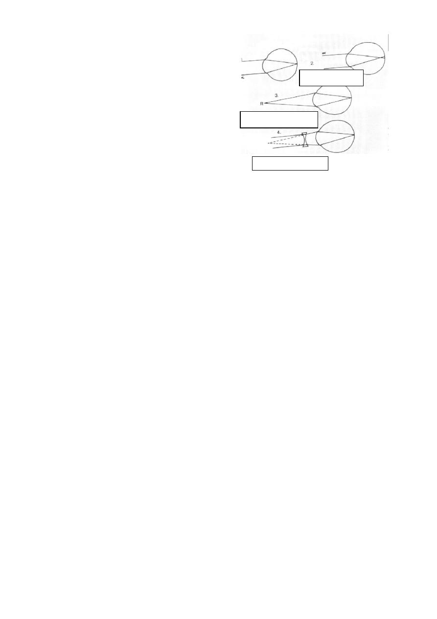
Myopia or short-sightedness:
That form of a refractive error where
parallel rays of light come to a focus in front
of the retina when the eye is at rest.
- As there is increase in the refractive
power of the eye, the near objects (closer
than 6m) will be seen normally, while far
objects (whom rays come parallel) will be
focused in front of retina.
Aetiological classification:
1- Axial myopia: anteroposterior length is longer than normal.
2- Curvature myopia: increased curvature of cornea, or one or both surfaces of
lens.
3- Index myopia: increased refractivity of lens, e.g. nuclear sclerosis (stage
before nuclear cataract), due increase the difference between refractive indices of
nucleus and cortex.
Clinical classification:
1- Simple (stationary): <6D, start after the age of 4y, stationary (stops before
20y), normal physiological variety and the VA can be corrected to 6/6.
2- Pathological (progressive): >6D, start before the age of 4y, progress even
after age of 20y, real pathological process and VA cannot be corrected to 6/6.
Symptoms:
1- Distance object "Blurred", near objects clear.
2- Headache due to sustained contraction of occipito-frontalis muscle in order to
make slit of palpebral fissure and increase vision and eye strain due to
dissociation of AC/A.
* A normal eye to see an object at distance of 1/2 meter needs 2D and this in
turn will produce 8 prism D convergence, while a eye with 2D myopia will not
need any accommodation to see this object, but there will convergence 8 prism D
to see this near object. Thus is in turn will produce 2 D accommodation which is
not needed by myopic patient. So any myopic should wear glasses for far and
near to create a normal relation between accommodative convergence and
accommodation.
3- general symptoms like fatigue and nausea.
Far & myopia
Near & myopia
By concave lens

Treatment:
- Concave lenses in spectacles.
- Contact lens.
-Radial keratotomy
- Excimer laser photorefractive
keratectomy. - Lasik.
- Pkakic IOL.
- Normal (clear) Lens extraction
8
Astigmatism
Astigmatism is that condition of refraction in which a point of focus of light
cannot be formed upon the retina. The optical condition is that instead of a
single focal point, there are two or more focal lines (depending on the type of
astigmatism), separated from each other by a focal interval.
Etiological classification:
1- Curvature astigmatism: Most commonly, it is corneal, less commonly it is
lenticular (induced by lens). The cornea or the lens having unequal radii of
curvature.
2- Decentering astigmatism: subluxation of lens.
3- Index astigmatism: due to cataract in one meridian more than other.
Clinical classification:
1- Regular: two principle meridians at right angle are involved (i.e. there is
difference in the refractive power of two meridians 90° in between):
a- Simple astigmatism: myopic or hypermetropic; one of the foci falls on
retina, other in front or behind it.
b- Compound astigmatism: neither of the two foci lies upon retina, but are
both placed in front or behind retina (myopic or hypermetropic respectively).
c- Mixed astigmatism: one focus in front and other behind retina.
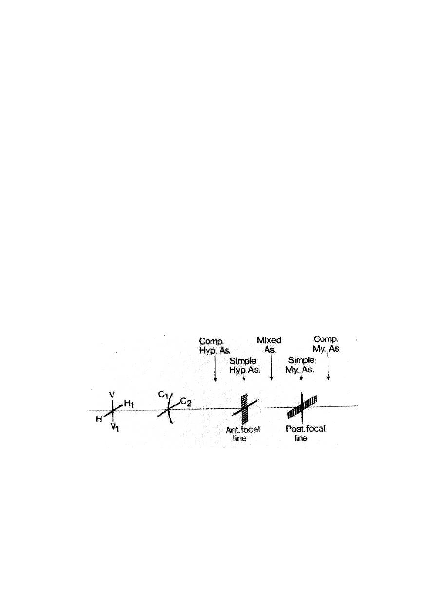
* This figure represents the appearance of the image in astigmatism (regular),
there are two perpendicular meridians; vertical (V-V
1
) and horizontal (H-H
1
),
and the refractive power of the vertical meridian is larger than that of
horizontal one as the curvature of the vertical meridian (C
1
) is larger than the
curvature of the horizontal meridian (C
2
).
In (1), the image of vertical meridian is focused as (A
1
) line on the retina
(normal), while the horizontal meridian image focused behind the retina
(hypermetropic) and blurred on retina as (B
1
) line, so it is called "Simple
hypermetropic astigmatism". In (2), vertical meridian image is focused in
front of the retina (myopic) and its image on retina is blurred (A
2
), while
3 1 5 2 4
A
1
A
2
B
1
B
2

9
horizontal meridian image (B
2
) is focused on the retina (normal), so it is
called "Simple myopic astigmatism". In (3) and (4) both image are focused
behind or in front of retina respectively and called "Compound
hypermetropic or myopic astigmatism". While in (5), one image focused in
front of and the other behind the retina and so it is called "Mixed
astigmatism".
2- Irregular astigmatism: refraction in different meridians is quite irregular.
Found in pathological condition of cornea; irregular healing after trauma or
inflammations or keratoconus.
Treatment:
- Cylindrical lenses in spectacles: only for simple astigmatism.
-Contact lenses: used for compound and mixed astigmatism, where we correct
one of the meridians by them, and then correct the other meridian by
cylindrical lenses (i.e. we switch it to simple and correct it accordingly).
- Photorefractive Excimer laser surgery: to correct one meridian.
-LASIK: like photorefractive Excimer laser surgery, but used for higher
refractive error.
- Phakic Toric IOL.
- Keratoplasty (corneal graft): for more than two meridians and central
corneal opacity.