College of Medicine University of Mosul /Department of: Microbiology
Subject: Microbiology-Immunology Stage: 3
rd
2021-2022
Lecturer: Dr. Ahmed Alharbi Date: 22/12/2021 No. 2
Pag
e
1
Hyper sensitivity reactions (Type II, III, and IV)
Objectives:
The main objectives of this lecture are to:
Understand the mechanism of TYPE II, III, and type IV HSR
Explain examples for diseases caused by the above 3 types of HSR.
Type II Hyper sensitivity reaction: (Tissue-specific,
or Cytotoxic hypersensitivity).
Mechanism of Type II HSR includes:
A.
Antibodies produced by the immune response bind to the antigens on the patient’s own cell
surfaces such as circulating red blood cells, and extracellular materials (basement membrane).
The resulting Ag-Ab complexes
EITHER:
1.
Activate complement (via the classical pathway), leading to cell lysis or extracellular tissue
damage, OR opsonization of the target cell via C3b.
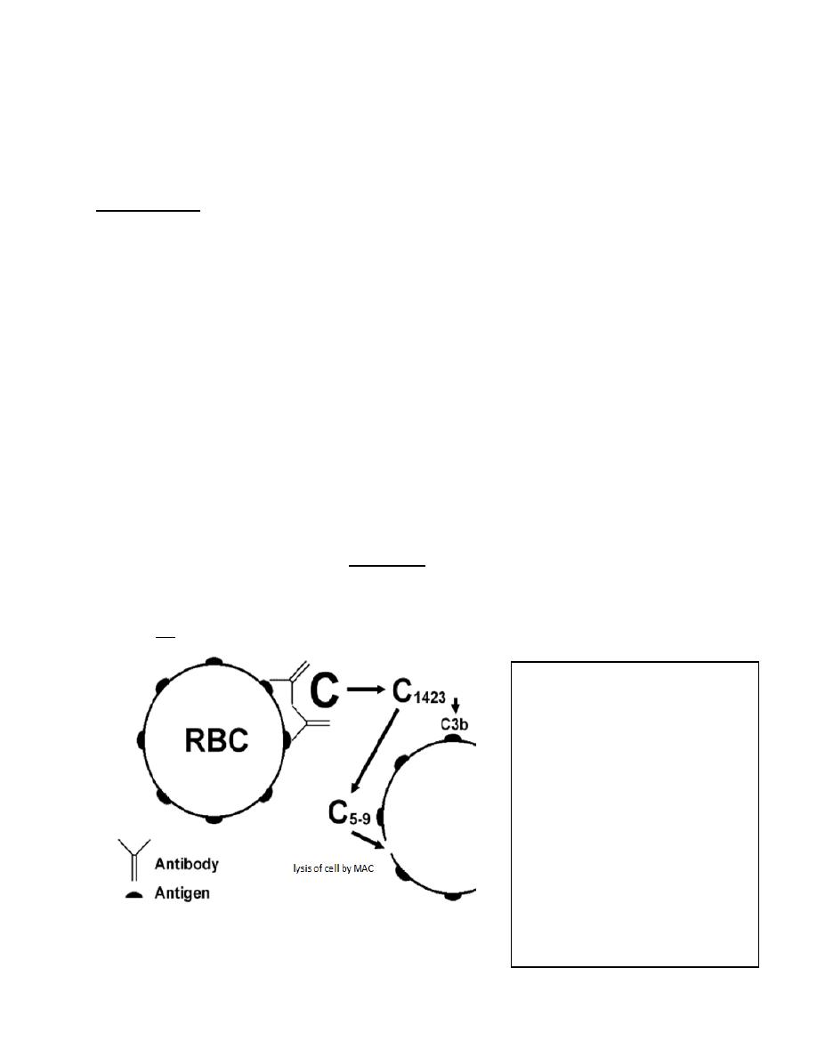
In this diagram, a red blood cell has
antigen fixed on its surface to
which antibody attaches. The
attached antibody activates the
complement cascade, which ends
with the formation of the
"Membrane Attack Complex“ MAC
of C5-9 which causes lysis of the
cell.
Other
complement
components may be generated,
such as the opsonin C3b Induces
phagocytosis (1).
College of Medicine University of Mosul /Department of: Microbiology
Subject: Microbiology-Immunology Stage: 3
rd
2021-2022
Lecturer: Dr. Ahmed Alharbi Date: 22/12/2021 No. 2
Pag
e
2
Diseases under this category include:
• Transfusion reactions: incompatibility reaction.
• Autoimmune hemolytic anemia: antibodies made against one's own RBC's.
• Erythroblastosis fetalis: due to Rh incompatibility between maternal and fetal blood.
• Goodpasture's syndrome: due to presence of anti-glomerular membrane antibodies.
2
.
The Ab-Ag reaction be independent from complement activation and dependent on cellular
activation and called (Antibody-dependent cell-mediated cytotoxicity (ADCC)):
Low concentrations of Abs coat target cells. Inflammatory cells as NK cells, monocytes, and
granulocytes then bind to the immunoglobulin Fc receptors and lyse, but do not phagocytize,
the target cells.
Diseases of ADCC include:
• Transplantation rejection
• Immune reactions against neoplasms
• Immune reactions against parasites
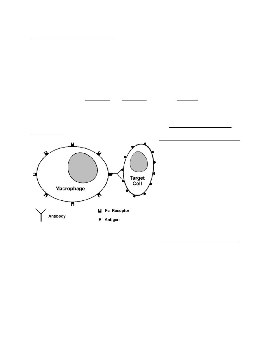
ADCC Complement independent
cytotoxic HSR.
In the given
diagram, a macrophage with Fc
receptors on its surface is able to
recognize a target cell coated with
antibody via the Fc receptor
portion of the attached antibody.
The
macrophage
can
then
demolish the targeted cell by
elaboration of proteases.
This reaction is complement
independent (1)
College of Medicine University of Mosul /Department of: Microbiology
Subject: Microbiology-Immunology Stage: 3
rd
2021-2022
Lecturer: Dr. Ahmed Alharbi Date: 22/12/2021 No. 2
Pag
e
3
B
. Anti-receptor antibodies:
IgG antibody is directed against receptors in target cells, either resulting in complement-
mediated destruction of the receptors, blocking or activation of receptors.
Diseases caused by this mechanism include:
Myasthenia gravis: acetylcholine receptor antibody (blocking Ab).
Graves’ disease (thyrotoxicosis): anti-TSH receptor antibody ( Activating Ab)
Pernicious anemia: anti-parietal cell antibody causing B12 deficiency
Complement activation in type II HSR:
• Activated C3 forms opsonin (C3b and C4b) recognized by phagocytes
• Formation of membrane attack complex (lytic enzymes)
• Formation of anaphylatoxins C3a and C5a.
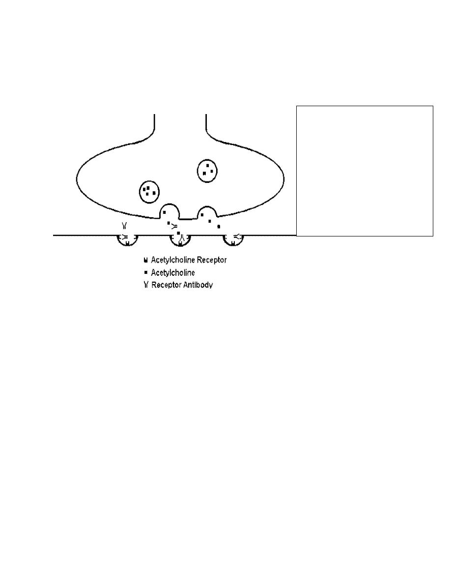
Anti-receptor Antibodies
In this diagram, antibody is directed
against acetylcholine receptors at the
motor end plate of a muscle, blocking
the receptors and diminishing the
muscular response. This is the
mechanism for muscle weakness in
myasthenia gravis (1).

College of Medicine University of Mosul /Department of: Microbiology
Subject: Microbiology-Immunology Stage: 3
rd
2021-2022
Lecturer: Dr. Ahmed Alharbi Date: 22/12/2021 No. 2
Pag
e
4
Type III HSR:
Immune Complex–Mediated (ICM HSR).
Soluble circulatory antigens (not bound to any cell surface) causing immune complex
mediated injury. They are either exogenous or endogenous Ags.
The reaction may occur 3 - 10 hours after exposure to the antigen.
Type III HSR can cause:
Localized reactions as in:
Hypersensitivity pneumonitis
Post streptococcal glomerulonephritis
Local vasculitis after vaccination ( Diphtheria and tetanus)
Systemic reactions as in:
Serum sickness
Systemic lupus erythematosus
Rheumatoid arthritis
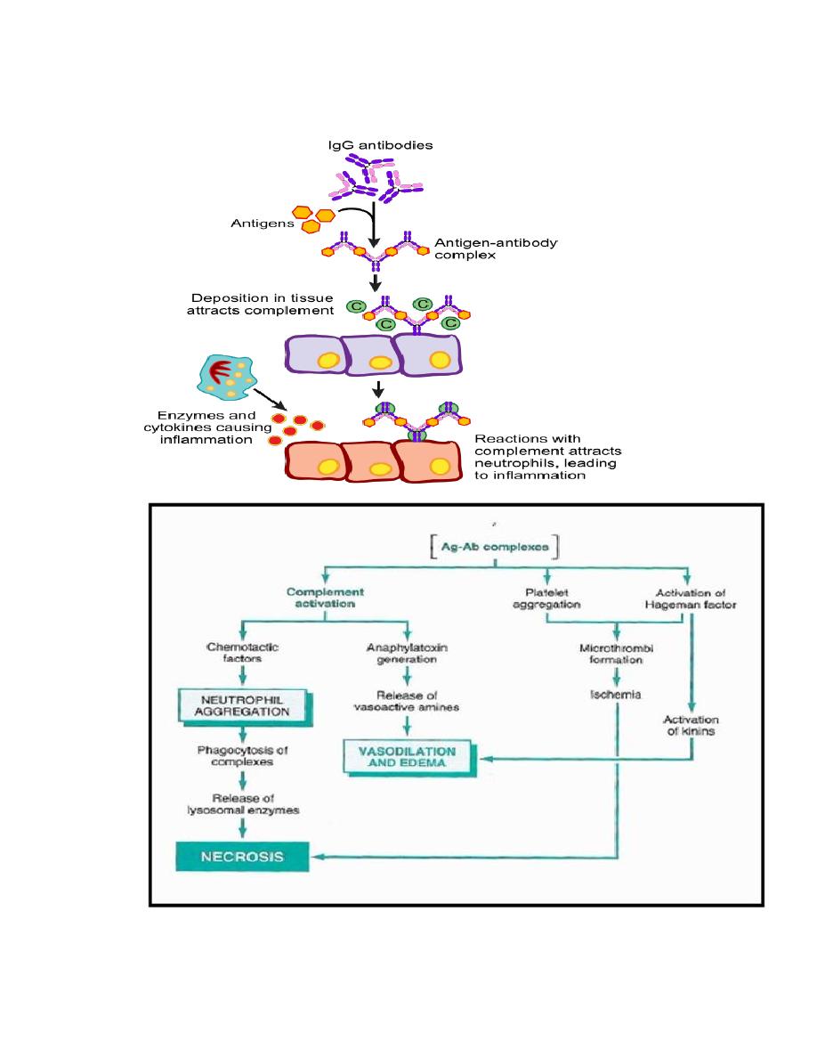
Mechanism of Type III Hypersensitivity:
Antigens bind to antibodies within circulation forming immune complexes.
The formed immune complexes will be deposited any where in the body organs.
Complement system will be activated and polymorph nuclear cells will be recruited
The activated complement system leads to:
Release of anaphylatoxins ( C3a and C5a)
Production of C3b and C4b which are opsonizing and chemotactic factors
MAC formation (membrane attack complex C5b-C9).
The recruitment of the Polymorph nuclear cells resulting in inflammation and tissue
injury.
Activation of platelets and Hagmen (Factor XII) leading to thrombosis of the micro
circulation and necrosis. FXII also can cause vasodilation through activation of kinins
College of Medicine University of Mosul /Department of: Microbiology
Subject: Microbiology-Immunology Stage: 3
rd
2021-2022
Lecturer: Dr. Ahmed Alharbi Date: 22/12/2021 No. 2
Pag
e
5
Diagram for mechanism of Type III HSR and role of Hagman factor (2)
College of Medicine University of Mosul /Department of: Microbiology
Subject: Microbiology-Immunology Stage: 3
rd
2021-2022
Lecturer: Dr. Ahmed Alharbi Date: 22/12/2021 No. 2
Pag
e
6
Local Reactions (
Arthus Reaction):
Inflammation caused by the deposition of immune complexes at a localized site.
Exogenous Ag may cause the Arthus reaction as in: Intrapulmonary Arthus-like
reactions in human which occurs due to inhaled antigen associated with farmer’s lung
and extrinsic allergic alveolitis.
Systemic form of Type III reaction (Serum Sickness): It may take place in these situations:
A. Introduction of:
1. Equine serum therapy, which is usually given as anti-toxin
2. Vaccines
3. Anti-lymphocyte globulins
4. Streptokinase
5. Hymenoptera venoms
6. Penicillin or other antibiotics.
B. After infections as bacterial infections, e.g. post-streptococcal glomerulonephritis or
after viral infections as Hepatitis B or post malarial infections.
C. It may occur after disseminated malignancies & autoimmune diseases
Clinical picture includes:
Fever
Lymphadenopathy
Splenomegaly
Arthritis
Glomerulonephritis
Endocarditis
Vasculitis
Urticarial rashes
Abdominal pain
Nausea and vomiting.

College of Medicine University of Mosul /Department of: Microbiology
Subject: Microbiology-Immunology Stage: 3
rd
2021-2022
Lecturer: Dr. Ahmed Alharbi Date: 22/12/2021 No. 2
Pag
e
7
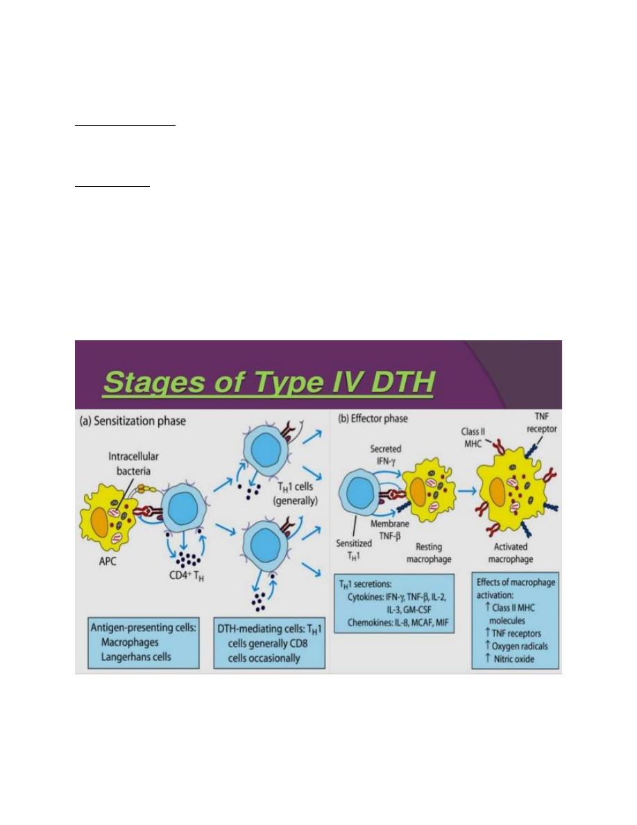
Type IV hypersensitivity reaction (Cell-mediated HSR,
Delayed type HSR):
It is mediated by antigen-specific effector T cells.
The term “delayed” refers to the cellular response that generally becomes apparent 48–
72 h after antigen exposure
Examples of type IV HSR:
Tuberculin and leishmanin tests.
Cellular responses to intracellular pathogens such as mycobacteria, fungi, and
intracellular parasites.
Graft rejection and graft versus host reactions.
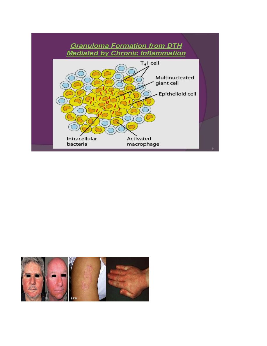
Granulomatous inflammation as occurs in Crohn’s disease and sarcoidosis.
Tumor immunity
Some autoimmune reactions
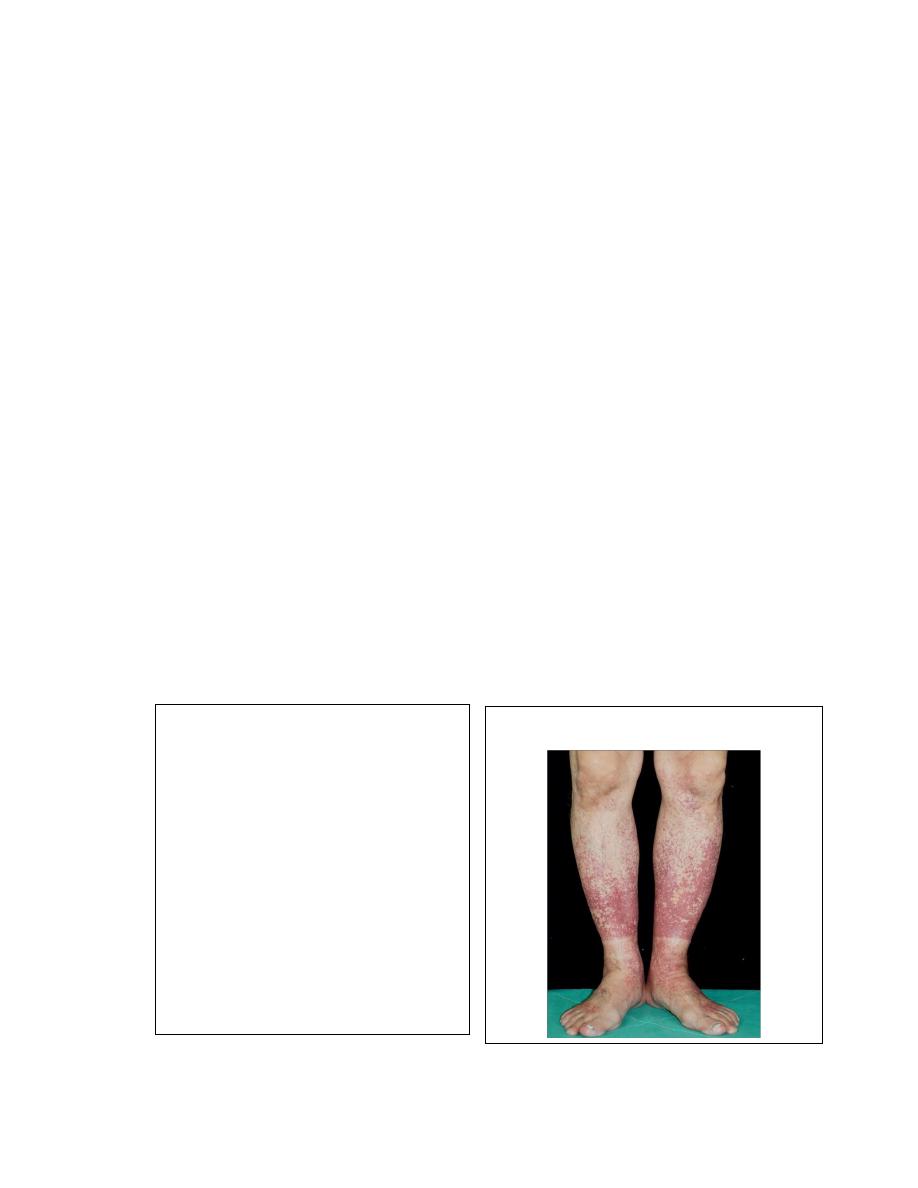
Contact allergy and allergic contact dermatitis: A well-known example of allergic contact
dermatitis is the reaction provoked by the lipid-soluble chemicals as urushiol oil , and
Ivy poison .
Intracellular microorganisms and contact Ags. associated with DTHSR:
Bacterial infections
Mycobacterium tuberculosis
Mycobacterium leprae
Listeria monocytogen
Brucella abortu
Fungal infections
Candida albicans
Pneumocyctis carinii
Histoplasma capsulatum
Cryptococcus neoformans
Viral infections
Herpes simplex virus
Small pox
Measles
Parasitic infections
Leishmania SPP
Toxoplasma gondii
Contact Ags
Hair dyes
Nickle SALTS
Poison Ivy
Poison oak
College of Medicine University of Mosul /Department of: Microbiology
Subject: Microbiology-Immunology Stage: 3
rd
2021-2022
Lecturer: Dr. Ahmed Alharbi Date: 22/12/2021 No. 2
Pag
e
8
Mechanism of Type IV HSR:
Sensitization stage:
A. Memory Th1 cells against DTH antigens are generated by dendritic cells during the
sensitization stage.
B. These Th1 cells can activate macrophages and trigger inflammatory response.
Effector stage:
Secondary contacts yield what is called DTH.
Th1 memory cells are activated and produce cytokines as :
• IFN –gamma and TNF, which cause tissue damage and inflammation.
• IL-2 activates T-cells and CTL.
• Chemokines act for macrophages recruitment.
• IL-3, GM-CSF increase the monocyte/macrophage.
Inflamed area becomes red and fluid filled.
Due to tissue damage there is activation of clotting cascades and tissue repair
Continued exposure to antigen can cause chronic inflammation and result in granuloma
formation
Mechanism of Type IV HSR (5)
College of Medicine University of Mosul /Department of: Microbiology
Subject: Microbiology-Immunology Stage: 3
rd
2021-2022
Lecturer: Dr. Ahmed Alharbi Date: 22/12/2021 No. 2
Pag
e
9
Abbreviations and their meaning:
MCAF : Monocyte chemotactic and activating factor
CTLs : cytotoxic T-lymphocytes
TH1 : T-helper 1
GM-CSF: granulocyte macrophage colony stimulating factor
MIF : Macrophage migration inhibitory factor
MHC : Major histocompatibility complex
APCs antigen presenting cells
Contact dermatitis
The response to poison oak is a classic type IV HSR.
Small molecules act as haptens and complex with skin proteins to be taken up by APCs
and presented to Th1 cells to get sensitization.
During secondary exposure Th1 memory cells become activated to cause DTH.
Contact dermatitis (5, 6)
College of Medicine University of Mosul /Department of: Microbiology
Subject: Microbiology-Immunology Stage: 3
rd
2021-2022
Lecturer: Dr. Ahmed Alharbi Date: 22/12/2021 No. 2
Pag
e
10
Time required by number of DTH examples to appear
Delayed Reaction
Maximal reaction time
Contact dermatitis 48-72 hours
Tuberculin test
48-72 hours
Granuloma formation at least 14 days
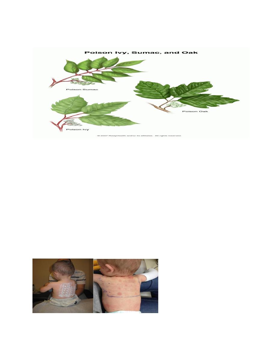
Patch test:
Any individual suspected of having allergic contact dermatitis may need patch test.
Patches with specific allergens will be applied on skin for 2-3 days.
Patch testing helps identify which substances may be causing a delayed-type allergic
reaction in a patient, and may identify allergens not identified by blood testing or skin
prick testing.
It is intended to produce a local allergic reaction on a small area of the patient's back,
where the diluted chemicals are planted. The chemicals included in the patch test kit are
the offenders in approximately 85–90 percent of contact allergic eczema, and include
chemicals, food, drink, preservatives----- etc.
Patch test (7)

College of Medicine University of Mosul /Department of: Microbiology
Subject: Microbiology-Immunology Stage: 3
rd
2021-2022
Lecturer: Dr. Ahmed Alharbi Date: 22/12/2021 No. 2
Pag
e
11
References:
1. webpath.med.utah.edu/IMMHTML/IMM102.
2. Robbins pathologic basis of diseases, 6
th
ed.
3. Jui-Hung Ko and Chung W(2012). Serum sickness. The Lancet. Clinical picture.
, E1, JANUARY 19, 2013
www.onlinebiologynotes.com/type-iv-hypersensitivity-reaction-or-delayed-type-
hypersensitivity-dth
5. Wkidoctors.com
6. Healthjade.net%2fallergic-contact-dermatitis
7.
https://tylercwithee.blogspot.com/2011/04/patch-test-results.html
8. Davidson’s principles and practice of medicine 23
rd
edition.
9. Kuby immunology 8th edition