Microbiology
Notes…
1
Immunology Lecture.3
The Specific Immune Response....Humeral Immunity
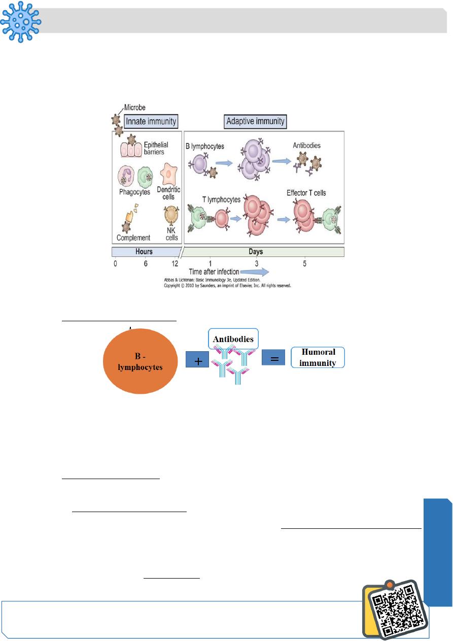
Types of Specific (adaptive) immunity
Specific immune response - humoral immunity
• B cell mediated immunity through the production of antibodies.
• Particularly effective against pathogens such as viruses and extracellular
bacteria in the blood or lymph and also against soluble pathogen products such
toxins
Humoral immunity: B- Lymphocytes
• Origin and Maturation: Bone marrow
- B- lymphocytes from the bone marrow are released into circulation in a resting state
and they do not secrete antibodies
- Instead, resting B-lymphocytes display membrane bound antibodies
(immunoglobulins) usually in the form of mIgD or mIgM
- After activation by antigen, B- lymphocyte divides (clonal expansion(
Some differentiated into plasma cells which secrete antibodies, die within 1- 2 weeks.
N
eed S
om
e H
el
p?
Microbiology
Notes…
2
Some change into memory cells- display same membrane bound antibodies as parent
cell.
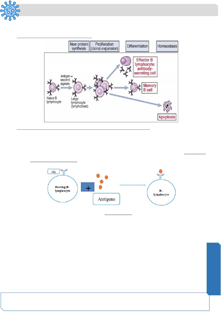
Phases of B-lymphocyte activation
Mechanism of humoral immune response by B – lymphocytes
• Resting B - lymphocyte is coated with membrane bound antibodies or
immunoglobulin (mIg) on the surface of the lymphocytes
• The first step in the initiation of the humoral immune response is the binding of
the antigen to the mIg
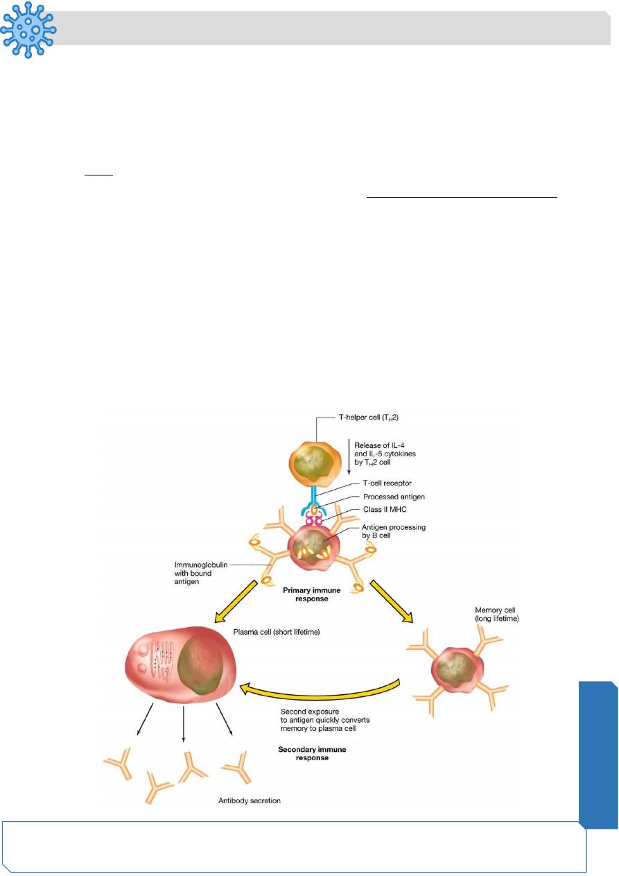
• The mIg- antigen complex is then endocytosed and complexed with MHC class II
molecule and then surface expressed
• Here , B - lymphocyte acts as APC where it presents the antigen-MHC class II
complex to TH cells
• Now, TH cells start to secrete cytokines ( IL-4 and IL-5) that stimulate B-
lymphocyte to divide (clonal expansion) and differentiate into plasma cells
(1 B cell --> 4,000 Ab-secreting cells --> ~1012 antibody molecules/hour)
• Plasma cells start to secrete antibodies (short half life, die in 1-2 weeks).
Microbiology
Notes…
3
• Some dividing B- lymphocytes change into memory cells where they display same
mIg as parent B- cell and change rapidly into plasma cells when encountering
same antigen for second time (secondary immune response.
• Primary immune response is usually mediated by IgM while the secondary
immune response is stronger and mediated by IgG.
• Note : In secondary immune response , memory cells convert immediately to
plasma cells and produce IgG in high amounts without the aid of helper T cells
Class II MHC proteins, helper T cells that stimulate antibody producing cells—the B cells
B cells are coated with antibodies that react with specific antigensWhen the antigen
binds to the antibody, the B cell first acts as an APC.
The bound antigen is endocytosed and complexed with MHC II and then surface
expressed
The surface expressed complex interacts with and activates TH cells that produce the
cytokines interleukin 4 & 5
IL4 and 5 stimulates the B cells to produce identical memory B cells and antibody
secreting plasma cells that secrete the same antibody
Microbiology
Notes…
4
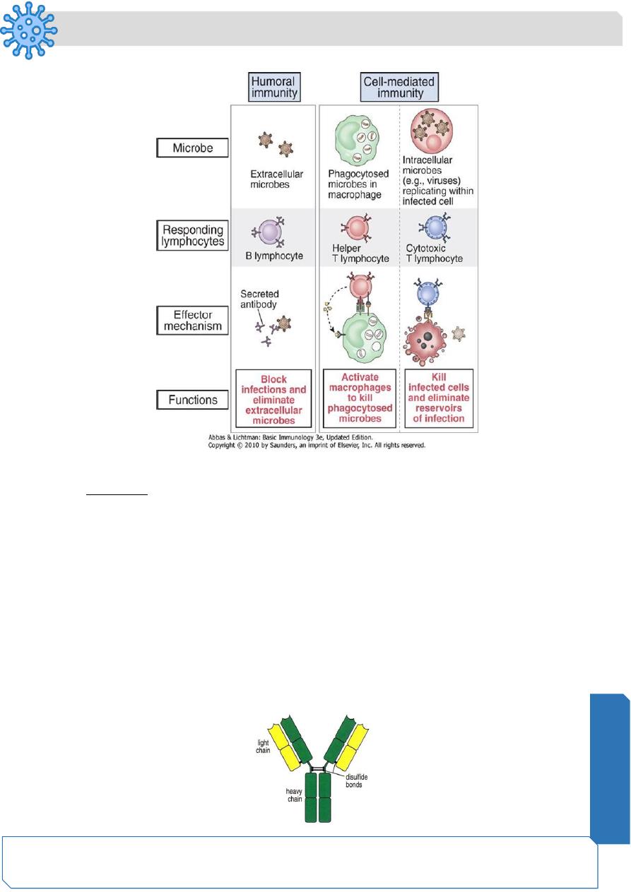
Specific immune response-Summary
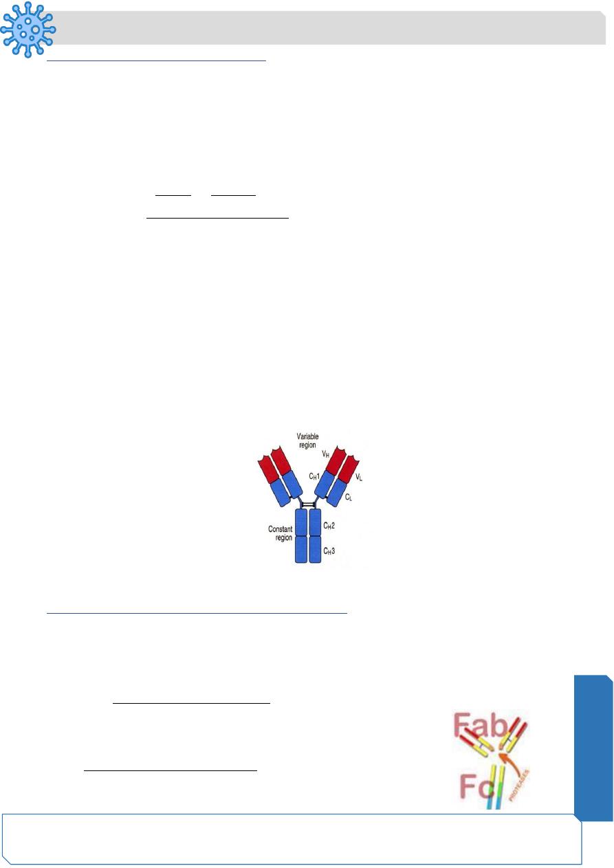
Antibody (Immunoglobulin) Structure
• 5 classes: IgG, IgM, IgA, IgD and IgE
• Common structure , four polypeptide chains:
- Two identical heavy (H) chains, each carrying covalently attached
oligosaccharide groups (50-70 kDa)
- Two identical, non-glycosylated light (L) chains (23kDa)
• Within the immunoglobulin, disulfide bonds join together:
- Two heavy chains
- Heavy chains to the light chains
• The disulfide bonds joining the antibody heavy chains are located in a flexible
region of the heavy chain known as the hinge region.
Microbiology
Notes…
5
Heavy chain determines the Ig class:
IgG : gamma HC
IgA: alpha HC
IgD: delta HC
IgM: mu HC
IgE:epsilon HC
Light chain either kappa or lambda irrespetive of Ig class
• Based on variability of amino acid sequences, both H and L chains can be divided
into: - VH and CH domains (variable and constant)
VL and CL domains (variable and constant)
The variable domains are attached to the constant domains.
• As the name implies, the variable domains vary in their amino acid sequence from
one antibody molecule to another, providing the vast diversity the immune system
needs to fight foreign invaders.
• The antigen binding site is formed where a heavy chain variable domain (VH) and
a light chain variable domain (VL) come close together. These parts show the
biggest difference among different antibodies.
Antigen binding site
Proteolytic treatment of Ig with protease enzymes
• When the immunoglobulin is treated with proteolytic enzymes (proteases), such
as pepsin or papain, it is broken at the hinge region into two fragments known as
Fab (Fragment for antigen binding) and Fc (Fragment Crystalizable(
• The immunoglobulin specificty is determined by the Fab fragment, as well as its
capability to react with the antigen.
• Fc) cannot bind with antigens, but is responsible for
biological effector functions like complement fixation,
binding to macrophages, natural killer cells and neutrophils.
Microbiology
Notes…
6
IgG
IgM
IgA
IgD
IgE
Structure
Monomer
Pentamer
Dimer
Monomer
Monom
er
Serum %
80%
5-10%
10-15%
0.2%
0.002%
Location
Blood,lymph,intest
ine
Blood,lymph
,B cells as
monomer
Secretion
s( tears,
milk),
blood,
lymph
Blood, lymph,
B cells
Mast
cells ,
basophil
s, blood
Placenta
transfer
Yes
No
No
No
No
Compleme
nt fixation
Yes
Yes
No
No
No
Function
Neutralize viruses
and toxins,
enhance
phagocytosis,
protect fetus
1ry immune
response
Localize
d
protectio
n on
mucous
surfaces
Serum
function not
known,initiati
on of immune
response on B
cells
Allergic
reaction
and
lysis of
parasiti
c worms
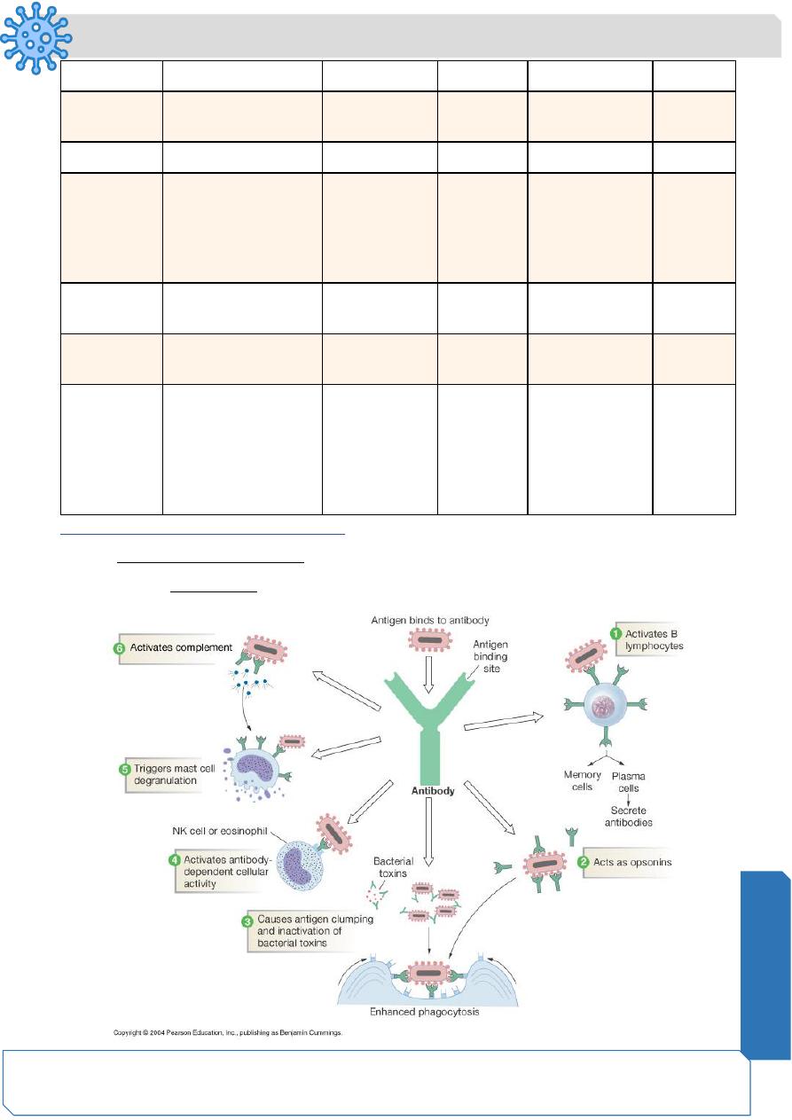
Antibody (Immunoglobulin) functions
1.
mIgs activate B- lymphocytes when comes in contact with antigen
2.
Secreted Ig neutralizes the effect of viruses , extracellular bacteria and toxins
Microbiology
Notes…
7
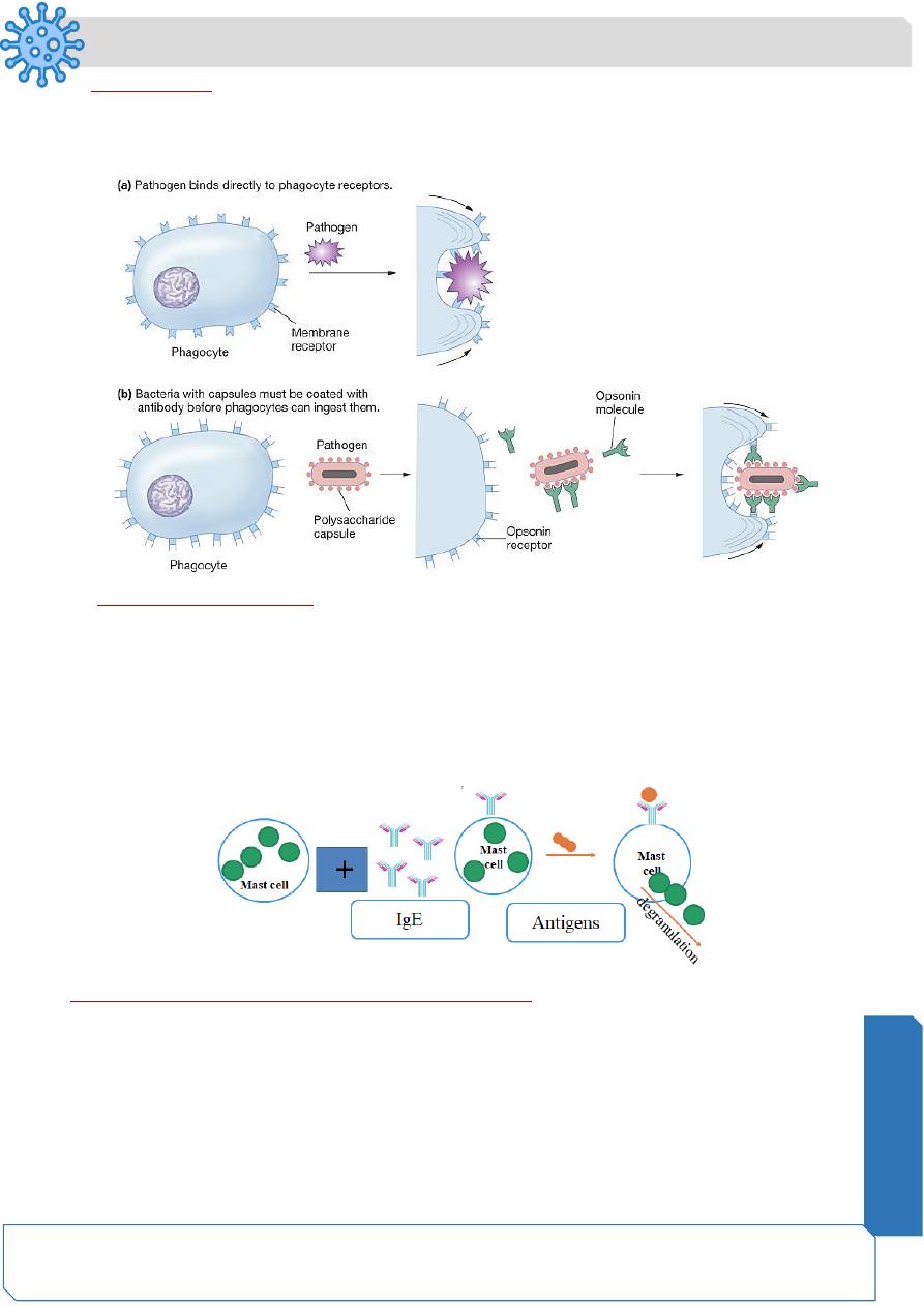
3. Opsonization:
bind pathogens for recognition by other immune cells (e.g. phagocytes)
Opsonins- are the tagging proteins that make unrecognizable particles into “food” for
phagocytes.
4. Mast cell degranulation:
• Mast cells contain histamine in intracellular granules
• Binding of IgE to cell surface receptors on a mast cell primes the cell to respond
to allergen
• Introduction of allergen and its subsequent binding to IgE stimulates the mast cell
to de-granulate and release of histamine
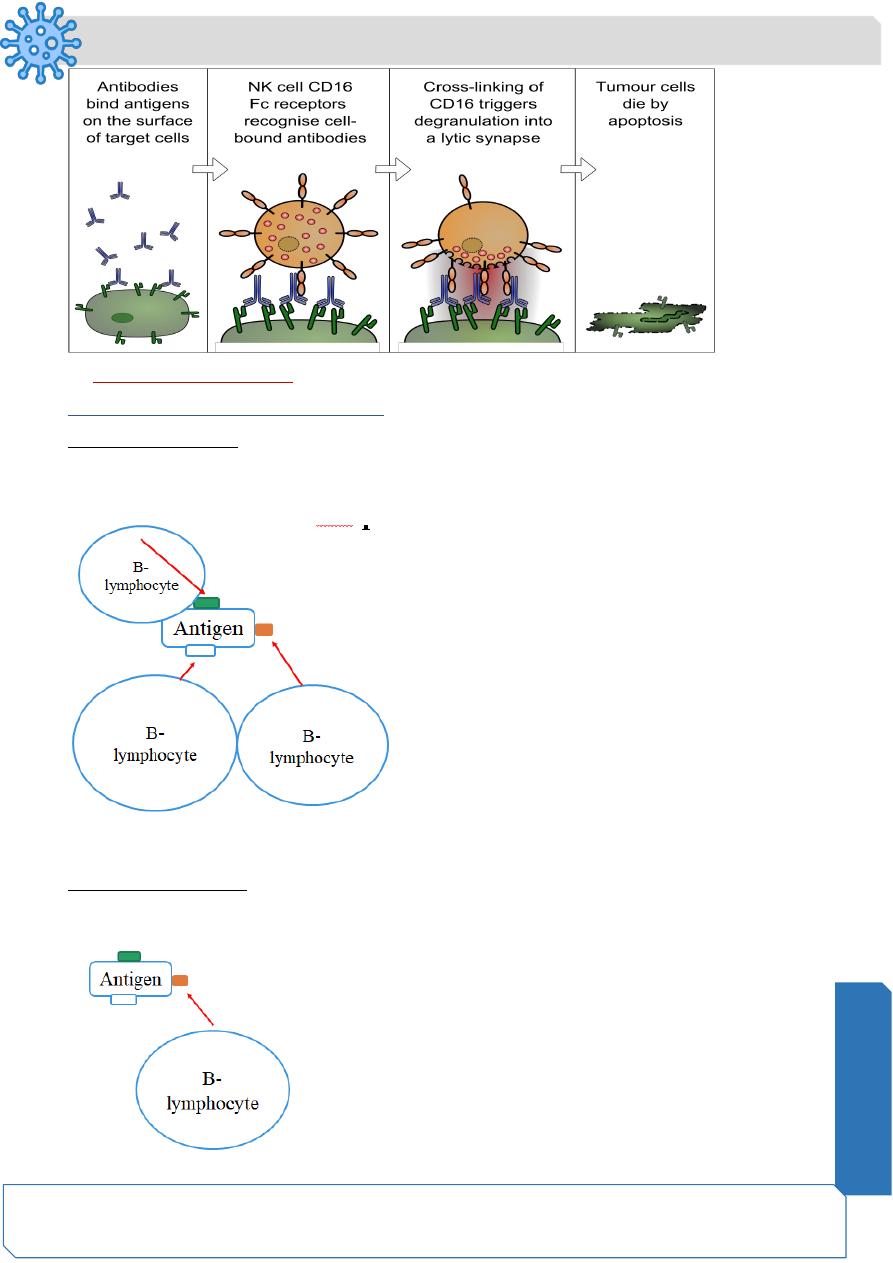
5. Antibody dependent-cellular cytotoxicity (ADCC)
- Classically mediated by NK, but also by eosinophils and neutrophils
- Part of the adaptive immune response (depend on antibodies)
Microbiology
Notes…
8
6. Complement activation
Will be discussed in details in next lecture
Monoclonal Vs polyclonal antibodies
Polyclonal antibody
Multiple clones from multiple B - lymphocytes each of which recognizes different
epitope on same antigen
Monoclonal antibody
Single clone from single B - lymphocyte recognizes single specific epitope on antigen