
Lower neck
Dr Firas Al-Hameed
M.B.Ch.B C.A.B.S MRCS (ENT) (England)
Thi-Qar Medical School
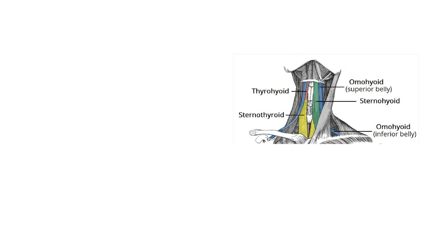
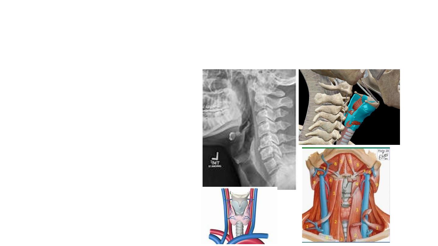
The infrahyoid muscles
They can be divided into two groups:
• Superficial plane – omohyoid and
sternohyoid muscles.
• Deep plane – sternothyroid and
thyrohyoid muscles.
• The arterial supply to the infrahyoid
muscles is via the superior and inferior
thyroid arteries, with venous drainage via
the corresponding veins.
Omohyoid
• The omohyoid is comprised of two muscle
bellies, which are connected by a muscular
tendon.
• It runs superomedially underneath the
sternocleidomastoid muscle.
Attachments:
• The inferior belly arises from the scapula.
• The superior belly ascends to attach to the
hyoid bone.
• The most lateral member of the infrahyoid
muscles, located lateral to both the
sternothyroid muscles and the thyrohyoid
muscles.
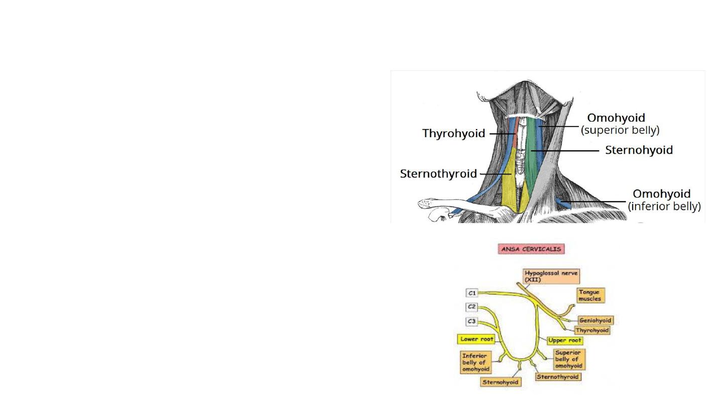
• Actions: Depresses the hyoid bone.
• Innervation: ansa cervicalis (anterior rami of
C1-C3)
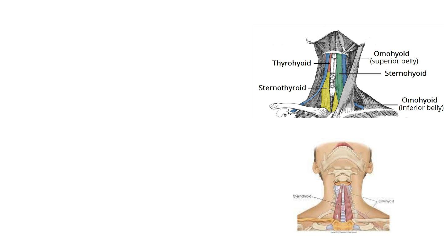
Sternohyoid
• Attachments: Originates from the sternum
and sternoclavicular joint. It ascends to insert
onto the hyoid bone.
• Actions: Depresses the hyoid bone.
• Innervation: ansa cervicalis (anterior rami of
C1-C3)
Sternothyroid
• It is below the sternohyoid muscle.
• It is wider and deeper than the sternohyoid.
• Attachments: Arises from the manubrium of
the sternum, and attaches to the thyroid
cartilage.
• Actions: Depresses the thyroid cartilage.
• Innervation: ansa cervicalis (anterior rami of
C1-C3)
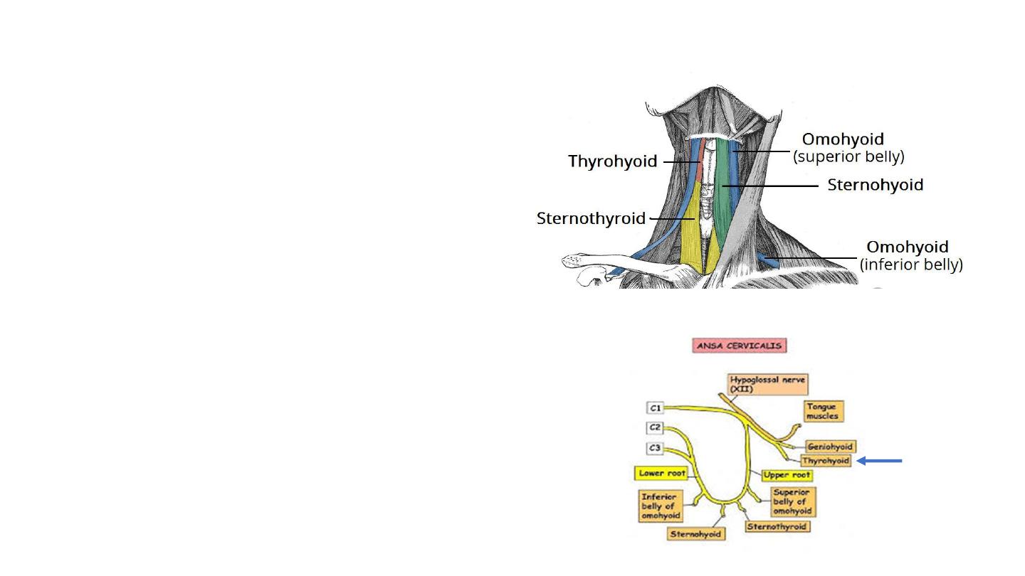
Thyrohyoid
• It is a short band of muscle, thought
to be a continuation of the
sternothyroid muscle.
• Attachments: Arises from the thyroid
cartilage of the larynx, and ascends
to attach to the hyoid bone.
• Actions: Depresses the hyoid. If the
hyoid bone is fixed, it can elevate the
larynx.
• Innervation: Anterior ramus of C1,
carried within the hypoglossal nerve.
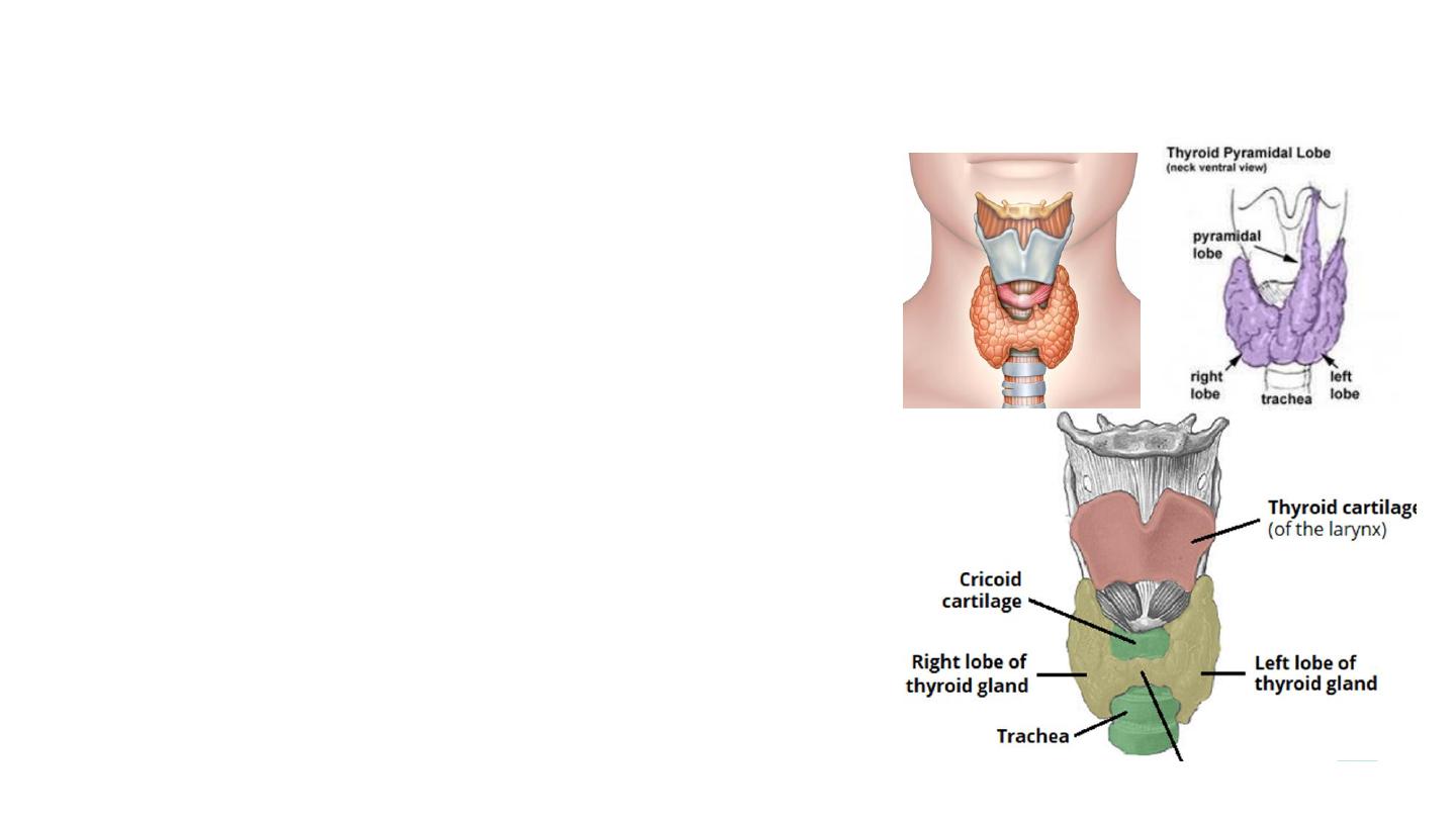
Thyroid gland
• A highly vascular, brownish-red gland.
• Located in the anterior neck.
• Its the largest endocrine gland in the body.
• The thyroid gland regulates the body’s metabolism
• Two lobes (left and right), which are connected by a
central isthmus anteriorly.
• The lobes are wrapped around the cricoid cartilage and
superior rings of the trachea. 50-60 mm long, 30 mm
wide, and 20 mm thick.
• Weight of thyroid gland: 25-30 g
• The isthmus is overlying the second to fourth tracheal
rings, with an average height of 12-15 mm. A pyramidal
lobe is often present, and it projects upward from the
isthmus, usually to the left of the midline.
Isthmus
Anatomical Relations
• Anteriorly – infrahyoid muscles
• Laterally – carotid sheath, containing the
common carotid artey, internal jugular vein
and vagus nerve
• Medially –
• Organs – larynx, pharynx, trachea and
oesophagus
• Nerves – external laryngeal and recurrent
laryngeal
• Pretracheal fascia.
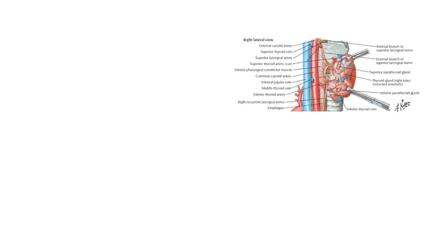
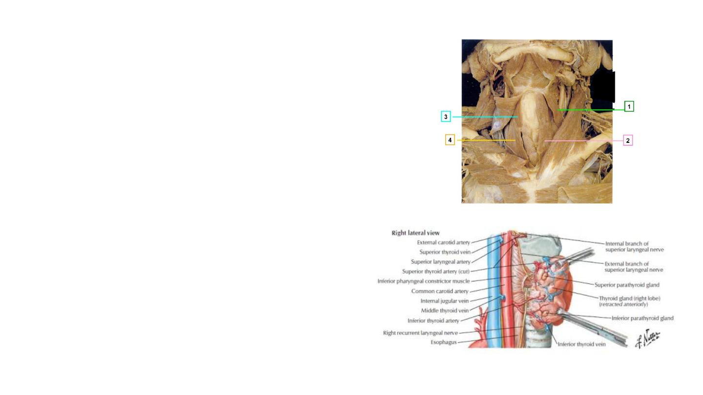
Arterial Supply
• Superior thyroid artery
– arises as the first branch of the
external carotid artery. It lies in close proximity to the external
branch of the superior laryngeal nerve (innervates the larynx).
• Inferior thyroid artery
– arises from the thyrocervical trunk (a
branch of the subclavian artery). It lies in close proximity to the
recurrent laryngeal nerve (innervates the larynx).
• Thyroid ima artery
In a small proportion of people (around
10%) there is an additional artery present . It arises from the
brachiocephalic trunk and supplies the anterior surface and
isthmus of the thyroid gland.
Venous Drainage
• Superior, middle and inferior thyroid veins.
• The superior and middle veins drain into the internal jugular
vein and the inferior empties into the brachiocephalic vein.

• Innervation
• The thyroid gland is innervated by branches derived from the sympathetic
trunk.
• These nerves do not control the secretory function of the gland – the release
of thyroid hormones is regulated by the pituitary gland.
• Lymphatic Drainage
• Paratracheal and deep cervical nodes.
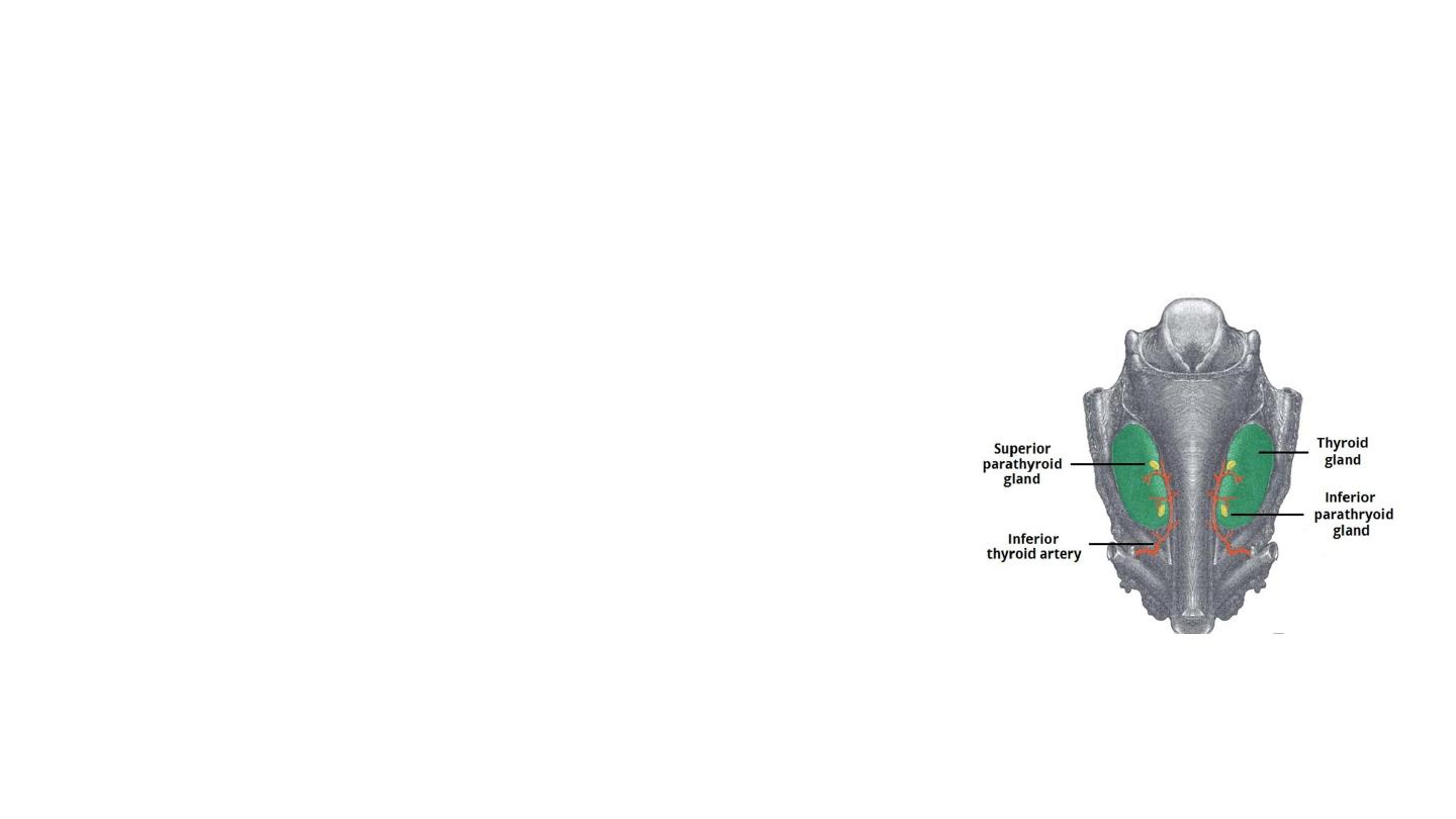
Parathyroid Glands
• Control calcium within the blood by secreting Parathyroid
hormone
• The parathyroid glands are usually located on the posterior aspect
of the thyroid gland. Situated external to the thyroid gland itself
but within the pretracheal fascia.
• Most individuals have four parathyroid glands, although variation
in number (from two to six) is common:
• Superior parathyroid glands (x2) – derived from the fourth
pharyngeal pouch. They are located at the middle of the posterior
border of each thyroid lobe.
• Inferior parathyroid glands (x2) – derived from the third
pharyngeal pouch. They are usually found near the inferior poles
of the thyroid gland.
• Can be found as far inferiorly as the superior mediastinum.

Vasculature
• The vascular supply is similar to that of the thyroid gland. Chiefly via
the inferior thyroid artery.
Venous drainage
is into the superior, middle, and inferior thyroid veins.
Lymphatics
• Paratracheal and deep cervical nodes.
Nerves
• Sympathetic nerves derived from thyroid branches of the cervical
ganglia.
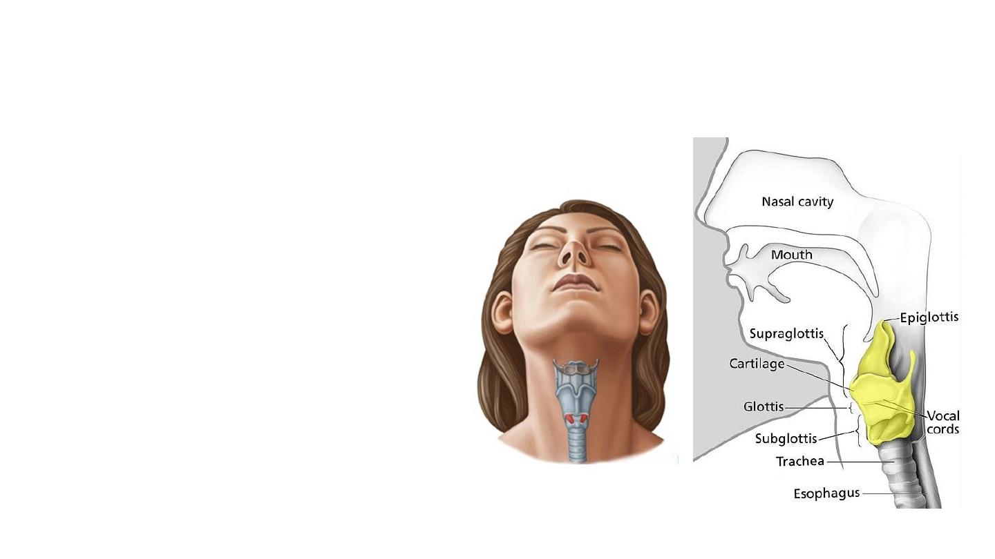
The larynx
• The larynx (voice box) is an organ
located in the anterior neck.
• It is a component of the
respiratory tract, and has several
important functions, including
phonation, the cough reflex, and
protection of the lower respiratory
tract.
• The structure of the larynx is
primarily cartilaginous, and is held
together by a series of ligaments
and membranes. Internally, the
laryngeal muscles move
components of the larynx for
phonation and breathing.
• Between C3 and C6.
• Suspended from the hyoid
bone
• It is continuous inferiorly
with the trachea, and
opens superiorly into the
laryngeal part of the
pharynx.
• Relations:
• infrahyoid muscles
• lobes of the thyroid gland
• major blood vessels of neck
• oesophagus.
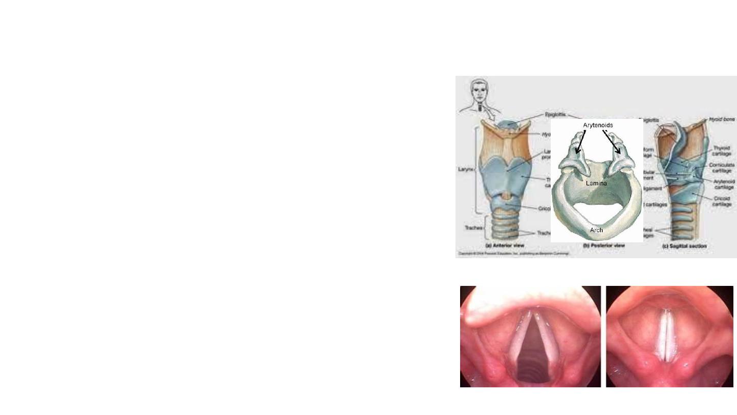
Laryngeal cartilages
• There are nine cartilages located within the larynx; three
unpaired, and three paired.
• Paired cartilages – the arytenoid, corniculate and cuneiform. They
are situated bilaterally in the larynx.
• The unpaired cartilages are the epiglottis, thyroid and cricoid
cartilages.
1.
Thyroid Cartilage
• The thyroid cartilage is a large, prominent structure which is easily visible
in adult males. It is composed of two sheets (laminae), which join
anteriorly to form the laryngeal prominence (Adam’s apple).
2.
Cricoid
• A complete ring of hyaline cartilage, consisting of a broad sheet
posteriorly and a much narrower arch anteriorly (said to resemble a
signet ring in shape).
3.
Epiglottis
• A leaf shaped plate of elastic cartilage which marks the entrance to the
larynx. During swallowing, the epiglottis flattens and moves posteriorly
to close off the larynx and prevent aspiration.
Vocal cords
: two folds of mucous membrane that extend across the
interior cavity of the larynx ( between arytenoids and thyroid
cartilage) and are primarily responsible for voice production

Vasculature
• Superior laryngeal artery
– a branch of the superior thyroid artery . It follows the internal branch
of the superior laryngeal nerve into the larynx.
• Inferior laryngeal artery
– a branch of the inferior thyroid artery. It follows the recurrent
laryngeal nerve into the larynx.
• Venous drainage is by the
superior and inferior laryngeal veins
. The superior laryngeal vein drains
to the internal jugular vein via the superior thyroid, whereas the inferior laryngeal vein drains to
the left brachiocephalic vein via the inferior thyroid vein.
Innervation
• The larynx receives both motor and sensory innervation via branches of the vagus nerve:
• Recurrent laryngeal nerve – provides sensory innervation to the infraglottis, and motor
innervation to all the internal muscles of larynx (except the cricothyroid).
• Superior laryngeal nerve – provides sensory innervation to the supraglottis, and motor
innervation to the cricothyroid muscle.