
THE SKULL
(3)
Dr. Firas Al-Hameed
M.B.CH.B C.A.B.S MRCS(ENT)(England)
Thi-Qar Medical School
Face
(Viscerocranium)
• Determine our facial appearance.
• 14 individual bones
• Bones fuse to house the orbits of the eyes, nasal
and oral cavities, as well as the sinuses.
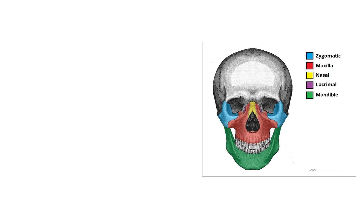
• The facial bones are:
• Zygomatic (2) – Forms the cheek bones of the face.
• Lacrimal (2) – The smallest bones of the face. They
form part of the medial wall of the orbit.
• Nasal (2)
• Inferior nasal conchae (2) – Located within the nasal
cavity.
• Palatine (2) – Situated at the rear of oral cavity, and
forms part of the hard palate.
• Maxilla (2) – Comprises part of the upper jaw and hard
palate.
• Vomer – Forms the posterior aspect of the nasal
septum.
• Mandible (jaw bone)

The maxilla
• Also known as the upper jaw.
This bone consists of five major parts:
• Body of the maxilla
:
• It contains the maxillary sinuses and contributes to
the floor of the orbit
• Infraorbital rim
• Infraorbital foramen
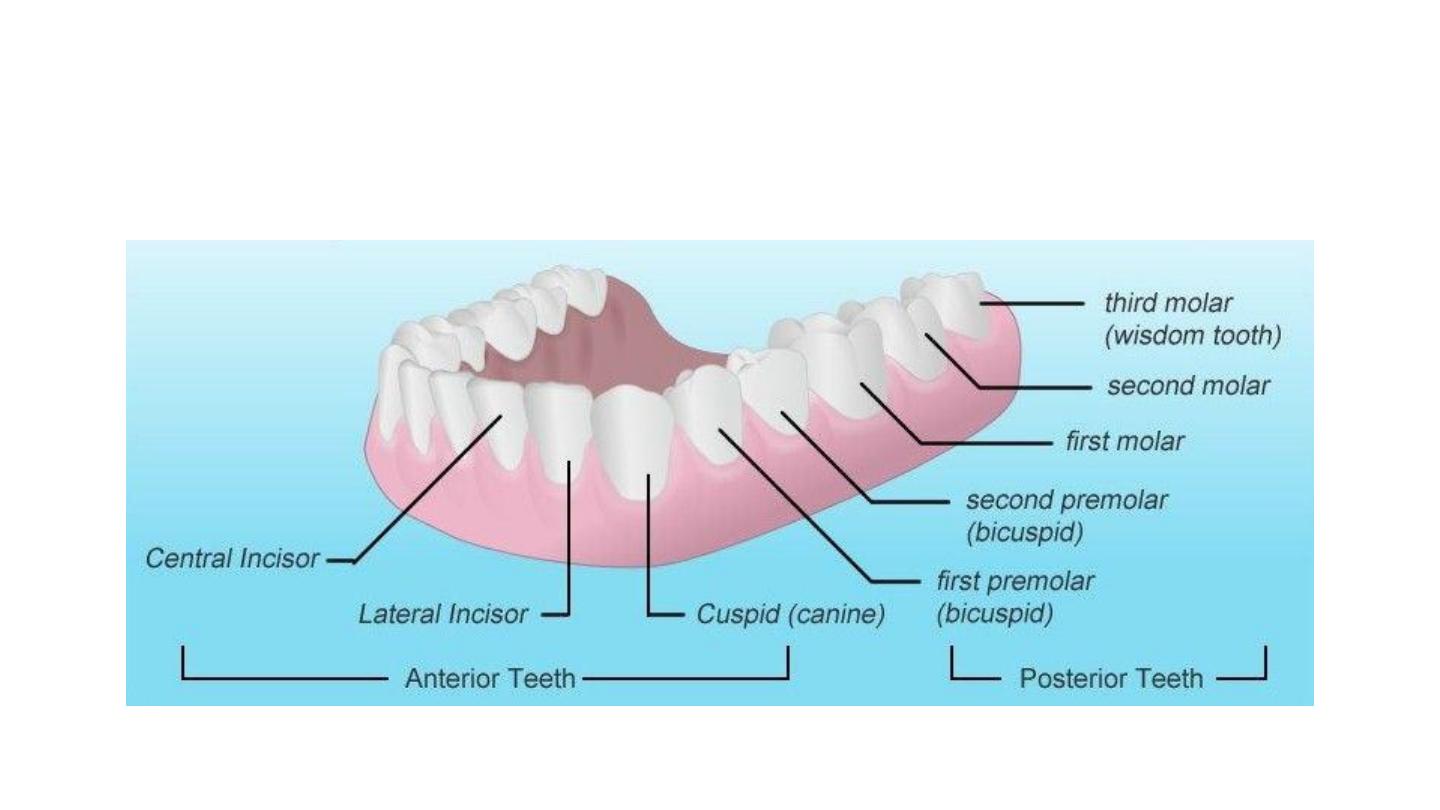
• Alveolar process
: forms the maxillary dental arch.
• Frontal process
: forms the medial border of the orbit.
• Zygomatic process
: contributes to the zygomatic arch
together with the zygomatic bone.
• Palatine process
: constitutes the roof of the mouth and
floor of the nasal cavity.
• Incisive foramen( nasopalatine/ anterior palatine)
• Nasopalatine nerve
• Greater palatine artery and vein
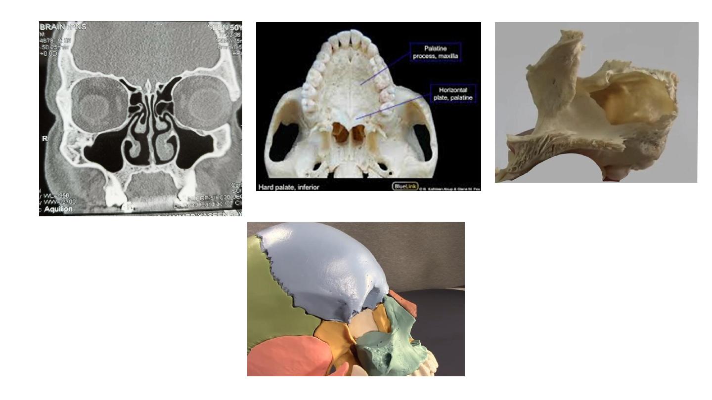
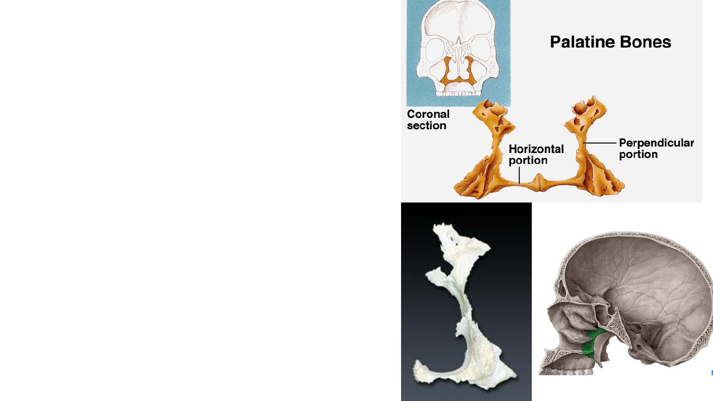
Palatine bone
located at the back of the nasal cavity, between the
maxillae and the sphenoid. Each bone consists of a
horizontal and perpendicular plate forming an L-
shape.
Sphenopalatine notch/ foramen
from the pterygopalatine fossa into the posterior
part of the superior meatus of the nose
sphenopalatine artery and vein a branch of nasal
nerve and nasopalatine nerves.
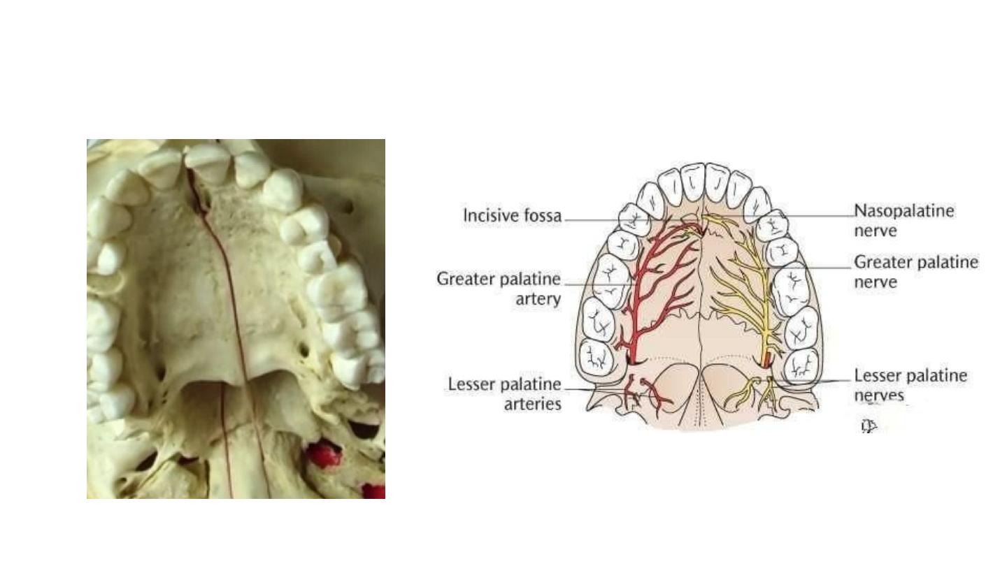
Greater and lesser palatine foramina
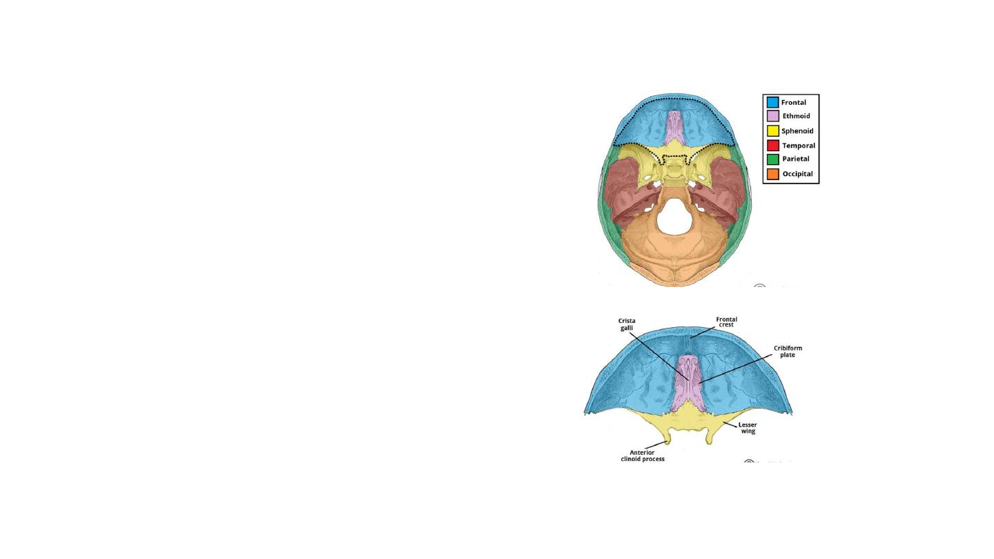
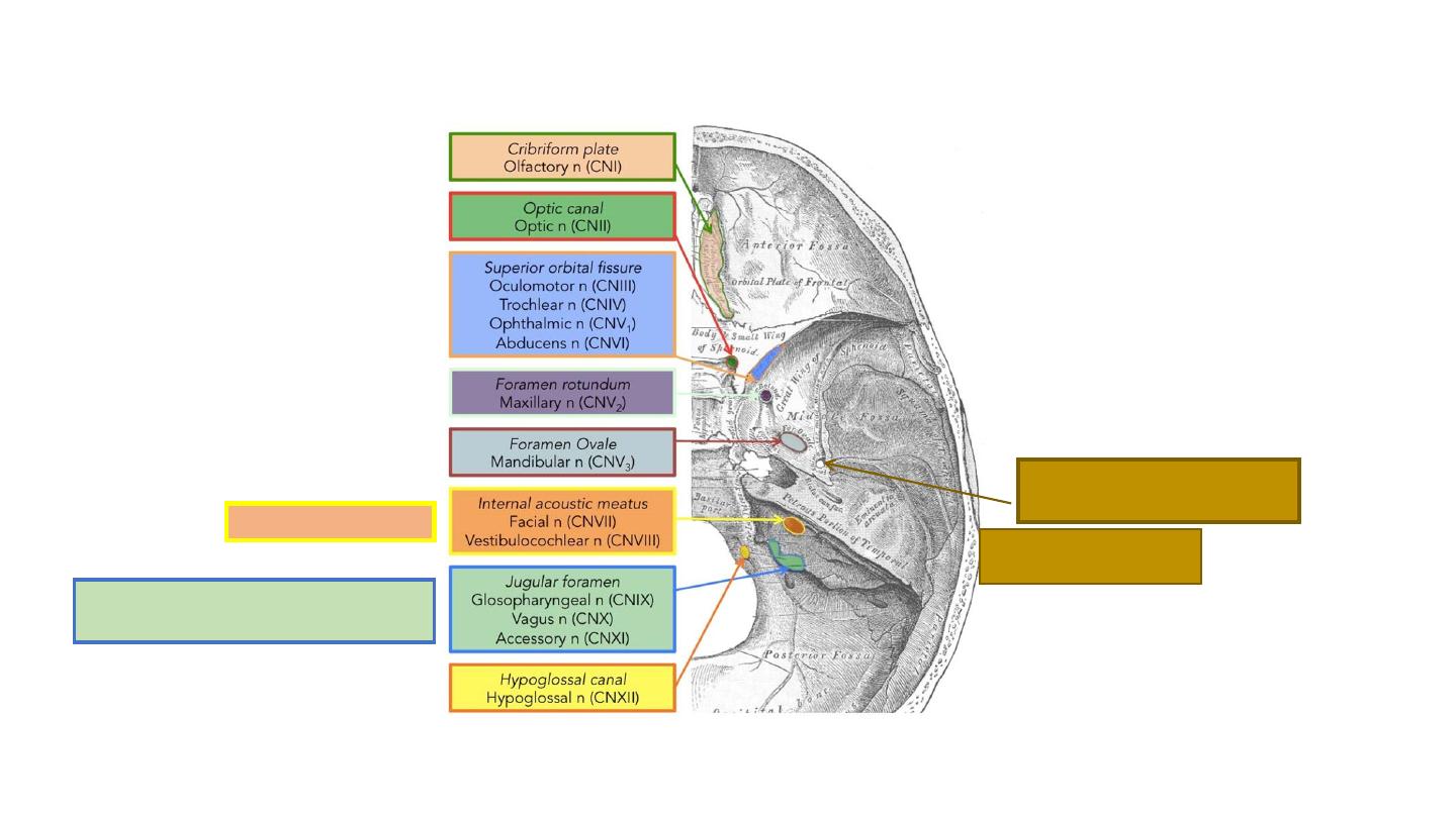
Cranial fossae
• The cranial cavity is divided into three
distinct depressions
The anterior cranial fossa is the most
shallow and superior of the three cranial
fossae.
• Three bones: the frontal bone, ethmoid
bone and sphenoid bone.
• It lies superiorly over the nasal and
orbital cavities. The fossa
accommodates the anteroinferior
portions of the frontal lobes of the
brain.
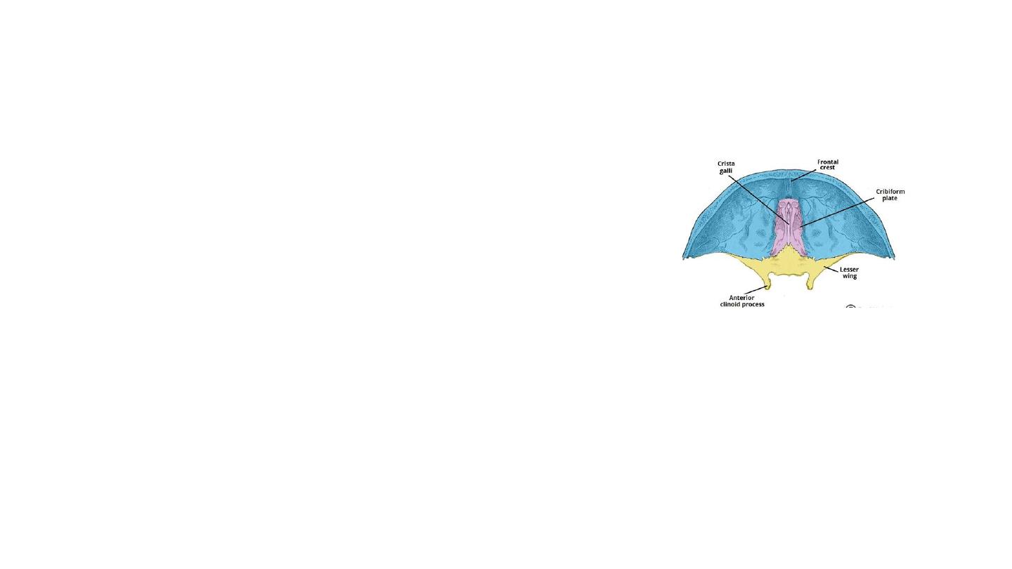
Contents of anterior cranial fossa
• The cribriform plate and crista galli
• Foramina for olfactory nerve
• Anterior and posterior ethmoidal foramina
• The anterior clinoid processes serve as a place of
attachment for the tentorium cerebelli (a sheet of
dura mater that divides the cerebrum from the
cerebellum).
• The lesser wings of the sphenoid bone also
separate the anterior and middle cranial fossae.
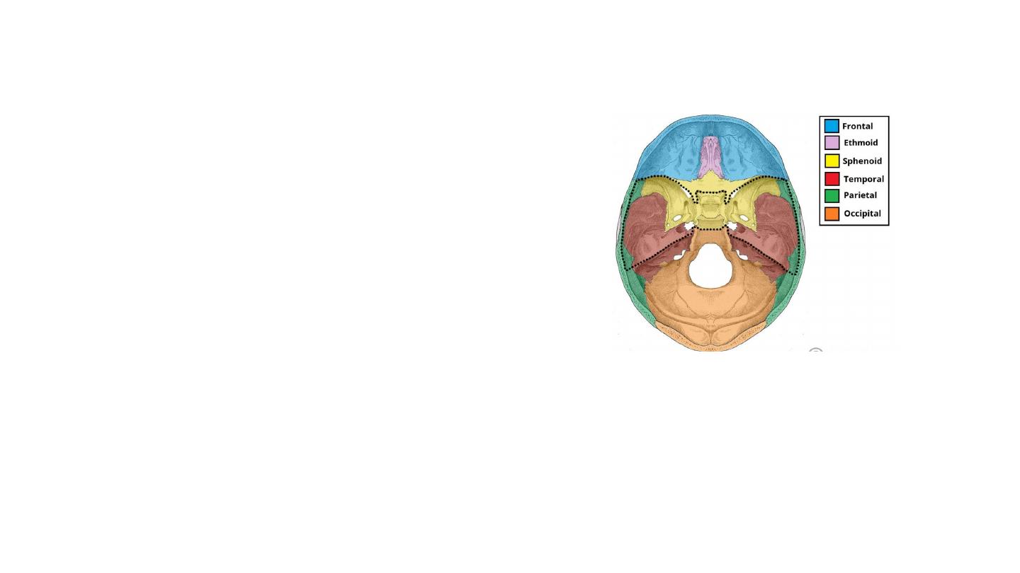
The Middle Cranial Fossa
• Located centrally in the cranial floor.
• Butterfly shaped
• Accommodating the pituitary gland
and two lateral parts accommodating
the temporal lobes of the brain.
• Three bones – the sphenoid bone and
the two temporal bones
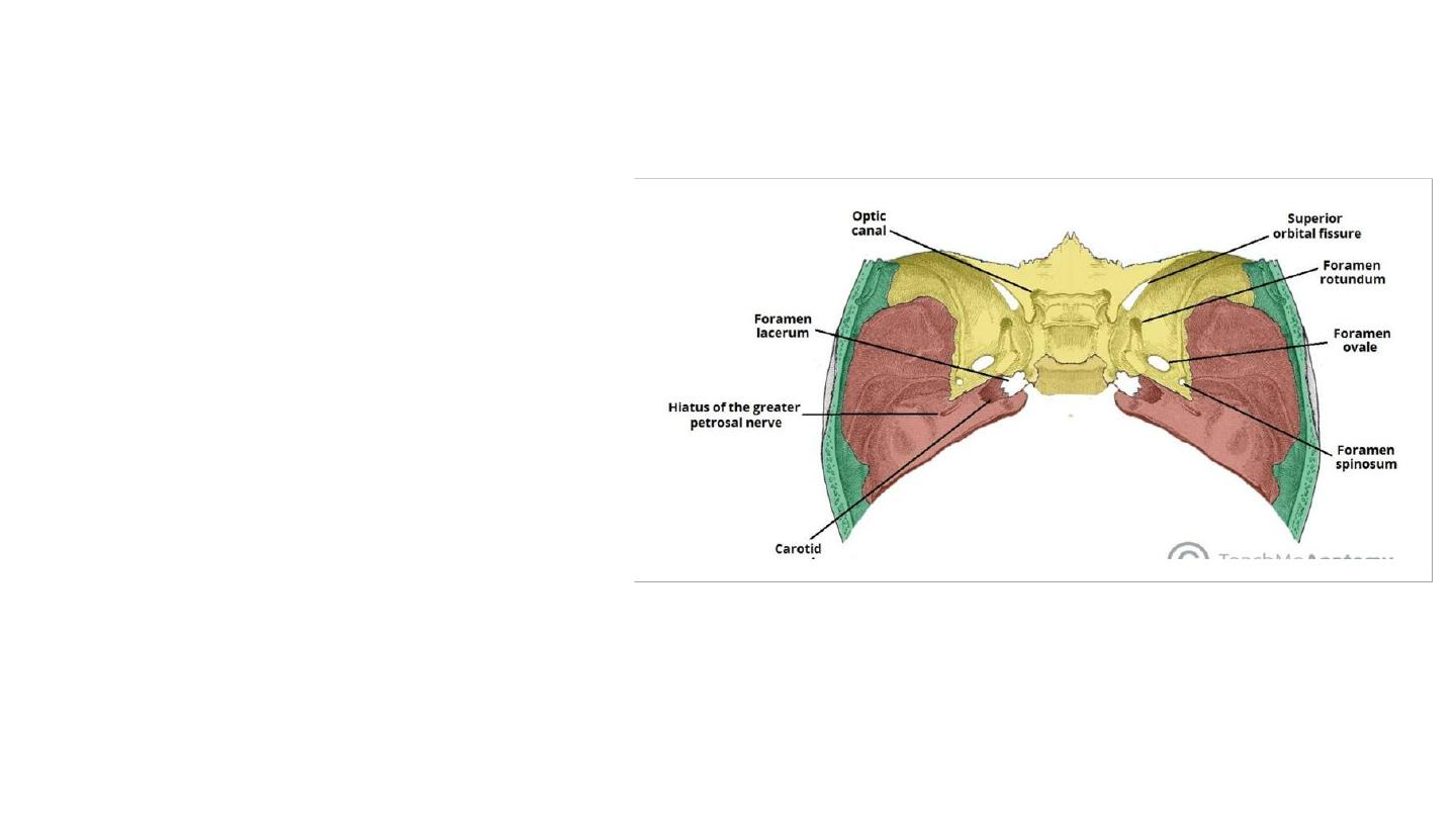
Foramina of the middle cranial fossa
Foramina of the Sphenoid Bone
• The
optic canals
: transmit the optic nerves
(CN II) and ophthalmic arteries into the
orbital cavities.
Immediately lateral to the central part of the
middle cranial fossa are four foramina:
• The
superior orbital fissure
opens
anteriorly into the orbit. It transmits the
oculomotor nerve (CN III), trochlear nerve
(CN IV), ophthalmic branch of the
trigeminal nerve (CN V1), abducens nerve
(CN VI), opthalmic veins and sympathetic
fibres.
• The
foramen rotundum
transmits the
maxillary branch of the trigeminal nerve
(CN V2).
• The
foramen ovale
transmits the
mandibular branch of the trigeminal nerve
(CN V3) and accessory meningeal artery.
• The
foramen spinosum
transmits the
middle meningeal artery, middle
meningeal vein and a meningeal branch of
CN V3.
Foramina of the Temporal Bone
• Carotid canal –
• located posteriorly and medially to the foramen
ovale.
• Transmits the internal carotid artery, internal
carotid venous plexus and sympathetic nerve
plexus from the neck into the cranial cavity.
• At the junction of the sphenoid, temporal and
occipital bones is the foramen lacerum.
• In life, this foramen is filled with cartilage, which
is pierced by small blood vessels
• The internal carotid artery passes from the
carotid canal in the base of the skull, emerging
and coursing superior to foramen lacerum as it
exits the carotid canal. The internal carotid
artery does not travel through foramen lacerum.
The segment of the internal carotid artery that
travels above foramen lacerum is called the
lacerum segment.
Carotid canal
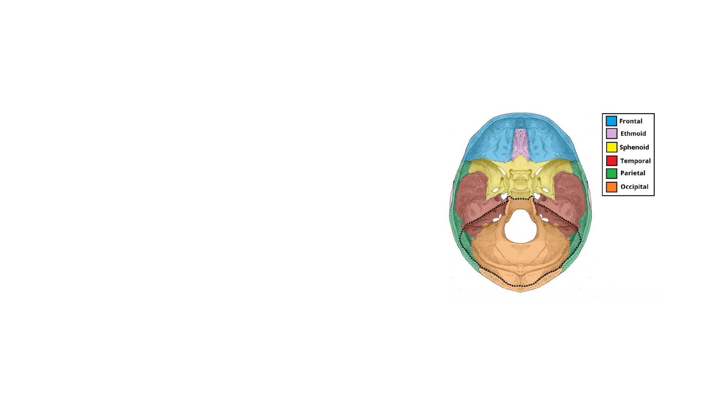
The posterior cranial fossa
• The most posterior and deep of the
three cranial fossae.
• It accommodates the brainstem and
cerebellum.
• Three bones: the occipital bone and the
two temporal bones.
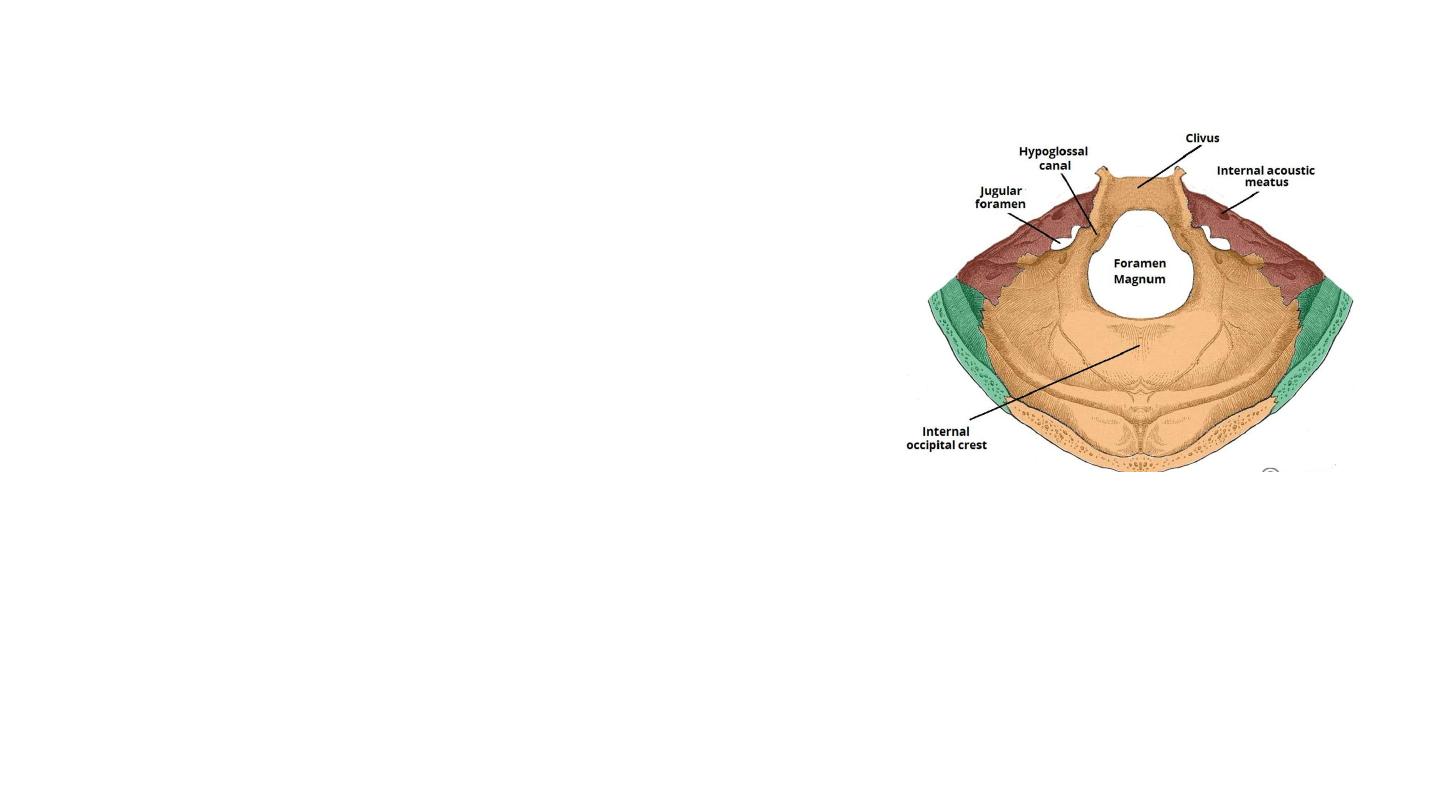
Foramina
Temporal Bone
• The internal acoustic meatus is an oval opening in the posterior
aspect of the petrous part of the temporal bone. It transmits the
facial nerve (CN VII), vestibulocochlear nerve (CN VIII) and
labyrinthine artery.
Occipital Bone
• Foramen magnum
, lies centrally in the floor of the posterior cranial
fossa.
• It is the largest foramen in the skull. It transmits the medulla of the
brain, meninges, vertebral arteries, spinal accessory nerve
(ascending), dural veins and anterior and posterior spinal arteries.
• The
jugular foramina
are situated either side of the foramen
magnum. Each transmits the glossopharyngeal nerve, vagus nerve,
spinal accessory nerve (descending), internal jugular vein, inferior
petrosal sinus, sigmoid sinus and meningeal branches of the
ascending pharyngeal and occipital arteries.
• Hypoglossal canal
: immediately superior to the anterolateral margin
of the foramen magnum . It transmits the hypoglossal nerve
through the occipital bone.
• Posterolaterally to the foramen magnum lies the cerebellar fossae.
These are bilateral depressions that house the cerebellum. They are
divided medially by a ridge of bone, the internal occipital crest.
Jugular bulb
Inferior petrosal and sigmoid sinuses
Labyrinthine artery
Middle meningeal artery
Middle meningeal vein
Foramen spinosum
Meningeal branch of CN V3
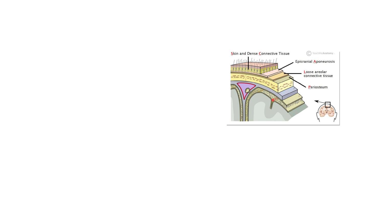
Scalp
• S
kin
– contains numerous hair follicles and
sebaceous glands (thus a common site for
sebaceous cysts).
• Dense
C
onnective tissue
– connects the skin to
the epicranial aponeurosis. It is richly
vascularised and innervated.
• The blood vessels within the layer are highly adherent to
the connective tissue.
• Epicranial
A
poneurosis
– a thin, tendon-like
structure that connects the occipitalis and
frontalis muscles.
• L
oose Areolar Connective Tissue
– a thin
connective tissue layer.
• It contains numerous blood vessels, including emissary
veins which connect the veins of the scalp to the diploic
veins and intracranial venous sinuses.
• Danger area
• P
eriosteum
– the outer layer of the skull bones.
Scalp
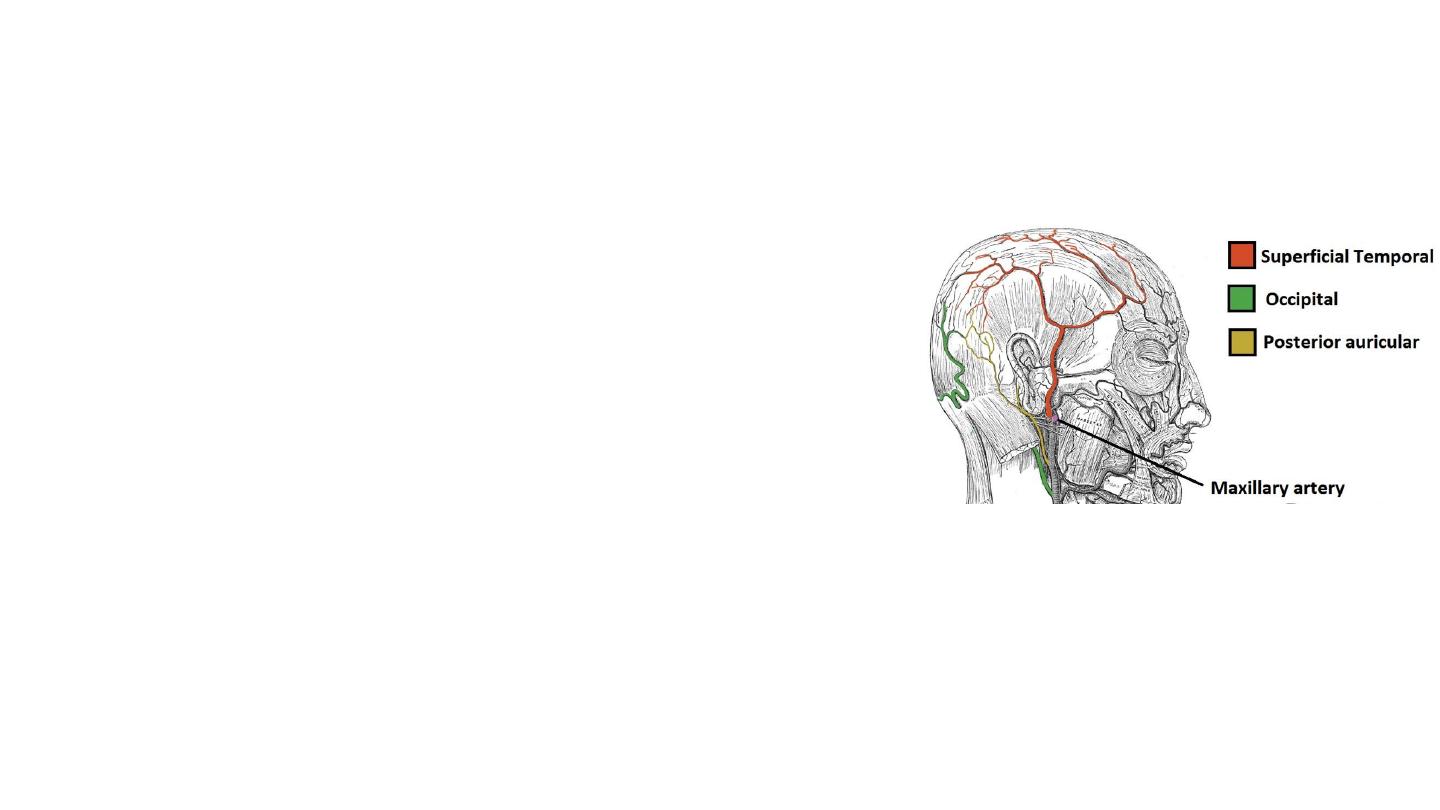
• Arterial Supply
• Branches of external carotid artery and the ophthalmic
artery (a branch of the internal carotid)
• Venous Drainage
• The superficial drainage follows the arterial supply
• The deep (temporal) region of the skull is drained by
the pterygoid venous plexus. Drain into the maxillary
vein.
• The veins of the scalp
connect to the diploic veins
of the skull via valveless emissary veins
. This
establishes a connection between the scalp and the
dural venous sinuses.
• Lymphatics: posterior half: occipital and posterior
auricular nodes. Anterior: parotid nodes
• Eventually: submandibular and deep cervical nodes.
• Innervation
• The scalp receives cutaneous innervation from branches
of the trigeminal nerve or the cervical nerve roots.
تابع لمحاضره سابقه
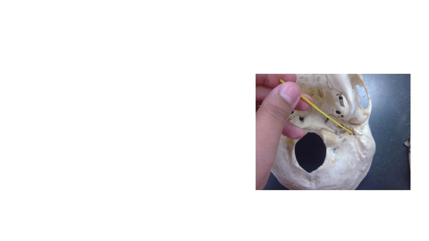
Stylomastoid foramen:
It is located on the inferior surface of the
petrous temporal bone, between the base of
the styloid process and the mastoid process of
the temporal bone. It transmits the facial nerve
and stylomastoid artery (branch of posterior
auricular artery).