
NERVOUS SYSTEM PHYSIOLOGY
Lecture one
Dr. Suroor Mohamed

Objective :
1. What is the physiologic anatomy of Nervous System & its parts?
2. Describe the peripheral nervous system ?
3. What is Effects of Sympathetic and Parasympathetic division of Autonomic system
on Specific Organs?
4. Whats mean by stress response ?

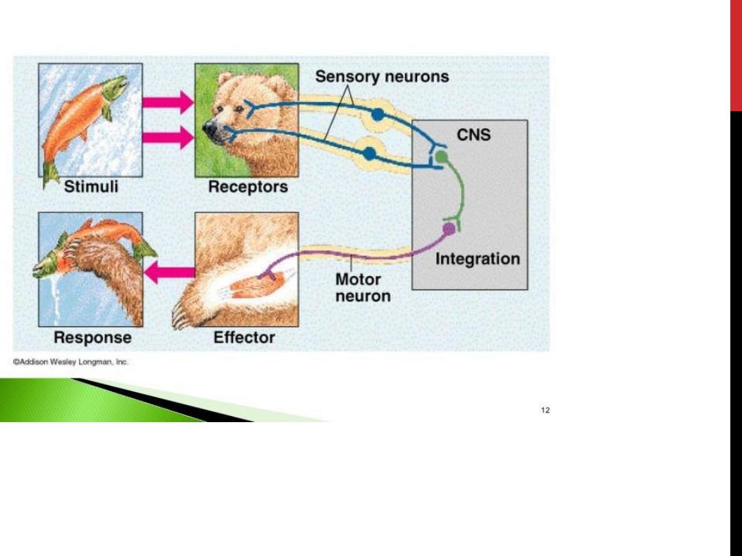
The
nervous system
is a complex network of nerves and cells
that carry messages to and from the brain and spinal cord to
various parts of the body.
The nervous system contains more than
100 billion
neurons and
consists of
Central nervous system and Peripheral nervous
system.
The Central nervous system is made up of the
brain and
spinal cord
and The Peripheral nervous system is made up of the
Somatic and the Autonomic
nervous systems
The Central Nervous System (CNS)
The
central nervous system is divided into two major parts:
the brain and the spinal cord.
The Brain
The brain lies within the skull and & consists of four principal parts:
the brain stem
the cerebrum
the cerebellum
the diencephalon

The brain has nerve cells called the
neurons and supporting cells
called the glia
.
خاليا عصبية وخاليا مساندة لها
There are two types of matter in the brain:
grey matter and white
matter
.
المادة الرمادية والبيضاء
Grey matter
receives and stores impulses. Cell bodies of neurons
and neuroglia are in the grey matter.
White matter
in the brain
carries impulses to and from grey matter. It consists of the nerve
fibers (axons).
The Spinal Cord
الحبل الشوكي
The spinal cord is along tube like structure which extends from the
brain.
The spinal cord is composed of a series of 31 segments. A pair of
spinal nerves comes out of each segment. The region of the spinal
cord from which a pair of spinal nerves originates is called the
spinal segment. Both motor and sensory nerves are located in the
spinal cord. It lies within the vertebral Column.


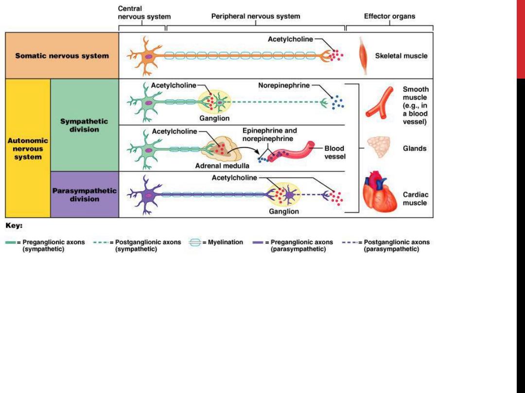
The Peripheral Nervous System
The Peripheral nervous system is made up of two parts:
1- Somatic nervous system
2- Autonomic nervous system
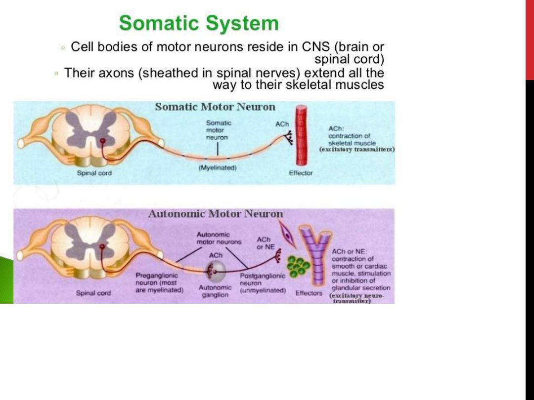
Somatic Nervous System
الحركي
The somatic nervous system consists of peripheral nerve fibers that
pick up sensory information or sensations from the peripheral or
distant organs (those away from the brain like limbs) and carry them
to the central nervous system.
These also consist of motor nerve fibers that come out of the brain
and take the messages for movement and necessary action to the
skeletal muscles. For example, on touching a hot object the sensory
nerves carry information about the heat to the brain, which in turn,
via the motor nerves, tells the muscles of the hand to withdraw it
immediately.
The whole process takes less than a second to happen. The cell body
of the neuron that carries the information often lies within the brain or
spinal cord and projects directly to a skeletal muscle.

Neurons in the Peripheral Nervous System
The smallest worker in the nervous system is the neuron. For each of
the chain of impulses there is
one preganglionic
neuron, or one before
the cell body or
ganglion, that is like a central controlling
body for
numerous neurons going out peripherally.
The preganglionic neuron is located in either the brain or the spinal cord.
In the autonomic nervous system this
preganglionic neuron projects to
an autonomic ganglion. The postganglionic neuron then projects to the
target organ.
In the somatic nervous system there is
only one neuron
between the
central nervous system and the target organ while the autonomic
nervous system uses
two neurons
.
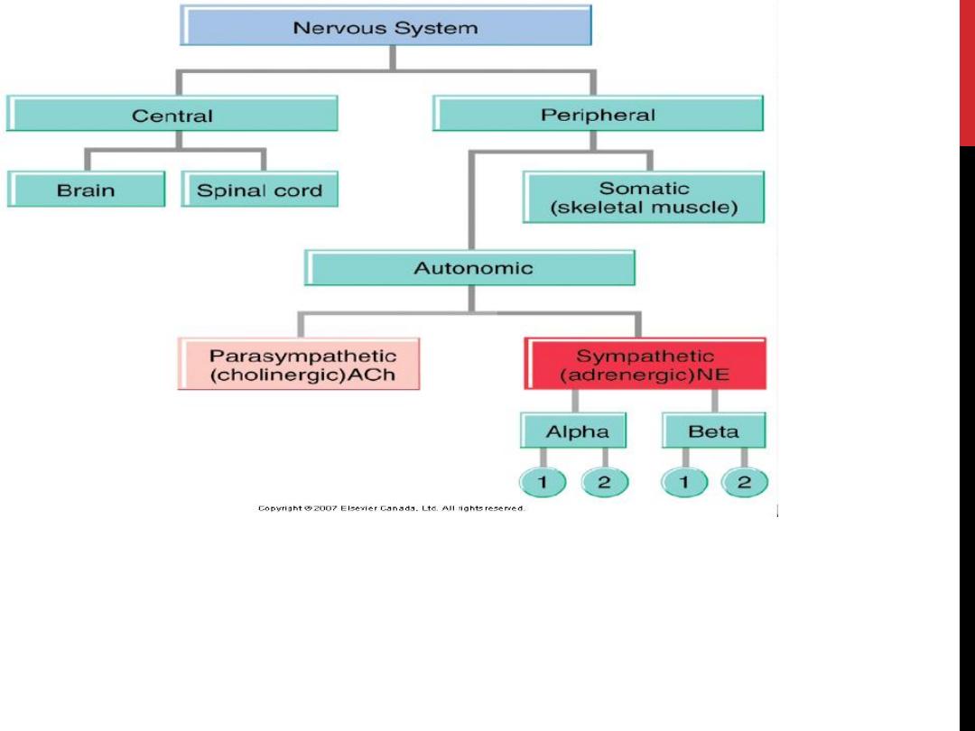

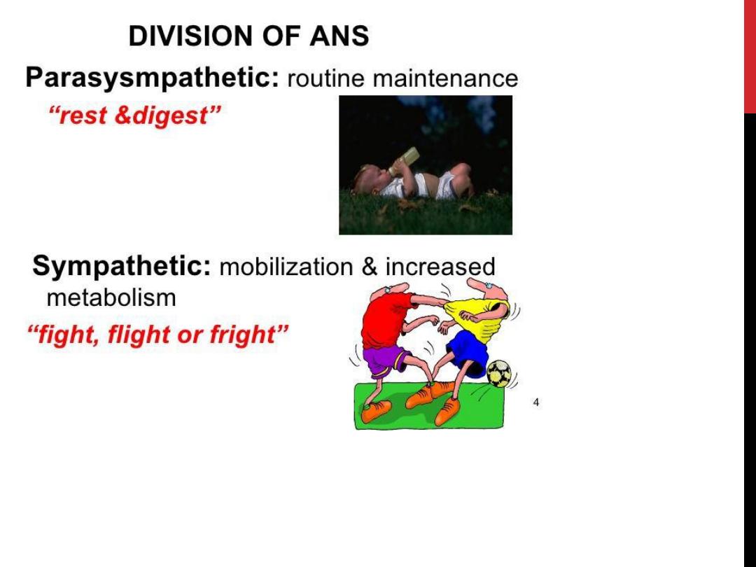
Autonomic Nervous System
الجهاز العصبي التلقائي
Another part of the nervous system is the Autonomic Nervous System. It has
three parts:
1- the sympathetic nervous system
2- the parasympathetic nervous system
3- the enteric nervous system
This nervous system controls the nerves of the inner organs of the body on
which humans have
no conscious control
. This includes the heartbeat,
digestion, breathing (except conscious breathing) etc.
The nerves of the autonomic nervous system enervate the smooth
involuntary
muscles of the (internal organs) and glands
and cause them to function and
secrete their enzymes etc.
The
Enteric nervous system
is the third part of the autonomic nervous system.
The enteric nervous system is a complex network of nerve fibers that innervate
the organs within the
abdomen like the gastrointestinal trac
t, pancreas, gall
bladder etc. It contains nearly 100 million nerves.

ANS
The autonomic nervous system (ANS) is the part of the nervous system that
is responsible for
homeostasis
(maintenance of nearly constant conditions in
the internal environment).
Except for skeletal muscle
, which gets its innervation from the
somatomotornervous system,innervation to all other organs is supplied
by the ANS.
Nerve terminals are located in
smooth muscle
(e.g., blood vessels, the
wall of the gastrointestinaltract, urinary bladder),
cardiac muscle
, and
glands
(e.g., sweat glands, salivary glands)
so, the A.N.S.
controls the involuntary functions
which are not under
our will &so controlled automatically & reflexly, because some of them
are too vital to allow any interference from our behavior.
One of the most striking characteristics of the autonomic nervous system
is the
rapidity and intensity
with which it can change visceral functions.
o
For instance, within 3 to 5 seconds it can increase the heart rate to twice
normal,
o
and within 10 to 15 seconds the arterial pressure can be doubled.

General organizationof the autonomic nervous system
The A.N. S often operates by
o
centers in the spinal cord (such as emptying of the urinary bladder),
o
brain stem (centers for control of B.pr., H.R. & respiration),
o
Hypothalamus (temperature regulation center & centers to control
hunger & thirst)
o
also portions of the cerebral cortex especially the limbic system.
Higher centers can transmit impulses to lower centers & influence
autonomic control.
o
For instance, stimulation in appropriate areas of the hypothalamus
can activate the medullary cardiovascular center control centers
strongly enough to increase the arterial B.P. to more than double
normal.
Cerebral cortex has connections with the hypothalamus. & brainstem.
ANS often operates by means of
visceral reflexes
. That is,
subconscious
sensory
signals from a visceral organ can enter the autonomic ganglia,
the brain stem, or the hypothalamus and then return subconscious
reflex responses directly back to the visceral organ to control its activities.
The efferent autonomic signals are transmitted to the various organs of the
body through two major subdivisions called the
sympathetic nervous
system
and the
parasympathetic nervous
system.

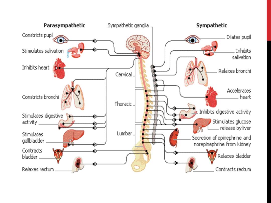
Sympathetic division
The sympathetic nerve fibers
originate
in the spinal cord along with spinal
nerves between cord segments
T-1 and L-2.
This is why the sympathetic nervous
system is sometimes called the
thoracolumbar division of the ANS.
Preganglionic neuron
o
The cell body
of each preganglionic neuron lies in the intermediolateral
horn of the spinal cord.
o
the axons
of the sympathetic preganglionic neurons leave the spinal cord
and exit via the ventral root.
o
They then separate from the ventral root and project to the adjacent
sympathetic
paravertebral ganglion
, where some of them end on the cell
bodies of the postganglionic neurons.
o
Some
preganglionic neurons
pass through the paravertebral ganglion chain
and end on postganglionic neurons located in
prevertebral ganglia (or
collateral)
ganglia close to the viscera
, including the celiac, superior
mesenteric, and inferior mesenteric ganglia.
o
There are also preganglionic neurons whose axons terminate directly on the
adrenal gland.
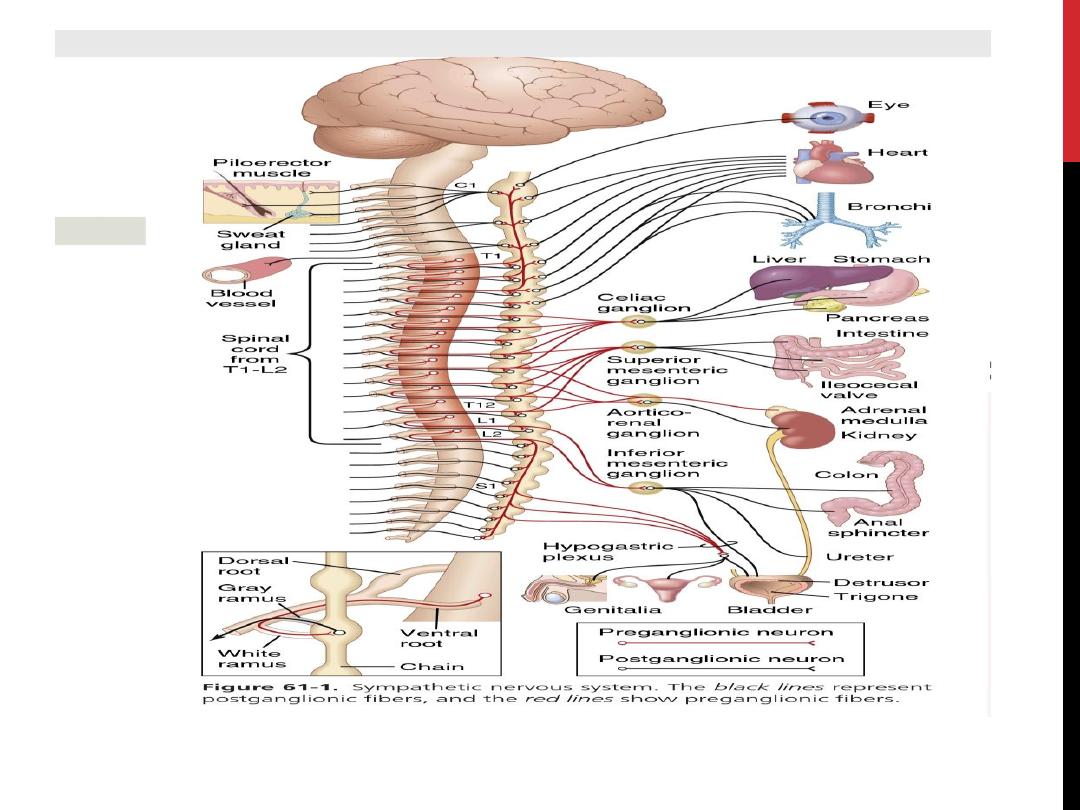

The ganglion
o
Paravertebral ganglia are
located
adjacent to each thoracic and upper lumbar spinal
segments; in addition, there are a few ganglia adjacent to the cervical and sacral spinal
segments.
o
The ganglia are
connected
to each other via the axons of preganglionic neurons that travel
rostrally or caudally to terminate on postganglionic neurons located at some distance.
o
Together these ganglia form the
sympathetic chain
bilaterally.
Adrenal medulla:
It is sympathetic ganglia in which the postganglionic cells loss their axons & become
specialized to secrete directly into the blood stream.
The adrenal medulla secretes catecholamine (epinephrine, nor epinephrine& dopamine)
directly into the blood.
Postganglionic neurons (Segmental Distribution of the Sympathetic Nerve Fibers)
The sympathetic pathways that originate in the different segments of the spinal cord are not
necessarily distributed to the same part of the body as the somatic spinal nerve fibers from the
same segments. Instead:
The postganglionic sympathetic to the head
originate in the superior, middle& Stellate
ganglia (cervical ganglia) in the cranial extension of the sympathetic ganglionic chain.
The post ganglionic sympathetic to the chest
originate in the above gang. & the
paravertebral sympathetic ganglia (T1-T4).
The postganglionic sympathetic to the abdominal viscera
originate in the collateral ganglia
(prevertebral ganglia, which include celiac, superior mesenteric. & inferior mesenteric
ganglia.) from the preganglionic. N. (greater splanichnic N.& small splanichnic N.) which
originate from (T4-L2).
Uterus & male genital tract
are innervated by a special system of short noradrenergic N. with cell
bodies in ganglia in or near these organs

Parasympathetic division (Cranio-sacral outflow)
parasympathetic fibers leave the central nervous system through:
a. Cranial nerves III, VII, IX, and X. (hint 1973)
1.
tenth cranial nerve:
About 75 percent of all parasympathetic nerve fibers are in the
vagus nerves (cranial nerve X), passing to the entire thoracic and abdominal regions
of the body.
2.
Parasympathetic fibers in the third cranial nerve
go to the pupillary sphincter and
ciliary muscle of the eye.
3.
Fibers from the seventh cranial nerve
pass to the lacrimal, nasal, and
submandibular glands,
4.
fibers from the ninth cranial
nerve go to the parotid gland.
b. The pelvic branches of the 2nd-4th sacral N.:
These fibers then distribute to the descending colon, rectum, urinary bladder, and lower
portions of the ureters& supplies nerve signals to the external genitalia to cause
erection.
In both outflow, the preganglionic division will end on a short postganglionic division on a
ganglion near the visceral structure.
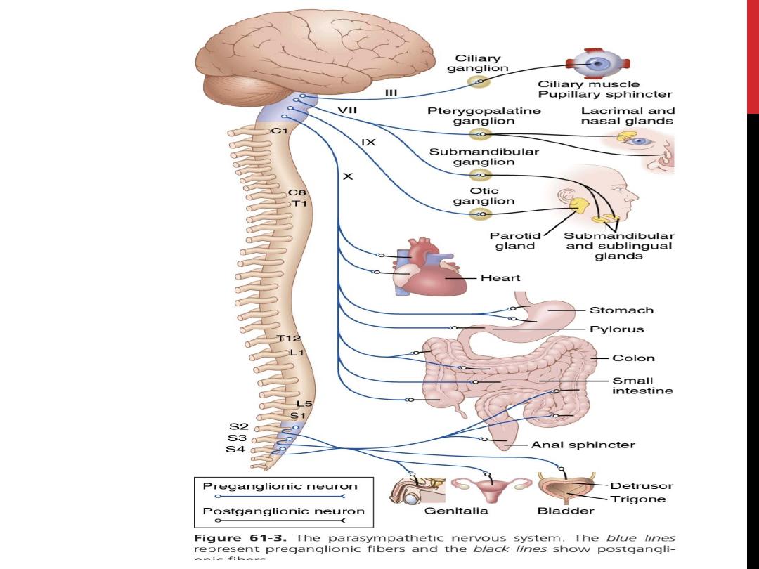
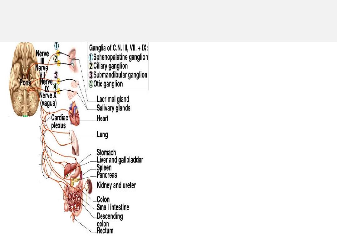
Parasympathetic Functions of Cranial Nerves
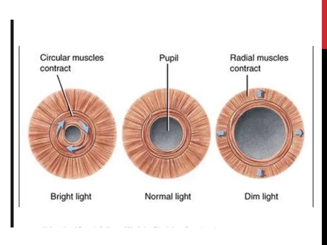
• Oculomotor nerve (III)
– narrows pupil & focuses lens
• Facial nerve (VII)
– regulates secretion of tear,
lacrimal & salivary glands
• Glossopharyngeal (IX)
– regulates parotid salivary gland
• Vagus nerve (X)
– muscles and glands of the viscera
as far inferiorly as the proximal
half of colon

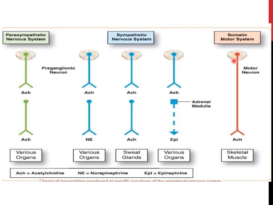
Chemical transmissionat autonomicjunctions
Transmissionat the synaptic junctions between preganglionicand postganglionicneurons and between
the postganglionic neurons and the autonomic effectors are chemically mediated. The principal
transmitter agents involved are
acetylcholine
and
norepinephrine
Chemical divisions of the A.N.S
Accordingto the type of neurotransmitter secreted,the A.N.S. can be divided into:
A. Cholinergic division: secret Ach.
The autonomic neurons that are
cholinergicare:
1) All preganglionic neurons
2) All parasympatheticpostganglionicneurons
3) Sympatheticpostganglionic neurons that innervate sweat glands
4) Sympatheticpostganglionic neurons that end on blood vessels in some skeletal muscles and
producevasodilation when stimulated (sympatheticvasodilator
nerves).
B. Adrenergicdivision: secret nor epinephrine
The remaining sympatheticpostganglionicneurons are noradrenergic.
The adrenal medulla is essentiallya sympatheticganglionin which the postganglioniccells have lost their axons
and secrete norepinephrine(20%) and epinephrine (80%) directlyinto the bloodstream
Cholinergic fibers secrete acetyl choline. Adrenergic fibers secrete norepinephrine (noradrenalin).
Non-adrenergic non-cholinergic (NANC)
1.
All preganglionic neurons (sympathetic & parasympathetic) cholinergic
2.
Postganglionic parasympathetic cholinergic
3.
Postganglionic sympathetic adrenergic except: postganglionic sympathetic to sweat glands and
few blood vessels cholinergic

Types of receptors
1. Cholinergic receptors
: which are stimulated by Ach. :
A. Nicotinic receptor: present in the autonomic ganglia. & at the neuromuscular
junction with skeletal muscle ,are ligand-gated ion channels
B. Muscarinic receptor: present in the organs on which cholinergic neurons will act, use
G proteins as their signaling mechanism
2. Adrenergic receptor
:
A. Alpha receptor:
o alpha1 & alpha2 which are linked to different G proteins
o Binding of an agonist to α 1 adrenoceptors leads an increase in intracellular Ca 2+.
o Binding of an agonist to α 2 -adrenoceptors causes dissociation of the inhibitory G protein to
inhibit adenylyl cyclase and decrease (cAMP).
B. Beta receptor:
beta1 , beta2 &beta3
(Binding of an agonist to β-adrenoceptors activates the G s - coupling protein
to activate adenylyl cyclase and increase cAMP.
Norepinephrine excites mainly alpha receptors but excites the beta receptors to a lesser extent as
well. Epinephrine excites both types of receptors approximately equally.
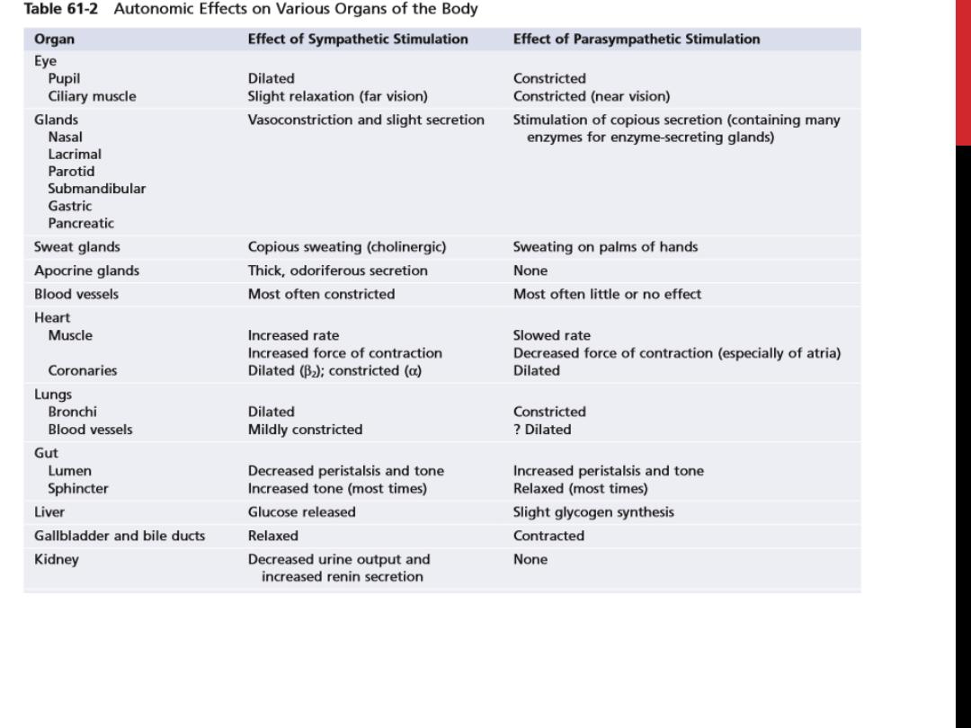

Basic Characteristics of Sympathetic and Parasympathetic Function
Cholinergic and Adrenergic Fibers— Secretion of Acetylcholine or Norepinephrine
The sympathetic and parasympathetic nerve fibers secrete mainly one or the other of two
synaptic transmitter substances, acetylcholine or norepinephrine. Those fibers that secrete
acetylcholine are said to becholinergic. Those that secrete norepinephrine are said to be
adrenergic, a term derived from adrenalin, which is an alternate name for epinephrine.
All preganglionic neurons are cholinergic in both the sympathetic and the parasympathetic
nervous systems. Acetylcholine or acetylcholine-like substances, when applied to the
ganglia, will excite both sympathetic and parasympathetic postganglionic neurons. Either
all or almost all of the postganglionic neurons of the parasympathetic system are also
cholinergic.
Conversely, most of the postganglionic sympathetic neurons are adrenergic. However, the
postganglionic sympathetic nerve fibers to the sweat glands, to the piloerector muscles of
the hairs, and to a very few blood vessels are cholinergic.
Thus, the terminal nerve endings of the parasympathetic system all or virtually all secrete
acetylcholine. Almost all of the sympathetic nerve endings secrete norepinephrine, but a few
secrete acetylcholine. These hormones in turn act on the different organs to cause respective
parasympathetic or sympathetic effects. Therefore, acetylcholine is called a parasympathetic
transmitter and norepinephrine is called a sympathetic transmitter.

Two Principal Types of Acetylcholine Receptors—Muscarinic and Nicotinic Receptors
Acetylcholine activates mainly two types of receptors. They are
called muscarinic and nicotinic receptors
.
Muscarinic receptors
are found on all effector cells that are stimulated by the postganglionic cholinergic neurons of
either the parasympathetic nervous system or the sympathetic system.
Nicotinic receptors
are found in the autonomic ganglia at the synapses between the preganglionic and postganglionic
neurons of both the sympathetic and parasympathetic systems. (Nicotinic receptors are also present at many non
autonomic nerve endings—for instance, at the neuromuscular junctions in skeletal muscle.
Adrenergic Receptors—Alpha and Beta Receptors
There are also two major types of adrenergic receptors,
alpha receptors and beta receptors
. (The beta receptors in turn
are divided into beta1 and beta2 receptors because certain chemicals affect only certain beta receptors. Also, there is a
division of alpha receptors into alpha1 and alpha2 receptors.)
Norepinephrine and epinephrine
, both of w hich are secreted into the blood by the adrenal medulla, have slightly
different effects in exciting the alpha and beta receptors. Norepinephrine excites mainly alpha receptors but excites the
beta receptors to a lesse rextent as well. Conversely, epinephrine excites both types of receptors approximately equally.
Therefore, the relative effects of norepinephrine and epinephrine on different effector organs are determined by the
types of receptors in the organs. If they are all beta receptors, epinephrine will be the more effective excitant.
Note that certain alpha functions are excitatory, whereas others are inhibitory. Likewise, certain beta functions are
excitatory and others are inhibitory. Therefore, alpha and beta receptors are not necessarily associated with excitation
or inhibition but simply with the affinity of the hormone for the receptorsin the given effector organ. .
Action potential → depolarization → increased
permeability
to
Ca
+2
→
secretion
of
neurotransmitter
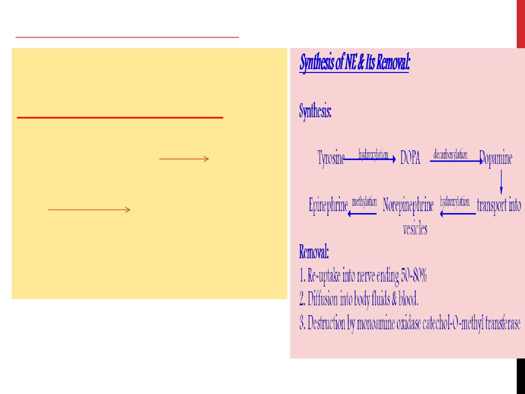
Synthesis of Ach & Its Destruction:
Synthesis:
Acetyl-CoA + choline
Cholineacetyl transferase
Ach
Axoplasm to vesicle
Destruction:
Ach
Acetylcholine esterase
Choline + acetate
Present in terminal nerve ending
receptor organ (tissue acetylcholine esterase)
serum acetylcholine
destruction in seconds
Mechanism of secretion of Ach & NE:

Autonomic Reflexes
Many visceral functions of the body are regulated by autonomic reflexes.
1.
Cardiovascular Autonomic Refle xes:
Several reflexes in the cardiovascular system help to control especially the arterial blood pressure and the heart rate. One of these is the
baroreceptor reflex. Briefly, stretch receptors called baroreceptors are located in the walls of several major arteries, including especially the
internal carotid arte ries and the arch of the aorta. When these become stretched by high pressure, signals are transmitted to the brain stem,
where they inhibit the sympathetic impulses to the heart and blood vessels and excite the parasympathetics; this allows the arterial pressure
to fall back toward normal.
2
.
Gastrointestinal Autonomic Refle xes.
The uppermost part of the gastrointestinal tract and the rectum are controlled principally by autonomic reflexes. For instance, the smell of
appetizing food or the presence of food in the mouth initiates signals from the nose and mouth to the vagal, glossopharyngeal, and salivatory
nuclei of the brain stem. These in turn transmit signals through the parasympathetic nerves to the secretory glands of the mouth and
stomach, causing secretion of digestive juices sometimes even before food enters the mouth.
Whe n fecal matter fills the rectum at the othe r end of the alimentary canal, se nsory impulses initiated by stretching the rectum are se nt to
the sacral portion of the spinal cord, and a reflex signal is transmitted back through the sacral parasympathetics to the distal parts of the
colon; these result in strong peristaltic contractions that cause defecation.
3. Other Autonomic Reflexes.
Emptying of the urinary bladde r is controlled in the same way as e mptying the rectum; stretching of the bladde r sends impulses to the sacral
cord, and this in turn causes reflex contraction of the bladder and relaxation of the urinary sphincters, thereby promoting micturition.
Also important are the sexual reflexes, which are initiated both by psychic stimuli from the brain and by stimuli from the sexual organs.
Impulses from these sources converge on the sacral cord and, in the male, result first in erection, mainly a parasympathetic function, and
the n e jaculation, partially a sympathe tic function

“Alarm” or “Stress” Response of the Sympathetic Nervous System
When large portions of the sympathetic nervous system discharge at the same time—that is, a mass discharge— this increases in many ways
the ability of the body to pe rform vigorous muscle activity. Let us summarize
these ways:
1. Increased arterial pressure
2. Increased blood flow to active muscles concurrent with decreased blood flow to organs such as
the gastrointestinal tract and the kidneys that are not needed for rapid motor activity
3. Increased rates of cellular metabolism throughout the body
4. Incre ased blood glucose concentration
5. Increased glycolysis in the liver and in muscle
6. Increased muscle strength
7. Increased mental activity
8. Increased rate of blood coagulation
The sum of these effects permits a person to perform far more strenuous physical activity than would otherwise be possible. Because either
mental or physical stress can excite the sympathetic system, it is frequently said that the purpose of the sympathetic system is to provide
extra activation of the body in states of stress: this is called the sympathetic stress response.

Thank for your listening