Slide 1
Oogenesis
Dr. Sumeya
Slide 2
Primary Oocyte
Secondary
oocyte
2N
1N
1N
1N
1N
1N
1N
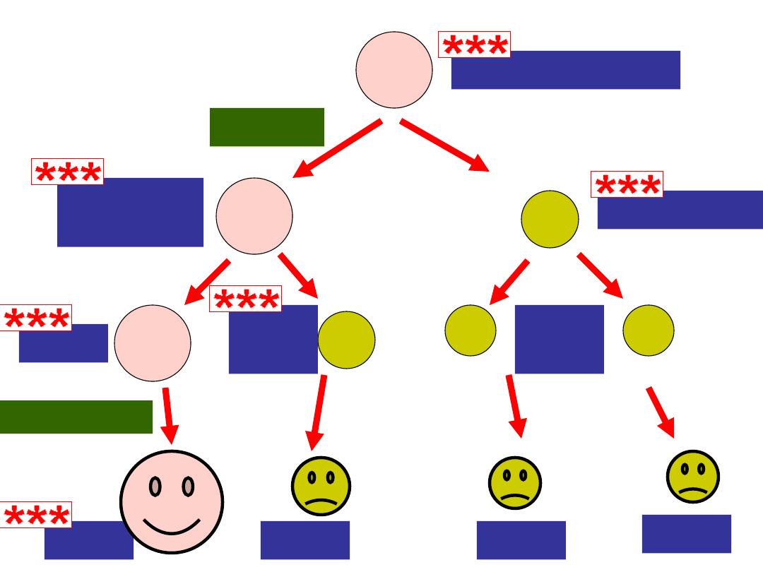
Meiosis
morphogenesis
Polar Body
Ootid
Polar
body
Polar
bodies
1N
Ovum
Die
Die
Die
Oogensis
Slide 3
Oogenesis
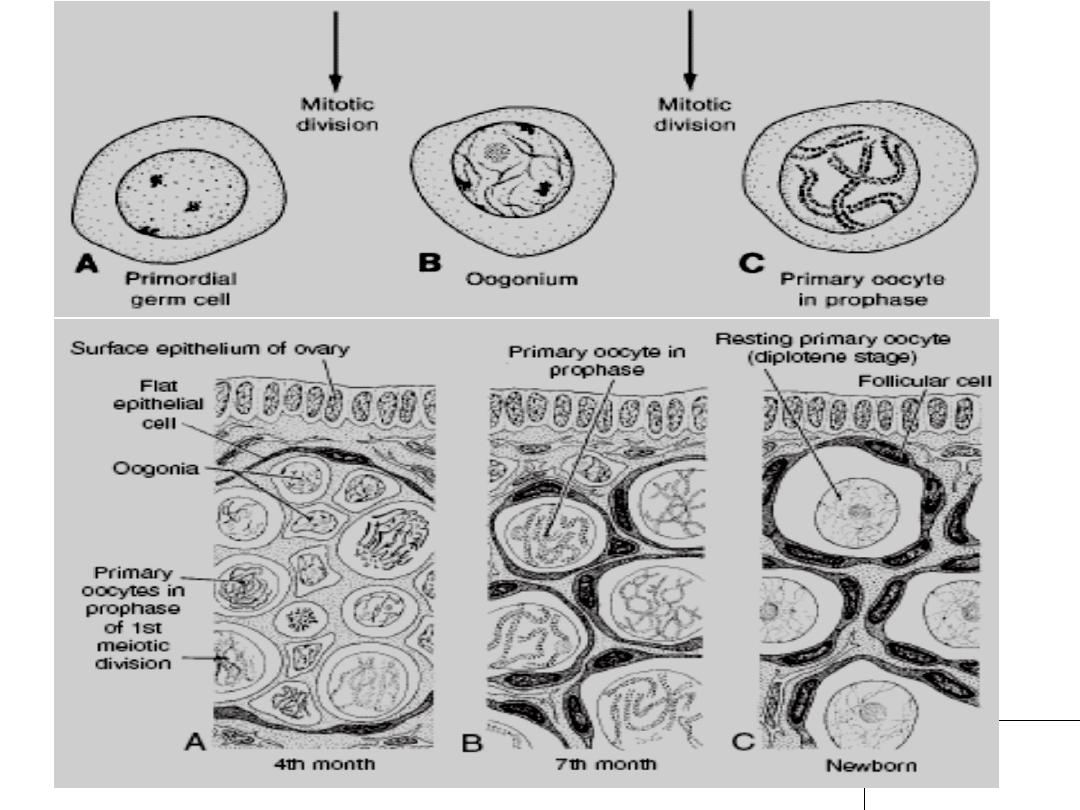
Is the process whereby oogonia differentiate into mature oocytes.
Maturation of Oocytes Begin Before Birth
•Once PGCs have arrived in the gonad of a genetic female, they
differentiate into oogonia .
•By the end of the third month: they are arranged in clusters surrounded
by a layer of flat epithelial cells(follicular cells) originate from surface
epithelium covering the ovary .
•All the oogonia in one cluster are properly derived from a single cell.
•Oogonia: majority: continue to devide by mitosis.
some: enter meiosis and arrest their cell division in prophase
MI forming primary oocytes .
Slide 4
• 5
th
month: total number of germ cells(oogonia + primary oocytes)
reach maximum(7 millions) .
• At this time: cell death begins leading to many oogonia and primary
oocytes degenerate and become atretic .
• 7th month: the majority of oogonia ----- degenerate except few near
a surface.
• All surviving primary oocytes ------ entered prophase of miosis 1 ,
and most of them are individually surrounded by a layer of flat
follicular epithelial cells.
• A primary oocyte, together with its surrounding flat epithelial cells, is
known as a
primordial follicle
Slide 5
Slide 6

Slide 7
Maturation of Oocytes
Continues at Puberty
• Near birth time: all primary oocytes ------- started prophase of
meiosis I, but instead of proceeding into metaphase, they
enter the
diplotene stage
, a resting stage during prophase that
is characterized by a lacy network of chromatin
• Primary oocytes remain arrested in prophase and do not fi nish
their fi rst meiotic division before puberty is reached.
• This arrested state is produced by
oocyte maturation
inhibitor (OMI),
a small peptide secreted by follicular cells.
• The total number of primary oocytes at birth is estimated to vary
from 600,000 to 800,000. During childhood, most oocytes
become atretic; only approximately 40,000 are present by the
beginning of puberty, and fewer than 500 will be ovulated.
Slide 8
• Some oocytes that reach maturity late in life have been
dormant in the diplotene stage of the fi rst meiotic
division
for 40 years
or more before ovulation-------
vulnerable to damage with age and increase
chromosomal abnormalities.
Slide 9

Slide 10
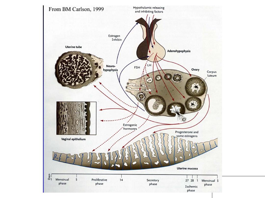
• At puberty, 15 to 20 primordial follicles monthly begin to
mature passing in 3 stages:1-Primary or preantral
2-Secondary or antral or vesicular
3-Preovulatory(Graafian follicle)
• Some of these die, while others begin to accumulate fluid
in a space called the antrum, thereby entering the
antral
or
vesicular stage
• Fluid continues to accumulate such that, immediately prior
to ovulation, follicles are quite swollen and are called
mature
vesicular follicles or Graffi an follicles
. The
antral stage is the longest, whereas the mature vesicular
stage encompasses approximately 37 hours prior to
ovulation.

Slide 11
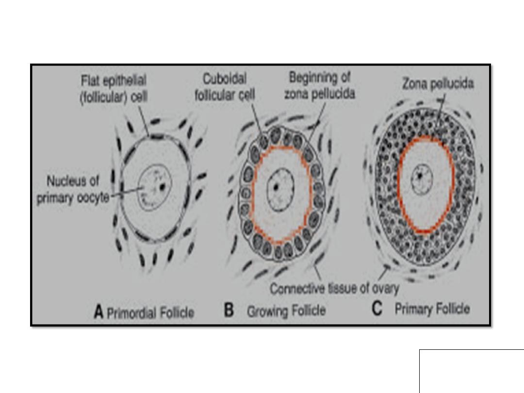
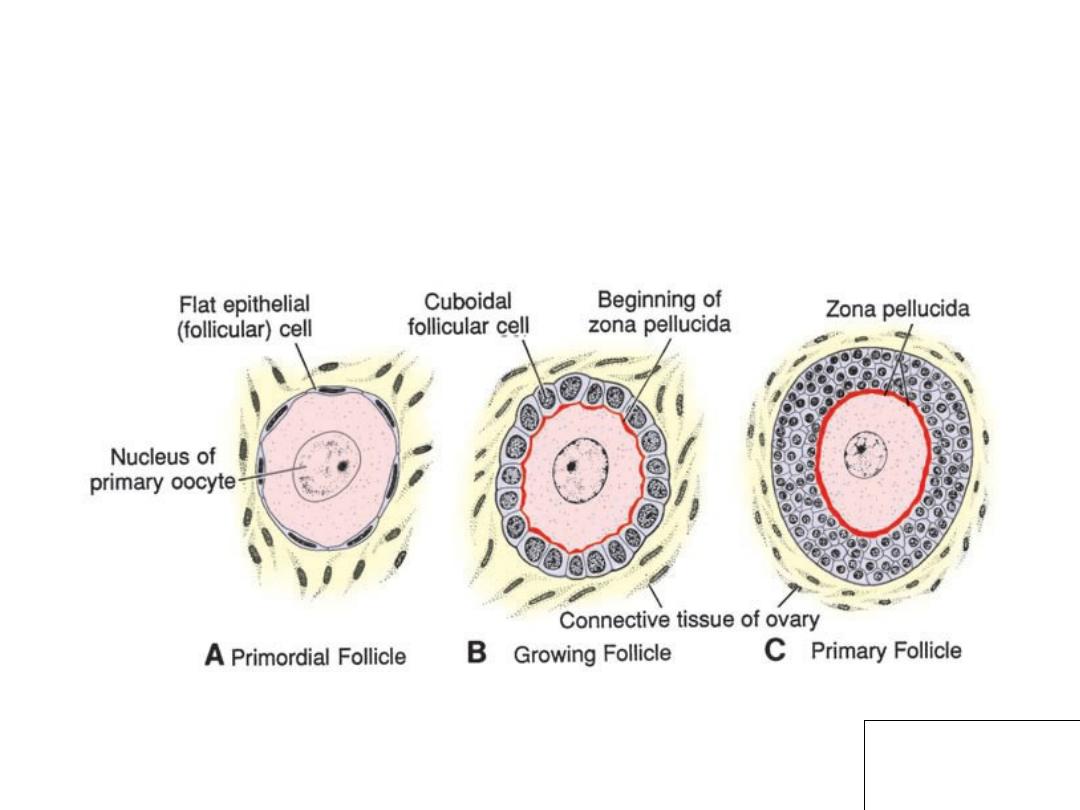
• As primordial follicles begin to grow, surrounding follicular
cells change from flat to cuboidal and proliferate to
produce a stratified epithelium of granulosa cells, and
the unit is called a
primary follicle .
• Granulosa cells: rest on a basement membrane
separating them from surrounding ovarian connective
tissue (stromal cells) that form the theca folliculi.
• Also, granulosa cells + the oocyte secrete a layer of
glycoproteins on the surface
of the oocyte, forming the
zona pellucida
.
Slide 12
• As follicles continue to grow, cells of the theca folliculi
organize into an inner layer of secretory cells, the
theca
interna
, and an outer fibrous capsule, the
theca
externa.
• Also, small, finger-like processes of the follicular
cells extend across the zona pellucida and
interdigitate with microvilli of the plasma
membrane of the oocyte. These processes are
important for transport of materials from
follicular cells to the oocyte.

Slide 13
• As development continues, fluid-filled spaces appear
between granulosa cells. Coalescence of these spaces
forms the
antrum, and the follicle is termed a vesicular
or an antral follicle
.
• Initially, the antrum is crescent-shaped, but with time, it
enlarges. Granulosa cells surrounding the oocyte remain
intact and form the
cumulus oophorus
.
• At maturity, the
mature vesicular (Graafian) follicle
may be 25 mm or more in diameter. It is surrounded by
the theca interna, which is composed of cells having
characteristics of steroid secretion, rich in blood vessels,
and the theca externa, outer fibrous capsule layer which
gradually merges with the ovarian connective tissue

Slide 14
• With each ovarian cycle, a number of follicles begin to
develop, but usually only one reaches full maturity. The
others degenerate and become atretic.
• When the secondary follicle is mature, a surge in
luteinizing hormone (LH) i
nduces the preovulatory
growth phase. Meiosis I is completed, resulting in
formation of two daughter cells of unequal size, each
with 23 double-structured chromosomes
Slide 15
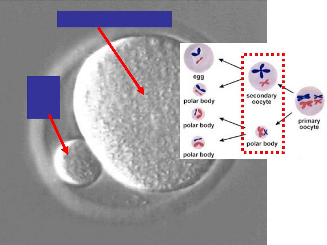
• One cell, the
secondary oocyte,
receives most of the
cytoplasm; the other, the
first polar
body, receives
practically
none. The fi rst polar body lies between the zona pellucida
and the cell membrane of the secondary oocyte in the
perivitelline space
The cell then enters meiosis II but arrests in metaphase
approximately 3 hours before ovulation. Meiosis II is
completed only if the oocyte is fertilized; otherwise, the
cell degenerates approximately 24 hours after ovulation.
The fi rst polar body may
undergo a second division
Slide 16
Slide 17
http://www.ucalgary.ca/UofC/eduweb/virtualembryo/PageMill_Images/image145.gif
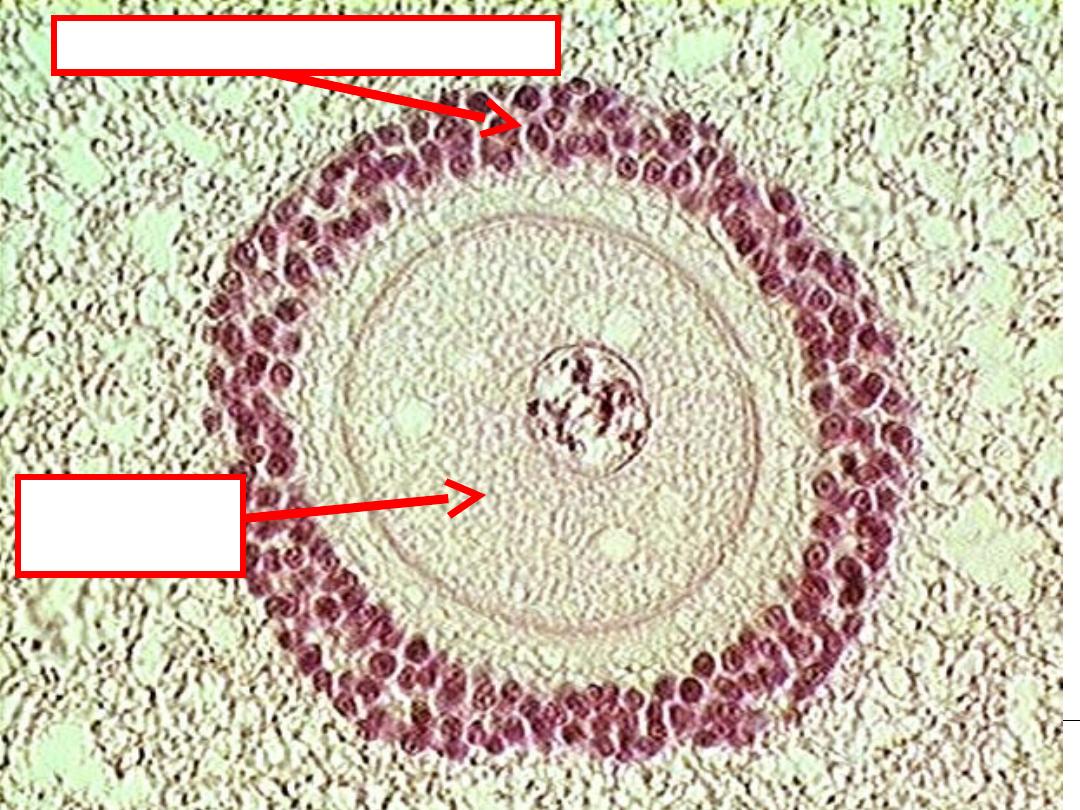
Secondary oocyte, 1N
Polar
body,
1N
Slide 18
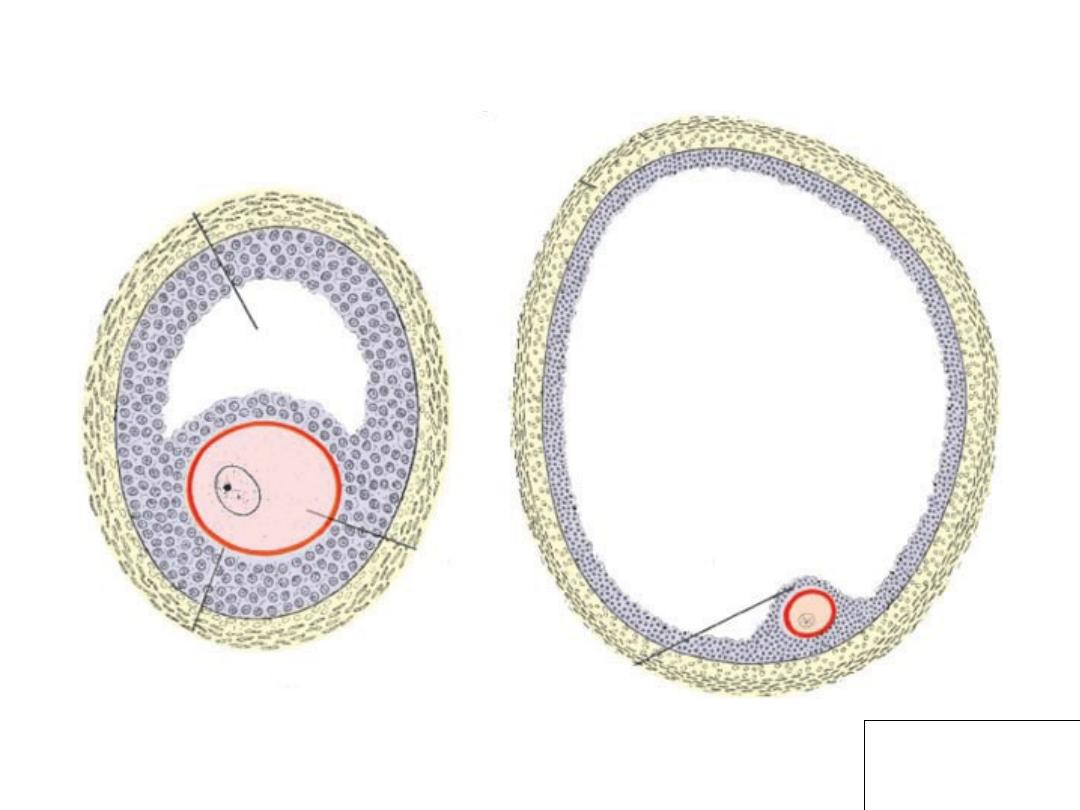
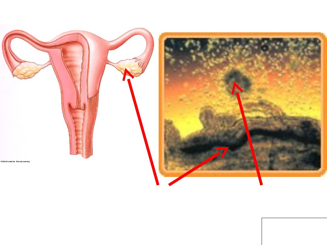
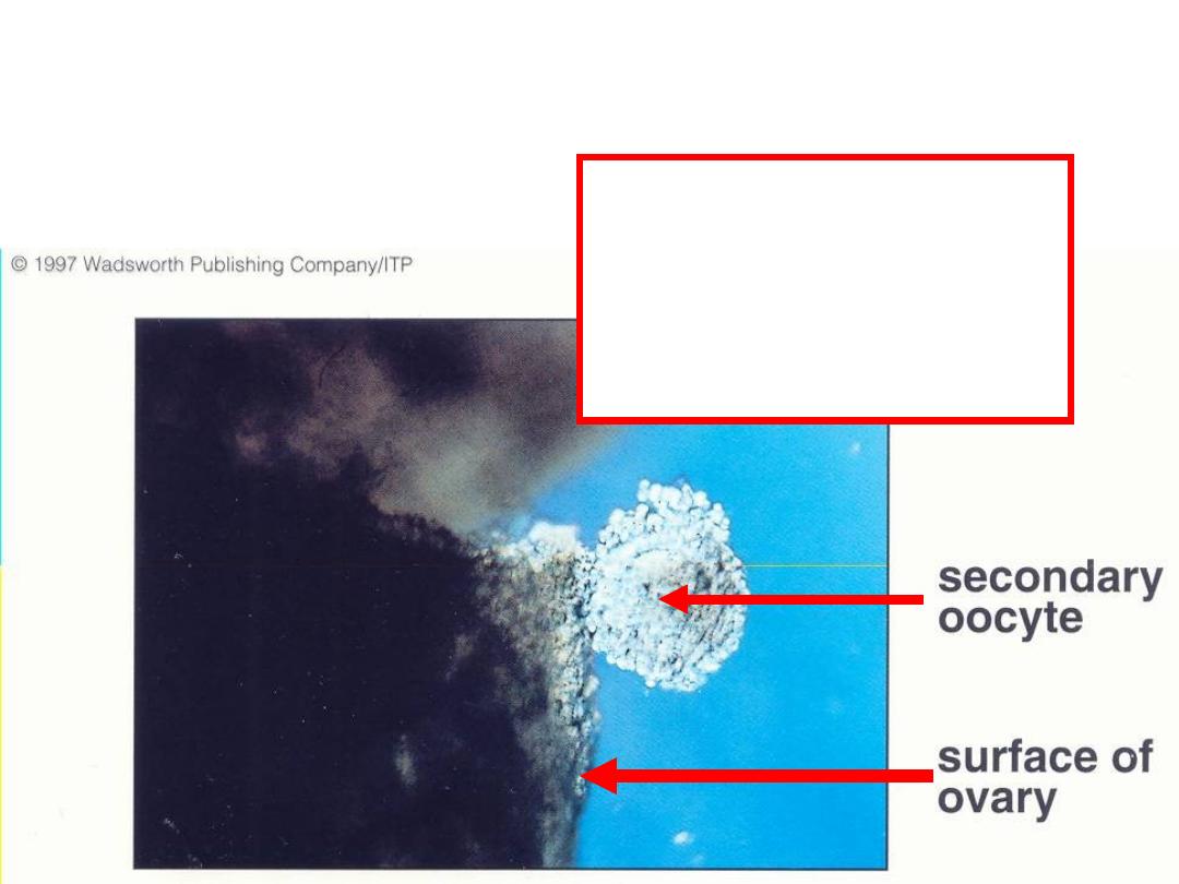
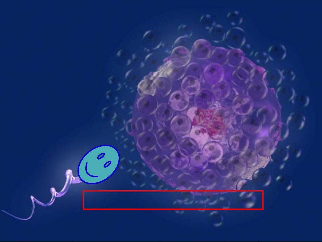
Ovary releases Secondary
Oocyte and Follicle Cells
Slide 19
Release of secondary
oocyte (1N)
surrounded by
follicle cells (2N)
Slide 20
Secondary
oocyte = 1N
Follicle Cells = 2N nurse cells
Slide 21
Slide 22
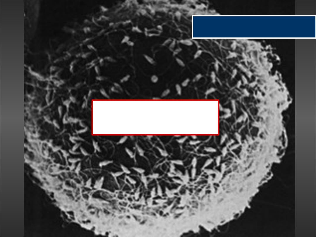
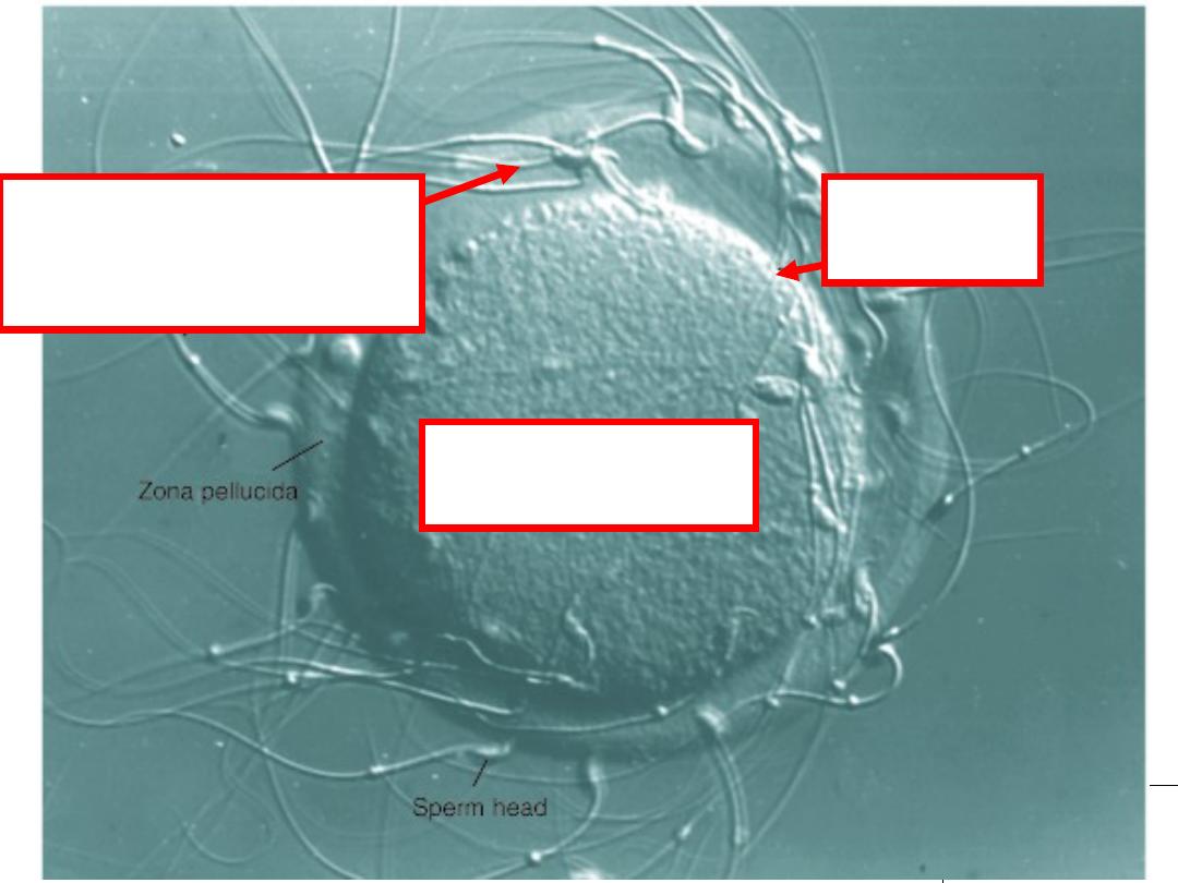
http://www.nature.com/ncb/journal/v3/n2/images/ncb0201_e59_F1.gif
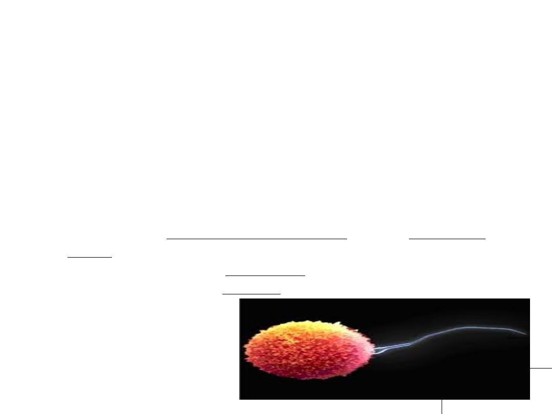
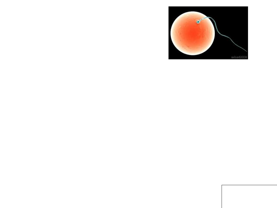
Egg (oocyte)
cytoplasm
Plasma
membrane
Sperm use acrosome
enzymes to penetrate
zona pellucida
Slide 23
THANK YOU