Dr- Suroor mohamed
Pulmonary function test lect.four –respiratory physiologyObjectives:
1- what are the pulmonary function tests?2-whats mean by pulmonary volumes & capacities?
3-The clinical application of these test.
• pulmonary function tests or PFTs used to evaluate how well your lungs work.
• The tests determine how much air your lungs can hold, how quickly you can move air in and out of your lungs, and how well your lungs add oxygen and remove carbon dioxide from your blood.• The tests can diagnose lung diseases and measure the severity of lung problems.
• Also used to assist the pre operation prepare to surgery( fit to anaesthesia or not)
Routine pulmonary function test.
–Reduced in restrictive lung disease (pulmonary fibrosis).–May be Normal in obstructive lung disease (asthma, chronic bronchitis
Spirometry
is the first lung function test done. It measures how much and how quickly you can move air out of your lungs.
For this test, you breathe into a mouthpiece attached to a recording device (spirometer).
The information collected by the spirometer may be printed out on a chart called a spirogram.
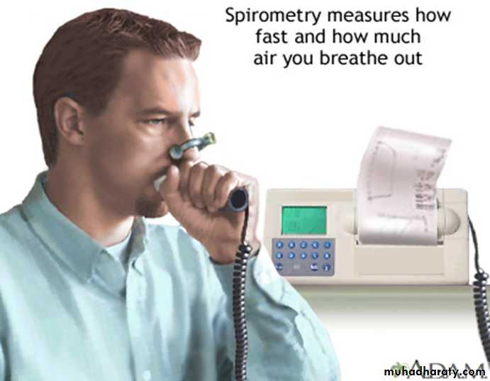
SPIROMETER: An instrument which measure the volume of air moved into or out of the lungs
It is a device that records changes in volume in relation to time
It can measure: All lung volumes & capacities [except residual volume & capacities including residual volume (FRC & TLC)]
You don’t need to memorize the normal numbers, just the definitions
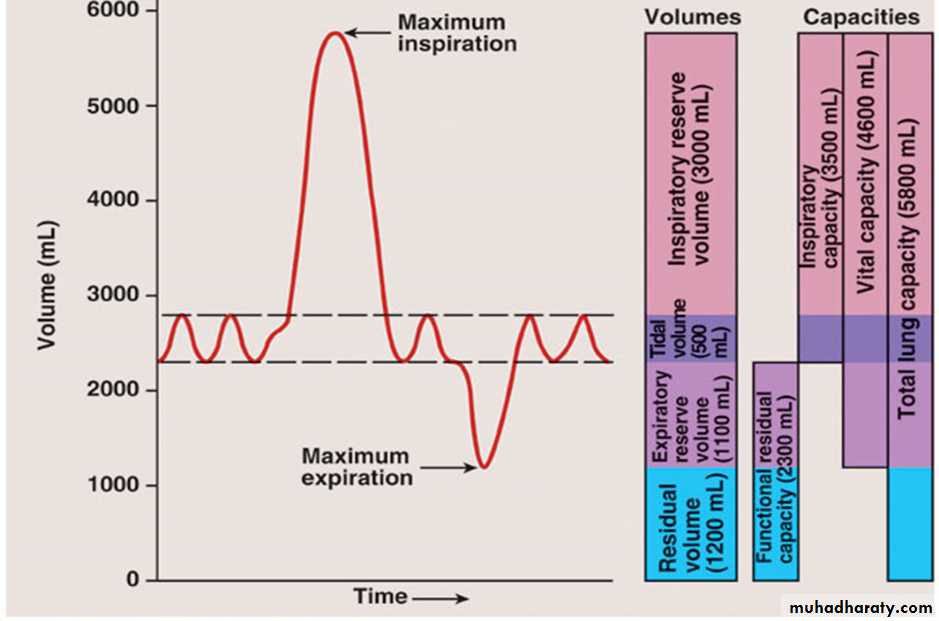
Respiratory Cycle: A single cycle of inhalation and exhalationRespiratory rate: number of breaths per minute (usually about 12-18; children higher 18-20).
Tidal Volume: normal breath in and out. Usually about 500 ml.
Inspiratory Reserve Volume: take in a normal breath, stop, now inhale as much more as you can. In other words, this is the amount of air that can be forcefully inhaled after a normal inhalation. This is your tidal volume in plus your inspiratory volume.
Expiratory Reserve Volume (Expiratory capacity): take a normal breath in, a normal breath out, then breathe out the most you can. In other words, this is the amount of air that can be forcefully exhaled after a normal exhalation. This is the air needed to perform the Heimlich maneuver. The maneuver decreases the thoracic cavity volume, causing increased pressure in lungs. That causes forced air with high pressure to be expelled from the lungs.
Residual volume: The amount of air left in your lungs after you exhale maximally. This air helps to keep the alveoli open and prevent lung collapse. This is estimated based on height and age
Vital capacity: The volume of air a patient can exhale maximally after a forced inspiration. Maximum deep breath in, then exhale as much as possible. It can be used to determine if problems are obstructive (normal) or restrictive (reduced). Vital capacity divided by expiratory reserve volume should be 80%.
Total Lung Capacity (TLC): the sum of all lung volumes
Inspiratory Capacity: amount of air for a deep breath in after normal exhalation
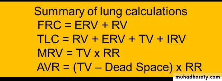
Functional residual capacity: amount of air left in your lungs after a normal exhale. You have to calculate this:
FRC = ERV + residual volume.
In COPD, their FRC increases.
They have a barrel chest
The lungs don’t have as much recoil, have decreased tidal volume, cannot exhale enough
Dead Space: Area where air fills the passageways and never contributes to gas exchange. Amounts to about 150 ml.
Minute Respiratory Volume (MRV): tidal volume x respiratory rate. This calculation does not take into account the volume of air wasted in the dead space. A more accurate measurement of respiratory efficiency is alveolar ventilation rate.
Alveolar Ventilation Rate (AVR)
AVR = (TV – Dead Space) x Respiratory Rate
Pulmonary volumes are:1-Tidal volume (VT): About 500 ml.2-Inspiratory reserve volume (IRV): About 3000 ml.3-Expiratory reserve volume (ERV): About 1100 ml.4-Residual volume (RV): ab. 1200 ml.Pulmonary capacities are:1-Inspiratory capacity (IC)= VT + IRV ≈ 3500 ml.2-Functional residual capacity (FRC) FRC = ERV + RV ≈ 2300 ml.3-Vital capacity (VC) = VT+ IRV+ERV ≈ 4600 ml.4-Total lung capacity (TLC) = VC + RV ≈ 5800ml.
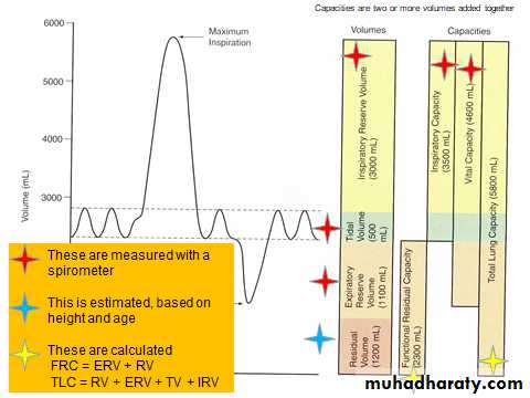
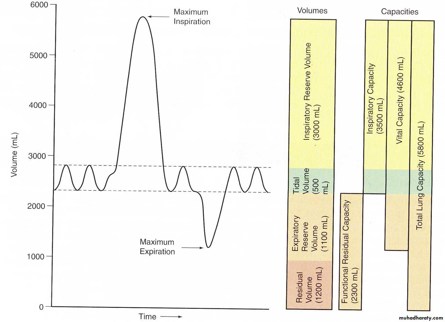
The tidal volume, vital capacity, inspiratory capacity and expiratory reserve volume can be measured directly with a spirometer.
Most air (80%) is exhaled during the first second of exhalation. You take a maximum inhale, then a maximum exhale (vital capacity). The pen moves down the paper, showing time.
You can calculate how much air you blew out (vital capacity), and the amount of air you blew out in one second (expiratory reserve volume in one second).
Expiratory reserve volume divided by vital capacity should be 80%. If you are less than 80%, it is suggestive of an obstructive pulmonary disorder.
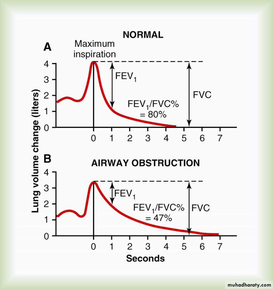
Someone with COPD takes longer than one second to exhale 80%.
FACTORS AFFECTING LUNG VOLUMES & CAPACITIES1•AGE : decrease in older age groups.
2•SEX : 25% greater in males.
3•Body build : tall thin subject had greater values than short obese one .
4•Athletics had greater values.
5•Body position :
PFT is maximum in standing, less in sitting and least in lying position because:
–Abdominal contents press on the diaphragm.
–About 500 ml of blood shifted to the pulmonary circulation.
classification of lung disorders
1•Obstructive lung diseases
2•Restrictive lung diseases
3•Gas diffusion diseases
Obstructive Diseases
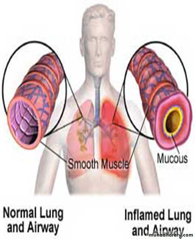
•Difficult to get air out of the lungs …Obstruct expirationObstructive lung diseases are characterized by inflamed and easily collapsible airways, obstruction to airflow.
•Examples:
–emphysema , chronic bronchitis , COPD & asthma .
Restrictive Diseases
•Difficult to get air in to the lungs ―Restrict‖ inspiration
resulting in a decreased lung volume (rapid, shallow breathing), an increased work of breathing, and inadequate ventilation and/or oxygenation. Decreased vital capacity.
•Examples:
–intersitial fibrosis –sarcoidosis –muscular diseases & chest wall deformities.
Gas Diffusion Diseases
•Inability of the tissue of the alveoli to move oxygen into a person's blood through the respiratory membrane•Examples: Pulmonary odema & Interstitial lung diseases
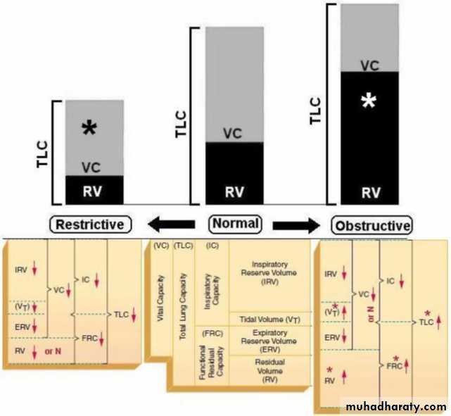
Restrictive Disease:
Decreased VCDecreased TLC, RV, FRC
So FEV1/VC ratio normal
Obstructive Disease:
Decreased VC
Increased TLC, RV, FRC.
FEV1/VC is less than 80%
Restrictive disorder:
◦ Vital capacity is reduced. (pulmonary fibrosis)
◦ FVC is normal.
● Obstructive disorder: Diagnosed by tests that measure the rate
of expiration.
⚫ VC is normal. In asthma , chronic bronchitits
⚫ FEV1 is < 80%.
Asthma:
⚫ Obstructive air flow through bronchioles, Caused by inflammation and mucus secretion.⚫ Inflammation contributes to increased airway responsiveness to agents that promote bronchial constriction.
, this inflammation causes recurrent episodes of wheezing, breathlessness, chest tightness and coughing, particularly at night or in the early morning.
Like : IgE, exercise , cold weathear ,trigger agent , food & drugs,,,
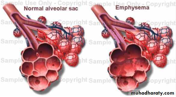
Emphysema:
⚫ Alveolar tissue is destroyed.
⚫ Decreases ability of bronchioles to remain open during expiration.
⚫ Cigarette smoking stimulates macrophages and leukocytes to
secrete protein digesting enzymes that destroy tissue.
Pulmonary fibrosis:
◦ Normal structure of lungs disrupted by accumulation of
fibrous connective tissue proteins.