
Clostridia
The clostridia are large anaerobic, gram-positive, motile rods. Many decompose proteins or form toxins, and some do both. Their natural habitat is the soil or the intestinal tract of animals and humans, where they live as saprophytes. Among the pathogens are the organisms causing botulism, tetanus, gas gangrene, and pseudomembranous colitis.
• Morphology & Identification
• Typical Organisms• Spores of clostridia are usually wider than the diameter of the rods in which they are formed. In the various species, the spore is placed centrally, subterminally, or terminally. Most species of clostridia are motile and possess peritrichous flagella.
• Clostridia are anaerobes; a few species are aero-tolerant and will also grow in ambient air. in general, the clostridia grow well on the blood-enriched media used to grow anaerobes and on other media used to culture anaerobes as well.
• Some clostridia produce large raised colonies (e.g, C perfringens); others produce smaller colonies (eg, C tetani). Some clostridia form colonies that spread on the agar surface. Many clostridia produce a zone of hemolysis on blood agar. C perfringens typically produces multiple zones of hemolysis around colonies.
• Growth Characteristics
• Clostridia can ferment a variety of sugars; many can digest proteins. Milk is turned acid by some and digested by others and undergoes "stormy fermentation" (ie, clot torn by gas) with a third group (eg, C perfringens). Various enzymes are produced by different species .Clostridium Botulinum
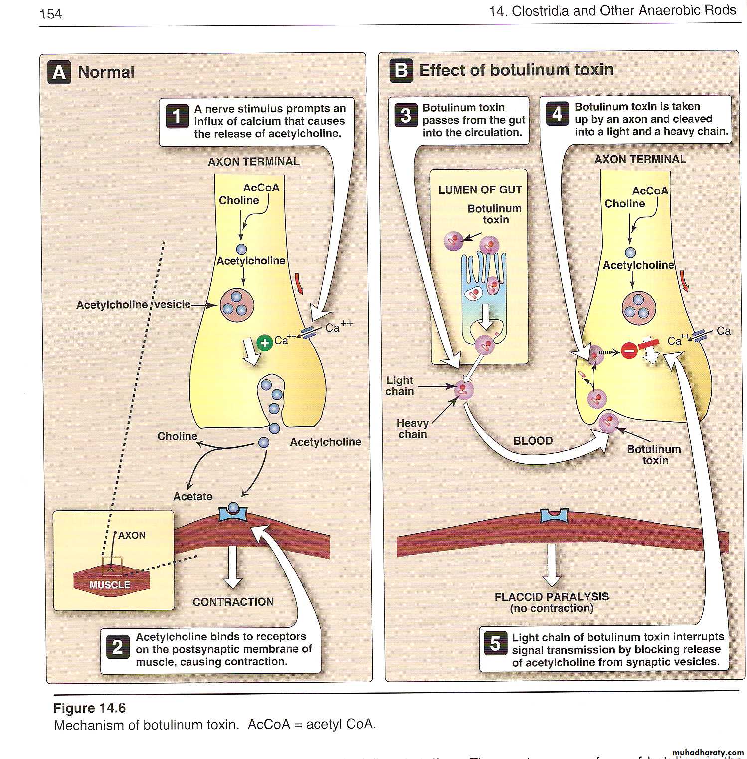
During the growth of C botulinum and during autolysis of the bacteria, toxin is liberated into the environment. . Botulinum toxin is absorbed from the gut and binds to receptors of motor neurons of the peripheral nervous system and cranial nerves. Proteolysis—by the light chain of botulinum toxin—of the target SNARE proteins ( synaptobrevin, SNAP 25, and syntaxin ) in neurons inhibits the release of acetylcholine at the synapse, resulting in lack of muscle contraction and paralysis. The lethal dose for a human is probably about 1–2 µg . So , it’s the most toxic substances known .
• Clinical Findings
• Symptoms begin 18–24 hours after ingestion of the toxic food, with visual disturbances (in coordination of eye muscles, double vision), inability to swallow, and speech difficulty; signs of bulbar paralysis are progressive, and death occurs from respiratory paralysis or cardiac arrest. There is no fever. The patient remains fully conscious until shortly before death. The mortality rate is high. Patients who recover do not develop antitoxin in the blood.• Diagnostic Laboratory Tests
• Toxin can often be demonstrated in serum from the patient . Mice injected intraperitoneally die rapidly. The antigenic type of toxin is identified by neutralization with specific antitoxin in mice. C botulinum may be grown from food remains and tested for toxin production . In infant botulism, C botulinum and toxin can be demonstrated in bowel contents but not in serum. Toxin may be detected by passive hemagglutination or radioimmunoassay.• Treatment
• Potent antitoxins , trivalent (A, B, E) antitoxin must be promptly administered intravenously with customary precautions. Adequate ventilation must be maintained by mechanical respirator . These measures have reduced the mortality rate from 65% to below 25%.• Clostridium Tetani
• Clostridium tetani, which causes tetanus, is worldwide in distribution in the soil and in the feces of horses and other animals. Several types of C tetani can be distinguished by specific flagellar antigens. All share a common O (somatic) antigen, which may be masked, and all produce the same antigenic type of neurotoxin, tetanospasmin.
• Toxin
• The vegetative cells of C tetani produce the toxin tetanospasmin , the toxin initially binds to receptors on the presynaptic membranes of motor neurons. It then migrates by the retrograde axonal transport system to the cell bodies of these neurons to the spinal cord and brain stem. The toxin diffuses to terminals of inhibitory cells, including both glycinergic interneurons and aminobutyric acid-secreting neurons from the brain stem. The toxin degrades synaptobrevin, a protein required for docking of neurotransmitter vesicles on the presynaptic membrane. Release of the inhibitory glycine and γ-aminobutyric acid is blocked, and the motor neurons are not inhibited. Hyperreflexia, muscle spasms, and spastic paralysis result. Extremely small amounts of toxin can be lethal for humans.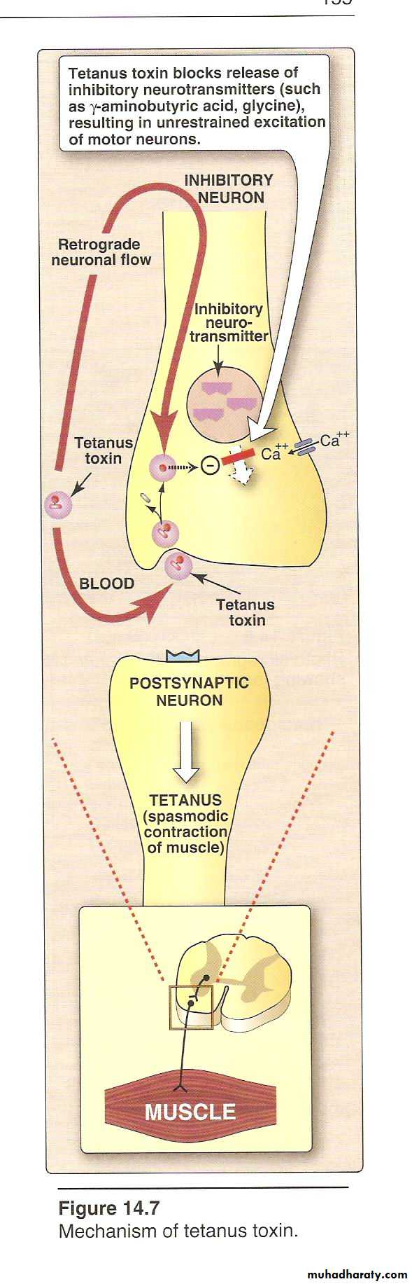
• Pathogenesis
• C tetani is not an invasive organism. The infection remains strictly localized in the area of devitalized tissue (wound, burn, injury, umbilical stump, surgical suture) into which the spores have been introduced. The volume of infected tissue is small, and the disease is almost entirely a toxemia. Germination of the spore and development of vegetative organisms that produce toxin are aided by (1) necrotic tissue, (2) calcium salts, and (3) associated pyogenic infections, all of which aid establishment of low oxidation-reduction potential.• Clinical Findings
• The incubation period may range from 4–5 days to as many weeks. The disease is characterized by tonic contraction of voluntary muscles. Muscular spasms often involve first the area of injury and infection and then the muscles of the jaw (trismus, lockjaw), which contract so that the mouth cannot be opened. Gradually, other voluntary muscles become involved, resulting in tonic spasms. Any external stimulus may precipitate a tetanic generalized muscle spasm. The patient is fully conscious, and pain may be intense. Death usually results from interference with the mechanics of respiration. The mortality rate in generalized tetanus is very high.• Diagnosis
• The diagnosis rests on the clinical picture and a history of injury, although only 50% of patients with tetanus have an injury for which they seek medical attention. The primary differential diagnosis of tetanus is strychnine poisoning. Anaerobic culture of tissues from contaminated wounds may yield C tetani, but neither preventive nor therapeutic use of antitoxin should ever be withheld pending such demonstration. Proof of isolation of C tetani must rest on production of toxin and its neutralization by specific antitoxin.• Prevention & Treatment
• The results of treatment of tetanus are not satisfactory. Therefore, prevention is all-important. Prevention of tetanus depends upon (1) active immunization with toxoids; (2) proper care of wounds contaminated with soil, etc; (3) prophylactic use of antitoxin; and (4) administration of penicillin.• Clostridia that Produce Invasive Infections
• Many different toxin-producing clostridia (Clostridium perfringens and related clostridia) can produce invasive infection (including myonecrosis and gas gangrene) if introduced into damaged tissue. About 30 species of clostridia may produce such an effect, but the most common in invasive disease is Clostridium perfringens (90%).
• Toxins
• The invasive clostridia produce a large variety of toxins and enzymes that result in a spreading infection. Many of these toxins have lethal, necrotizing, and hemolytic properties. The alpha toxin of C perfringens type A is a lecithinase, which splits lecithin (an important constituent of cell membranes) to phosphorylcholine and diglyceride. The theta toxin has similar hemolytic and necrotizing effects but is not a lecithinase. DNase and hyaluronidase, a collagenase that digests collagen of subcutaneous tissue and muscle, are also produced. Its produce enterotoxin that cause GIT menifestations .• Pathogenesis
• In invasive clostridial infections, spores reach tissue either by contamination of traumatized areas (soil, feces) or from the intestinal tract. The spores germinate at low oxidation-reduction potential; vegetative cells multiply, ferment carbohydrates present in tissue, and produce gas. The distention of tissue and interference with blood supply, together with the secretion of necrotizing toxin and hyaluronidase, favor the spread of infection. Tissue necrosis extends, providing an opportunity for increased bacterial growth, hemolytic anemia, and, ultimately, severe toxemia and death.• Clinical Findings
• From a contaminated wound (eg, a compound fracture, postpartum uterus), the infection spreads in 1–3 days to produce crepitation in the subcutaneous tissue and muscle, foul-smelling discharge, rapidly progressing necrosis, fever, hemolysis, toxemia, shock, and death. Treatment is with early surgery (amputation) and antibiotic administration. Until the advent of specific therapy, early amputation was the only treatment. At times, the infection results only in anaerobic fasciitis or cellulitis.• Diagnostic Laboratory Tests
• Specimens consist of material from wounds, pus, tissue. The presence of large gram-positive rods in Gram-stained smears suggests gas gangrene clostridia; spores are not regularly present.• Material is inoculated into chopped meat-glucose medium and thioglycolate medium and onto blood agar plates incubated anaerobically. The growth from one of the media is transferred into milk. A clot torn by gas in 24 hours is suggestive of C perfringens. Once pure cultures have been obtained by selecting colonies from anaerobically incubated blood plates, they are identified by biochemical reactions (various sugars in thioglycolate, action on milk), hemolysis, and colony form. Lecithinase activity is evaluated by the precipitate formed around colonies on egg yolk media. Final identification rests on toxin production and neutralization by specific antitoxin.
• Treatment
• The most important aspect of treatment is prompt and extensive surgical debridement of the involved area and excision of all devitalized tissue, in which the organisms are prone to grow. Administration of antimicrobial drugs, particularly penicillin, is begun at the same time. Hyperbaric oxygen may be of help in the medical management of clostridial tissue infections.Clostridium Difficile
Pseudomembranous colitis is diagnosed by detection of one or both C difficile toxins in stool and by endoscopic observation of pseudomembranes or microabscesses in patients who have diarrhea and have been given antibiotics. Plaques and microabscesses may be localized to one area of the bowel.
The diarrhea may be watery or bloody, and the patient frequently has associated abdominal cramps, leukocytosis, and fever. Although many antibiotics have been associated with pseudomembranous colitis, the most common are ampicillin and clindamycin. The disease is treated by discontinuing administration of the offending antibiotic and orally giving either metronidazole or vancomycin.
Administration of antibiotics results in proliferation of drug-resistant C difficile that produces two toxins. Toxin A, a potent enterotoxin that also has some cytotoxic activity, binds to the brush border membranes of the gut at receptor sites. Toxin B is a potent cytotoxin.