Digestive System-part -1-Dr: Ezdehar Nassif Ali
objectivesDivisions of the gut tube
Mesenteries
Pharyngeal gut
Foregut
Divisions of the gut tube
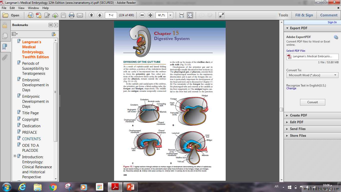
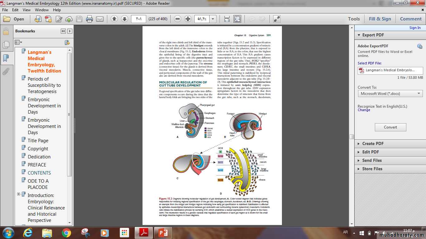
The gut system extends from the oropharyngeal membrane to the cloacal membrane and is divided into the pharyngeal gut, foregut, midgut, and hindgut. The mid gut, remains temporally connected to the yolk sac by means of the vitelline duct or yolk stalk.
The epithelium of the digestive system and the parenchyma of its derivatives originate in the endoderm. The connective tissue, muscular components, and peritoneal components originate in the mesoderm.
Mesenteries
Portions of the gut tube and its derivatives are suspended enclose an organ and connect it to the body wall from the dorsal and ventral body wall by mesenteries, double layers of peritoneum that enclose an organ and connect it to the body wall. Such organs are called intra peritoneal, whereas organs that lie against the posterior body wall and are covered by peritoneum on their anterior surface only (e.g., the kidneys) are considered retroperitoneal.
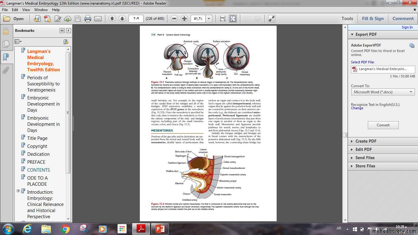
Mesenteries provide pathways for vessels, nerves, and lymphatics to and from abdominal viscera .
Dorsal mesentery which extends from the lower end of the esophagus to the cloacal region of the hindgut.
In the region of the stomach, it forms the dorsal mesogastrium or greater omentum, in the region of the duodenum, dorsal mesoduodenum).
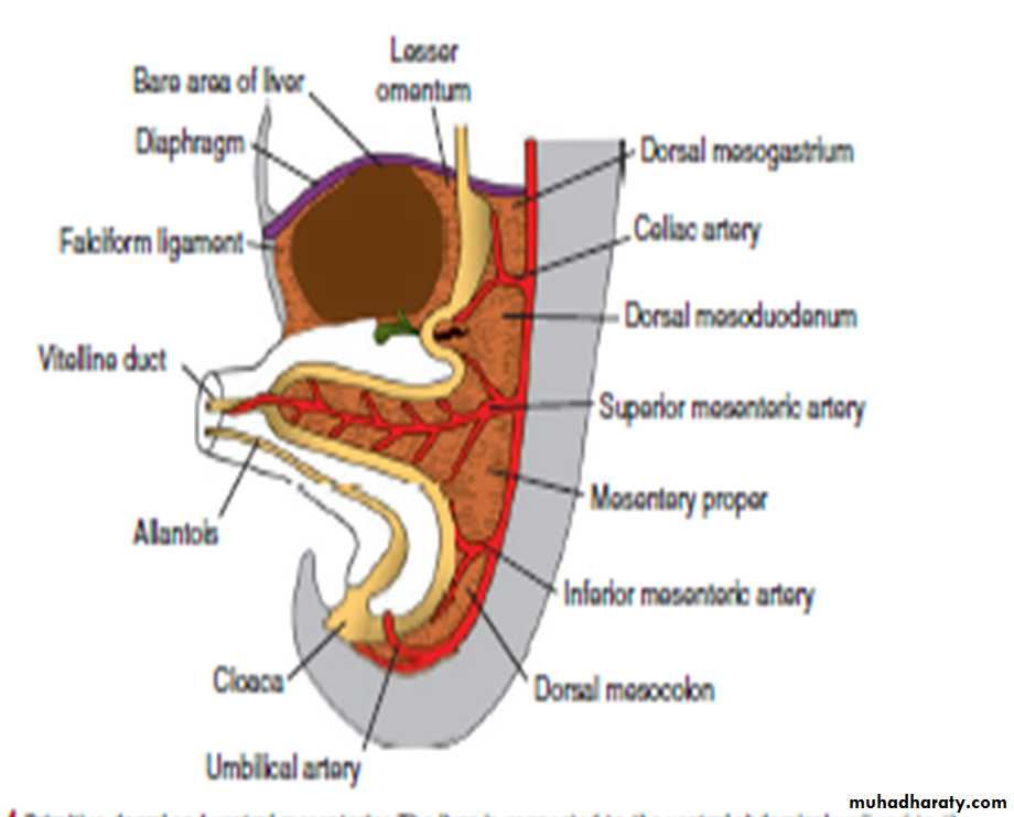
In the region of the colon, it forms the dorsal mesocolon.
Dorsal mesentery of the jejunal and ileal loops forms the mesentery proper.Ventral mesentery, which exists only in the region of the terminal part of the esophagus, the stomach, and the upper part of the duodenum.
Growth of the liver bud divides the ventral mesentery into
(a) lesser omentum, extending from the lower portion of the esophagus, the stomach, and the upper portion of the duodenum to the liver(b) falciform ligament, extending from the liver to the ventral body wall .
Pharyngeal gut
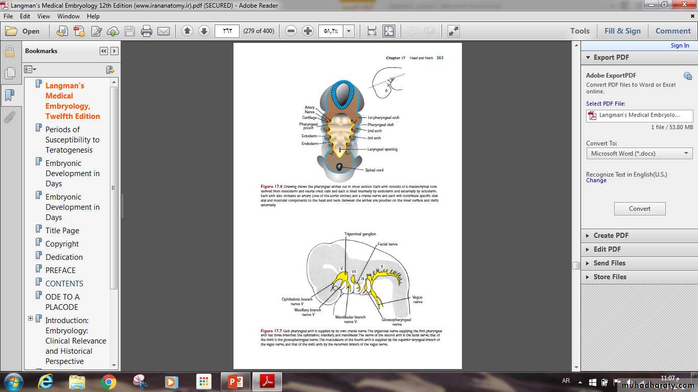
It extends from the oropharyngeal membrane to the respiratory diverticulum (part of the foregut).this section is important for development of the head and neck.
The most distinctive feature in development of the head and neck is the presence of pharyngeal arches, clefts and pouches.
Foregut
The foregut gives rise to the esophagus, trachea and lung buds, the stomach, and the duodenum .In addition, the liver, pancreas, and biliary apparatus.
Esophagus
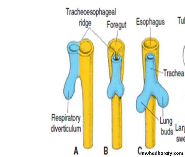
When the embryo is approximately 4 weeks old, the foregut divides into a ventral portion ,the respiratory diverticulum and a dorsal portion ,the esophagus.
At first, the esophagus is short, but with descent of the heart and lungs, it lengthens rapidly.
STOMACH
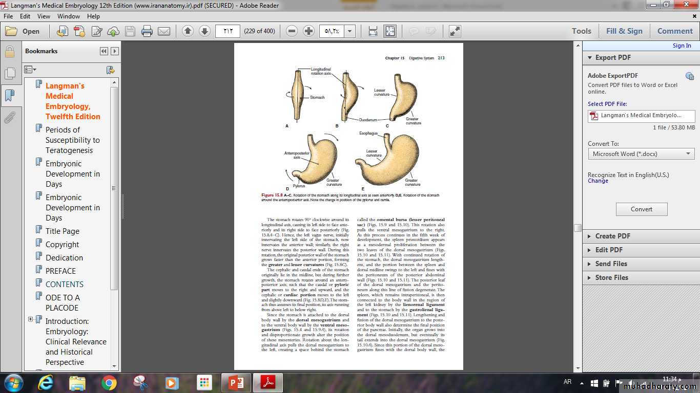
The stomach appears as fusiform dilation of the foregut in the fourth week of development. Positional changes of the stomach are easily explained by assuming that it rotates around a longitudinal and an anteroposterior axis.
During this rotation, the original posterior wall of the stomach grows faster than the anterior portion, forming the greater and lesser curvatures
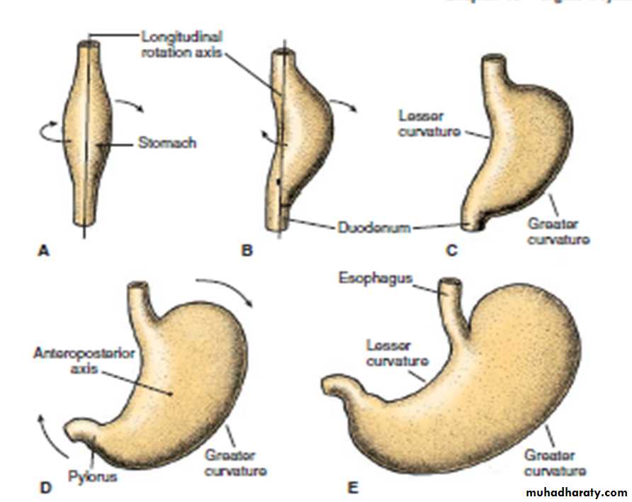
The stomach rotates around an anteroposterior axis, such that the caudal or pyloric part moves to the right and upward, and the cephalic or cardiac portion moves to the left and slightly downward .
Esophageal Abnomalities
Esophageal atresia and –or tracheoesophageal fistulaEsophageal stenosis
Stomach Abnomalities
One of the most common abnormalities of the stomach in infants, Pyloric stenosis was previously believed to develop only during fetal life ,despite the fact that most cases present 3 to 5 days after birth.
THANK YOU