URINARY SYSTEM
Dr.Sumeya

Functionally, the urogenital system can bedivided into two
entirely different components:
the urinary system and the genital system.
Embryologically and anatomically, however, they are
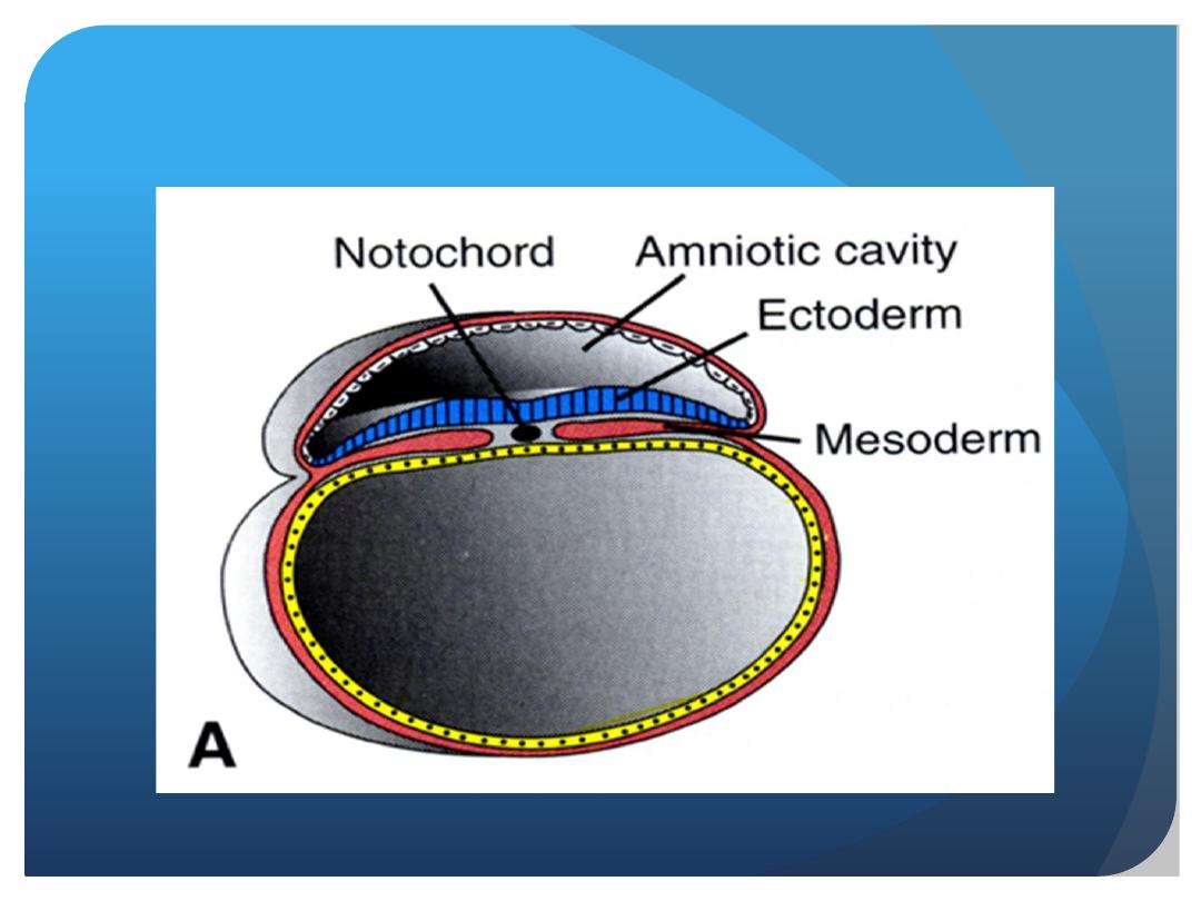
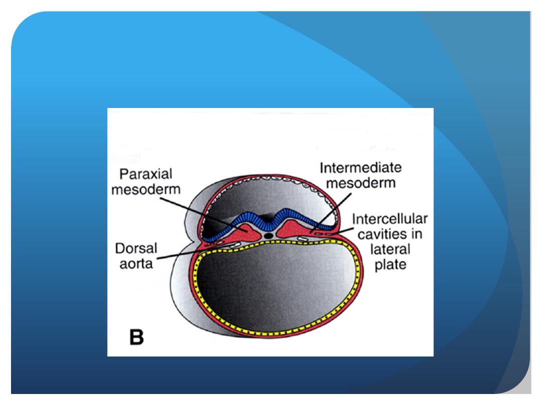
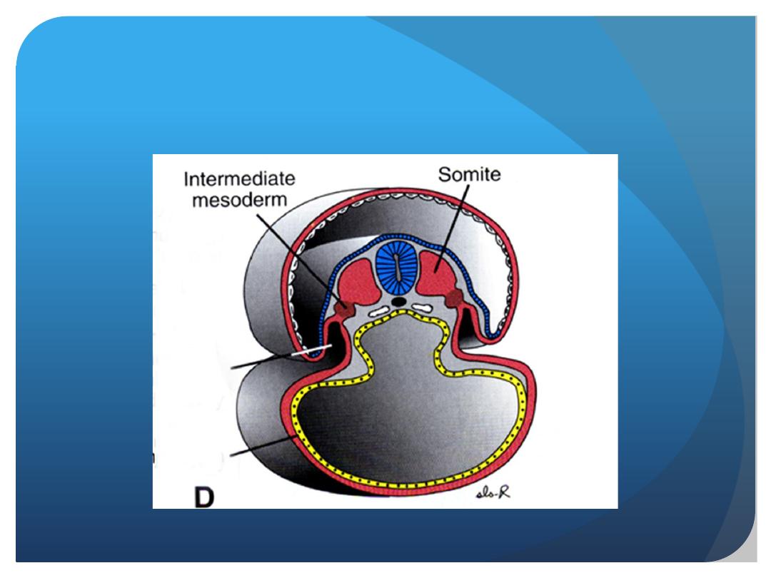
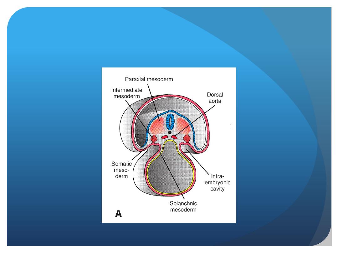
intimately interwoven. Both develop from a common
mesodermal ridge (intermediate mesoderm) along the
posterior wall of the abdominal cavity, and initially, the
excretory ducts of both systems enter a common cavity,
the cloaca.

URINARY SYSTEM
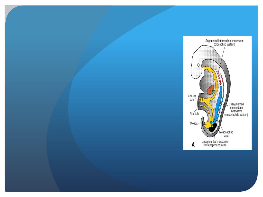
Kidney Systems
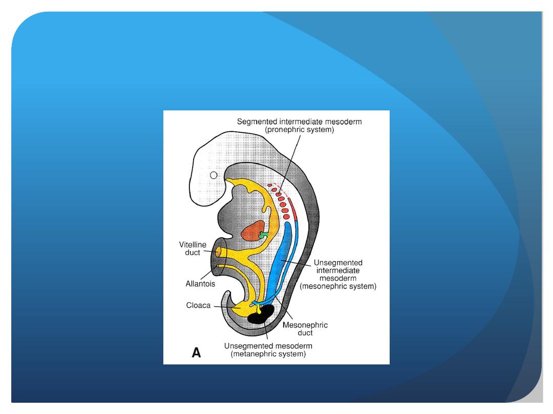
Three slightly overlapping kidney systems are formed in a
cranial-to-caudal sequence during intrauterine life in
humans:
the pronephros, mesonephros, and metanephros.
The first of these systems is rudimentary and
nonfunctional; the second may function for a short time
during the early fetal period; the third forms the
permanent kidney.

The development of
Urinary System

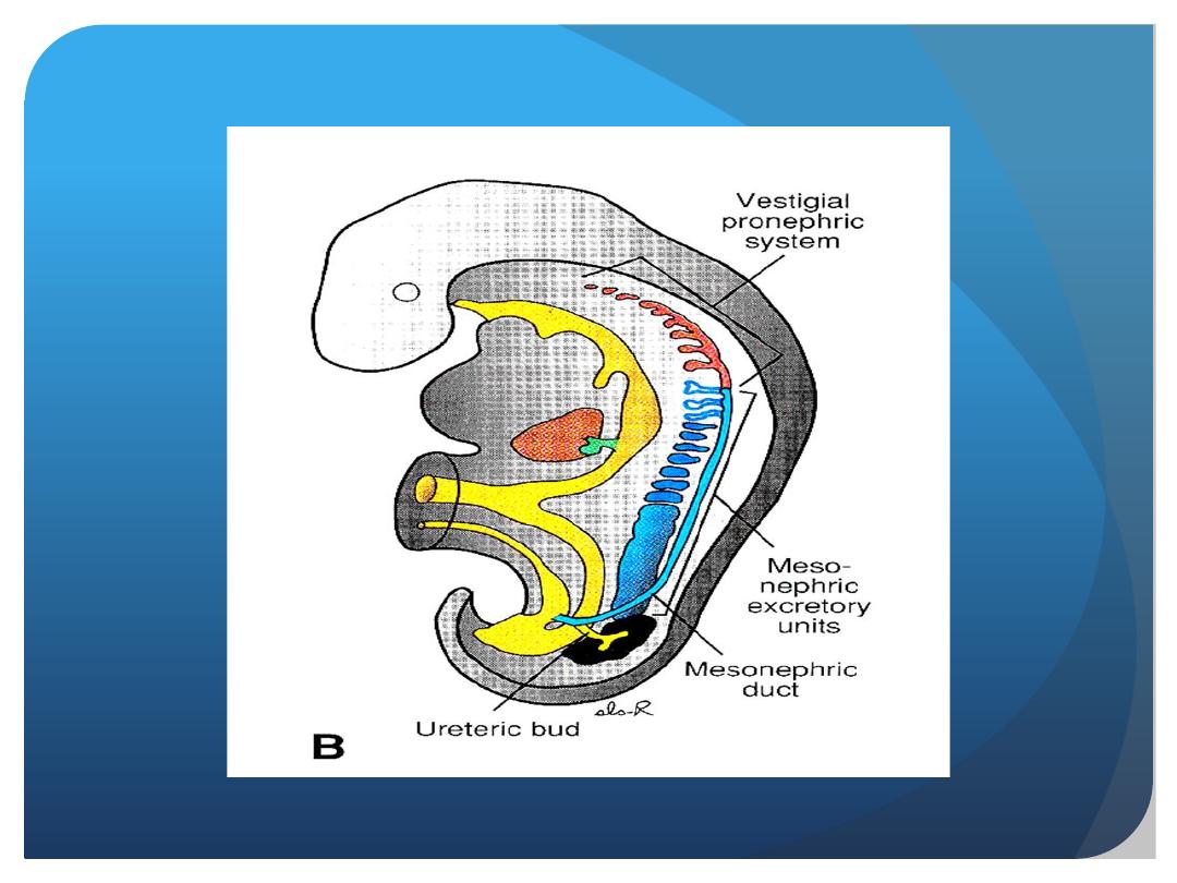
Pronephros
At the beginning of the fourth week, the pronephros
is represented by 7 to 10 solid cell groups
in the cervical region.
These groups form vestigial excretory units,
nephrotomes, that regress before more caudal
ones are formed. By the end of the fourth week,
all indications of the pronephric system have
disappeared.

Mesonephros
The mesonephros and mesonephric ducts are
derived from
intermediate mesoderm
from
upper thoracic to upper lumbar (L3) segments.
Early in the fourth week of development,
during regression of the pronephric system,
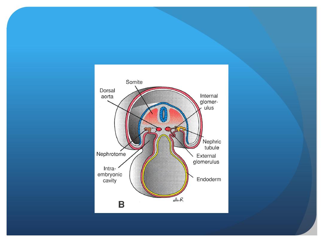
the fi rst excretory tubules of the mesonephros
appear. They lengthen rapidly, form an S-shaped
loop, and acquire a tuft of capillaries that will
form a glomerulus at their medial extremity

Around the glomerulus, the tubules
form Bowman’s capsule, and together these structures
constitute a renal corpuscle.
Laterally,the tubule enters the longitudinal collecting duct
known as the mesonephric or wolffi an duct .
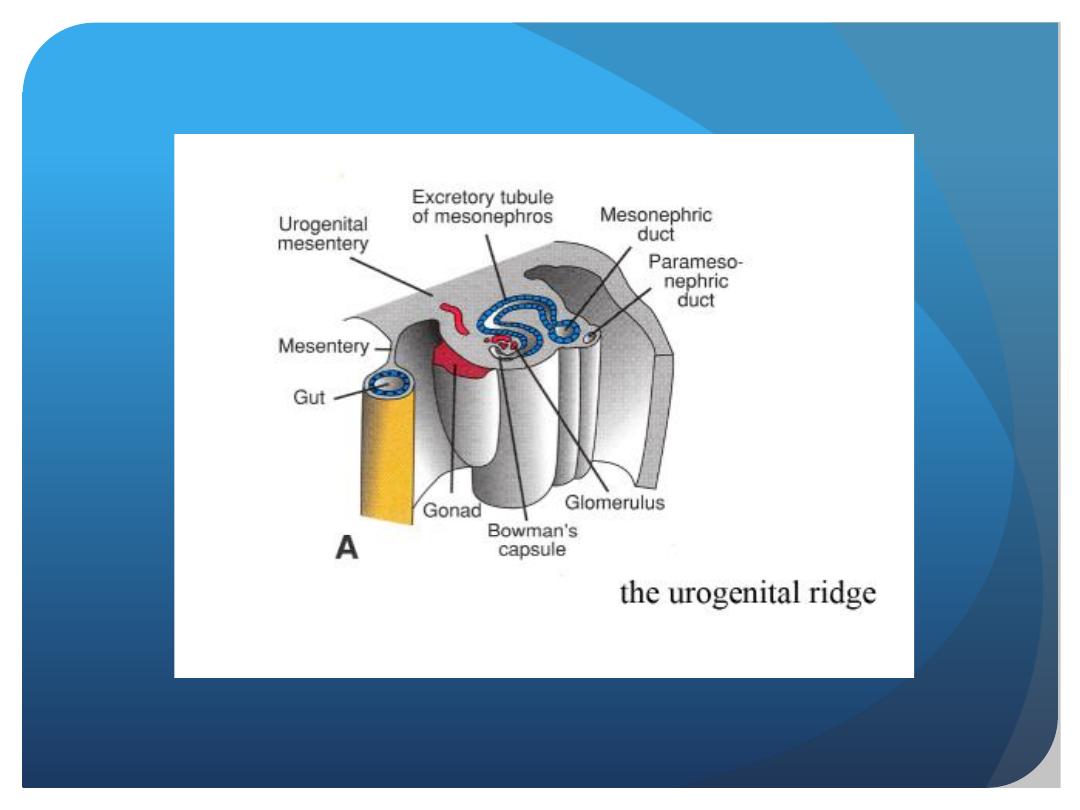
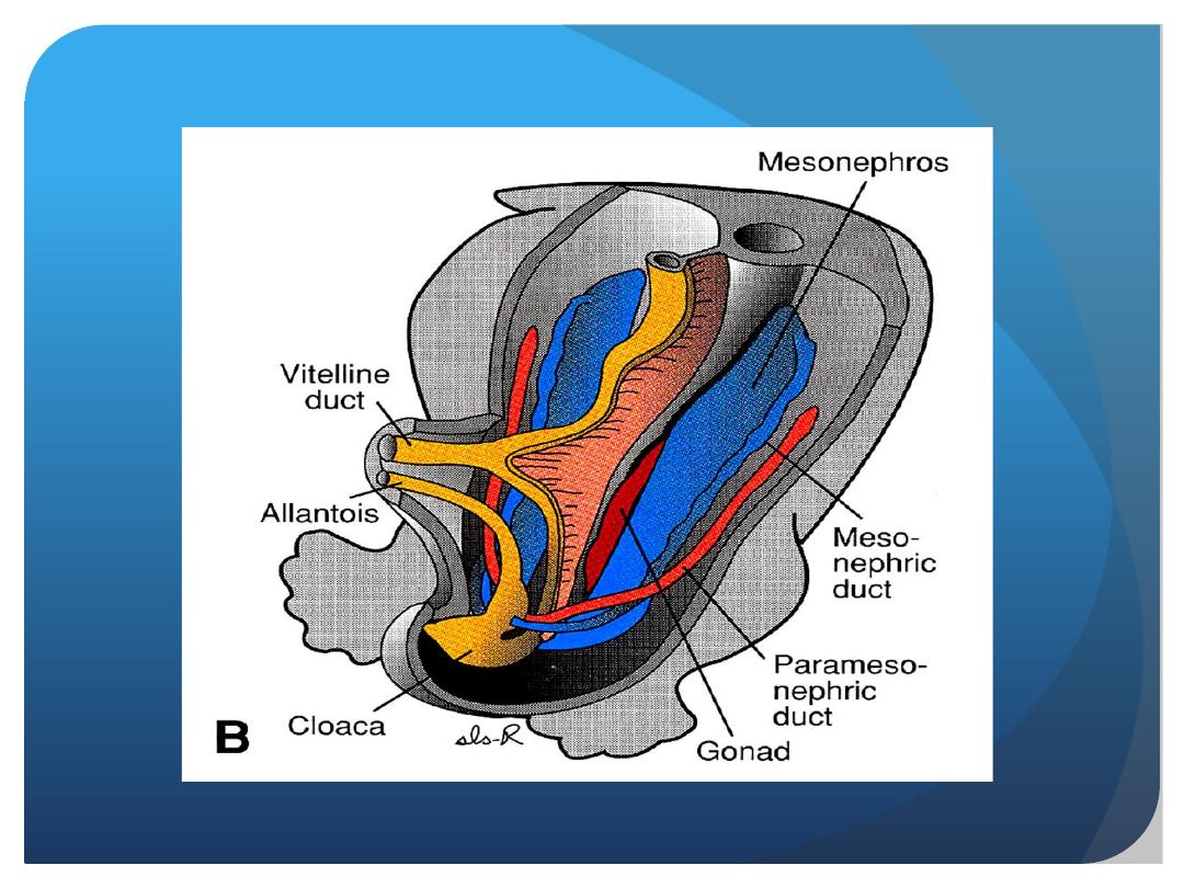
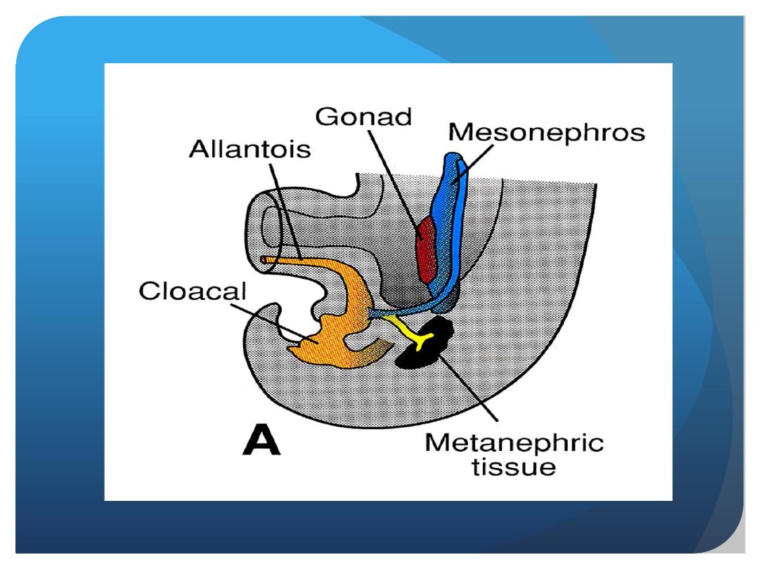
In the middle of the second month, the mesonephros
forms a large ovoid organ on each side
of the midline . Since the developing
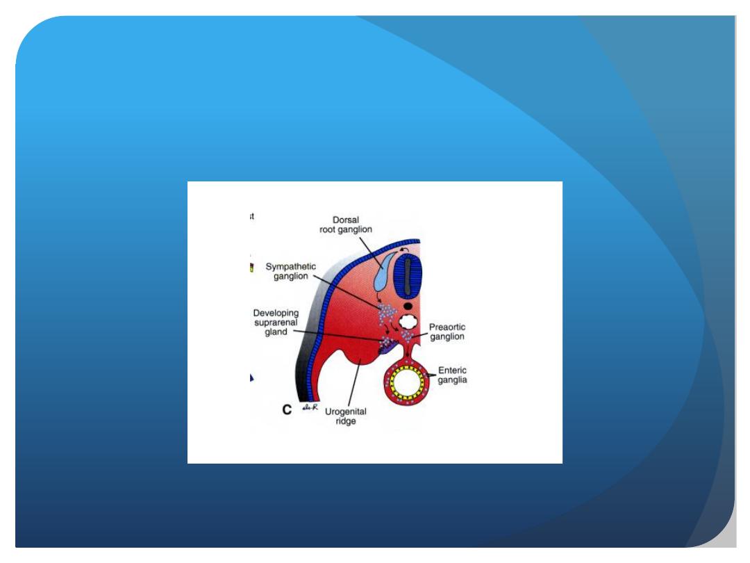
gonad is on its medial side, the ridge formed by
both organs is known as the urogenital ridge

While caudal tubules are still differentiating,
cranial tubules and glomeruli show
degenerative changes, and by the end of the second
month, the majority have disappeared. In the
male, a few of the caudal tubules and the mesonephric
duct persist and participate in formation
of the genital system, but they disappear in the
female.

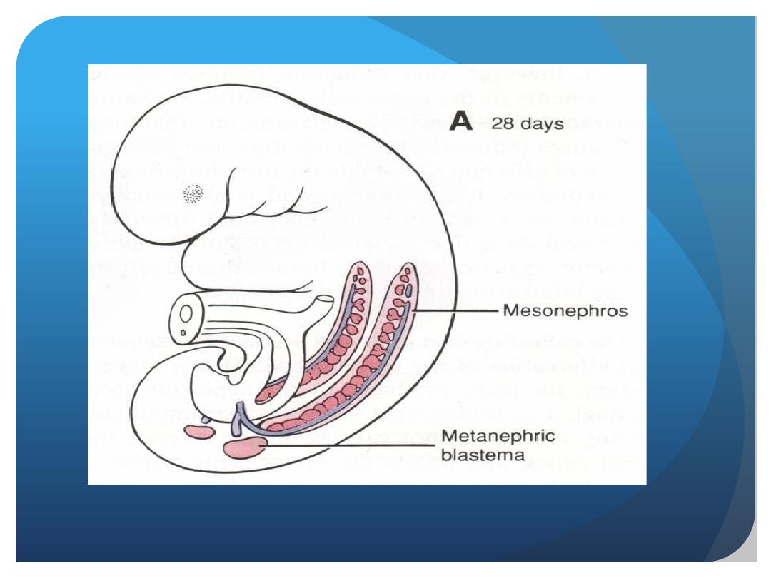
Metanephros: The Definitive
Kidney
Metanephros: The Definitive Kidney
The third urinary organ, the metanephros or
permanent kidney, appears in the fi fth week.
Its excretory units develop from metanephric
mesoderm in the same manner as in
the mesonephric system. The development of the
duct system differs from that of the other kidney
systems.

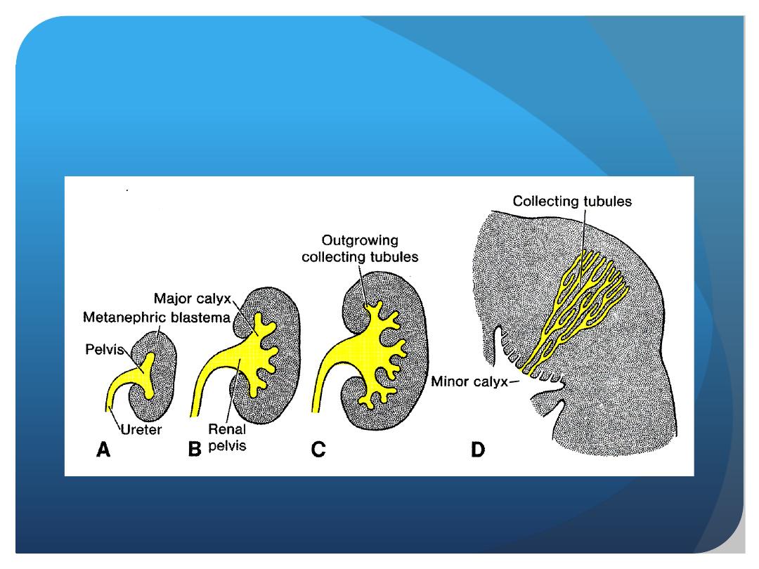
Collecting System
Collecting ducts of the permanent kidney develop
from the
ureteric bud,
an outgrowth of the
mesonephric duct close to its entrance to the
cloaca . The bud penetrates the metanephric
tissue, which is molded over its distal end
as a cap .Subsequently, the bud dilates,
forming the
primitive renal pelvis
, and splits
into cranial and caudal portions, the future major
calyces

Each calyx forms two new buds while penetrating
the metanephric tissue. These buds continue
to subdivide forming the minor calyces of the renal pelvis.

During further development, collecting tubules of the fi
fth and successive generations elongate considerably and
converge on the minor calyx, forming the renal pyramid.
The ureteric bud gives rise to the
ureter, the renal
pelvis, the major and minor calyces, and approximately
1 to 3 million collecting tubules.

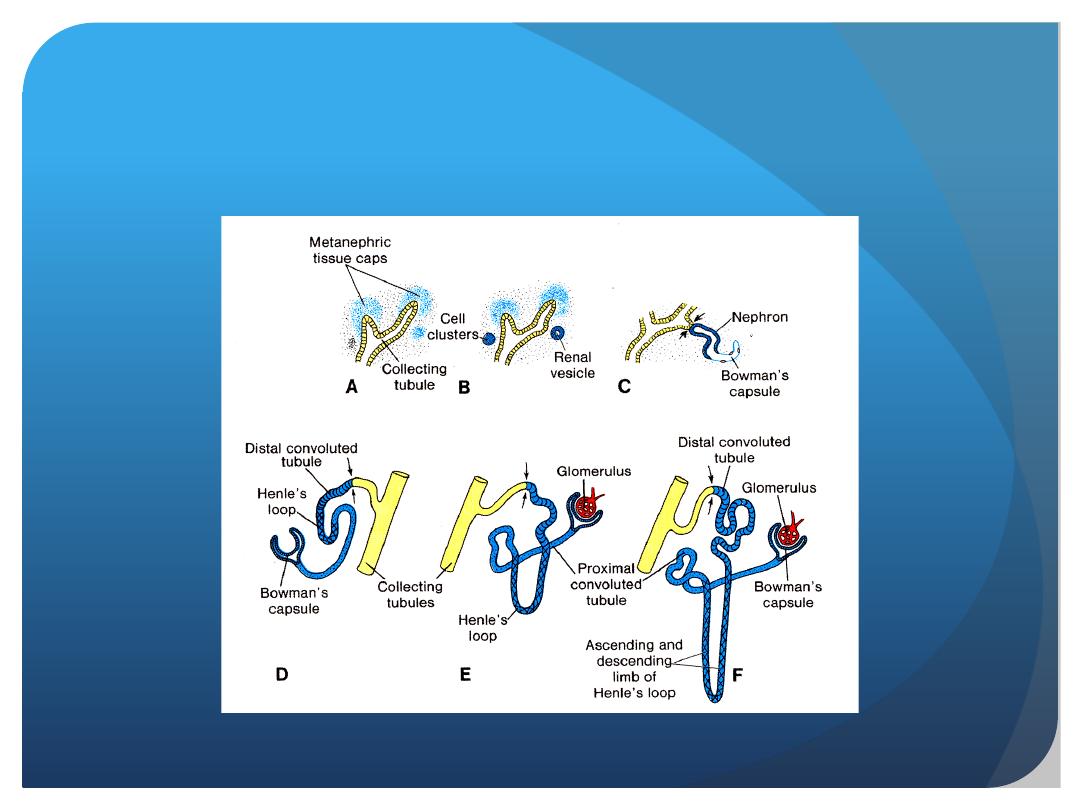
Excretory System
Each newly formed collecting tubule is covered
at its distal end by a
metanephric tissue
cap
. Cells of the tissue cap form small
vesicles,
the renal vesicles, which in turn give
rise to small
S-shaped tubules.
Capillaries
grow into the pocket at one end of
the S and differentiate into
glomeruli.

These tubules, together with their glomeruli, form
nephrons, or excretory units.
The proximal
end of each nephron forms
Bowman’s capsule,
which is deeply indented by a glomerulus.
The distal end forms an open connection
with one of the collecting tubules, establishing
a passageway from Bowman’s capsule to
the collecting unit. Continuous lengthening of
the excretory tubule results in formation of the

proximal convoluted tubule, loop of Henle,
and distal convoluted tubule
.
Hence, the kidney develops from two sources:
(1)
metanephric mesoderm
, which provides
excretory units and (2)
the ureteric bud
, which
gives rise to the collecting system.

Nephrons are formed until birth, at which
time there are approximately
1 million in each
kidney
. Urine production begins early in gestation,
soon after differentiation of the glomerular
capillaries, which start to form by the
10th week
.
At birth, the kidneys have a lobulated appearance,
but the lobulation disappears during infancy as a
result of further growth of the nephrons, although
there is no increase in their number.

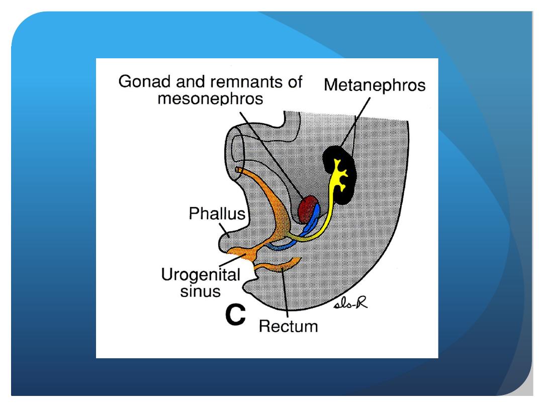
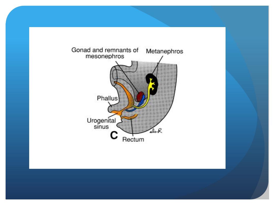
POSITION OF THE KIDNEY
The kidney, initially in the pelvic region,
later shifts to a more cranial position in the
abdomen. This ascent of the kidney is
caused by diminution of body curvature
and by growth of the body in the lumbar
and sacral regions.
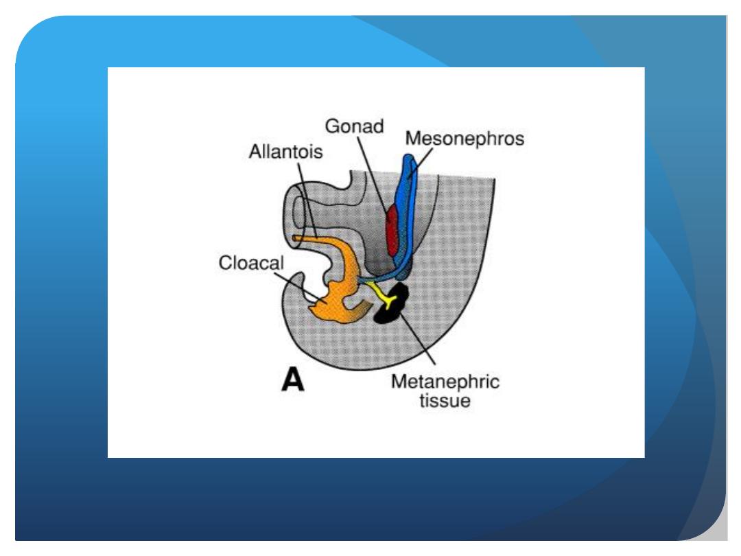

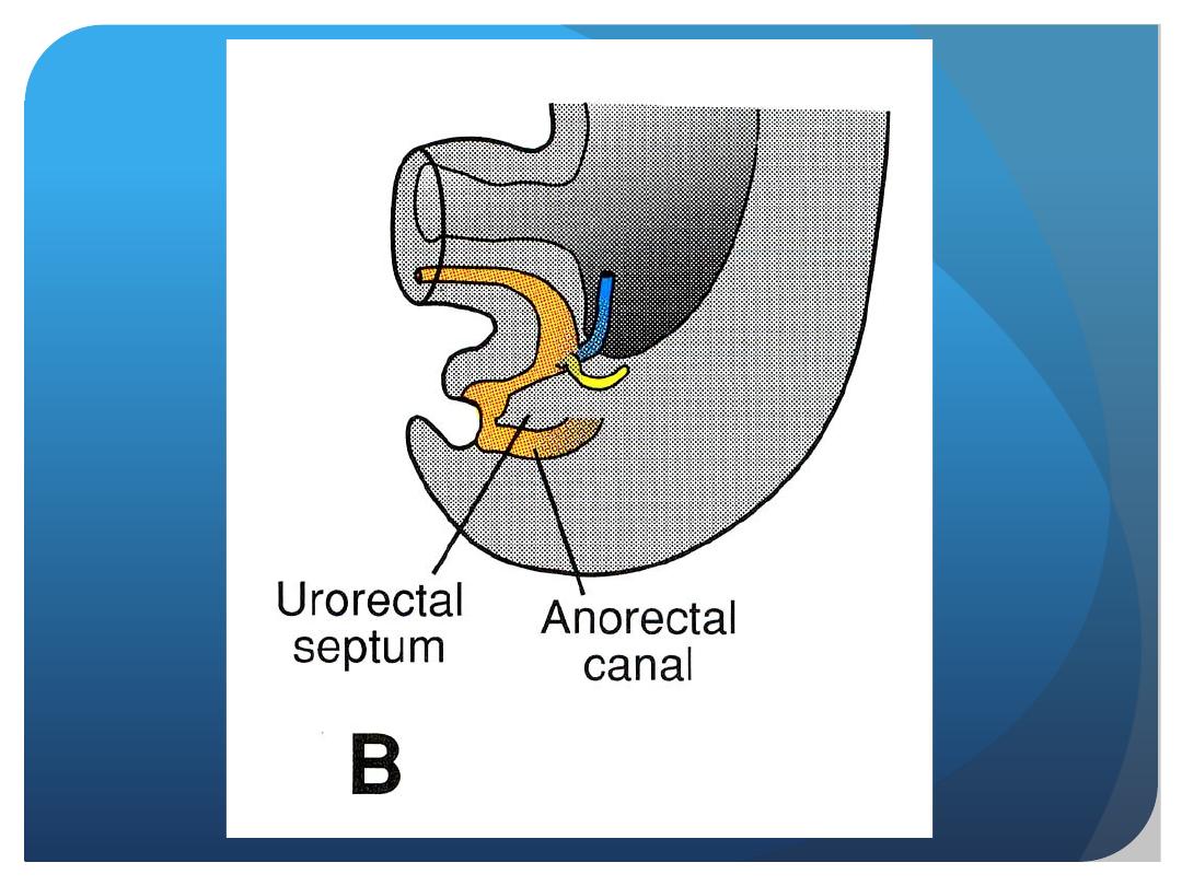
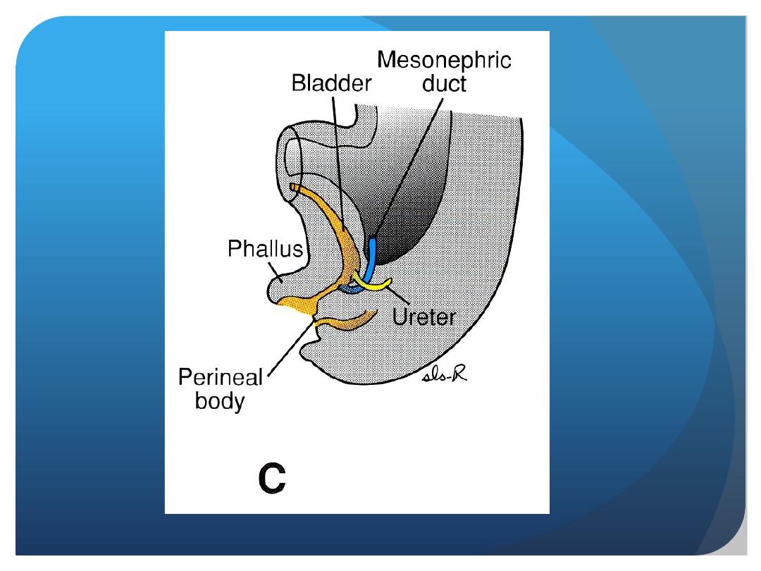
BLADDER AND URETHRA
Urinary bladder and urether are derived
from the cloaca.
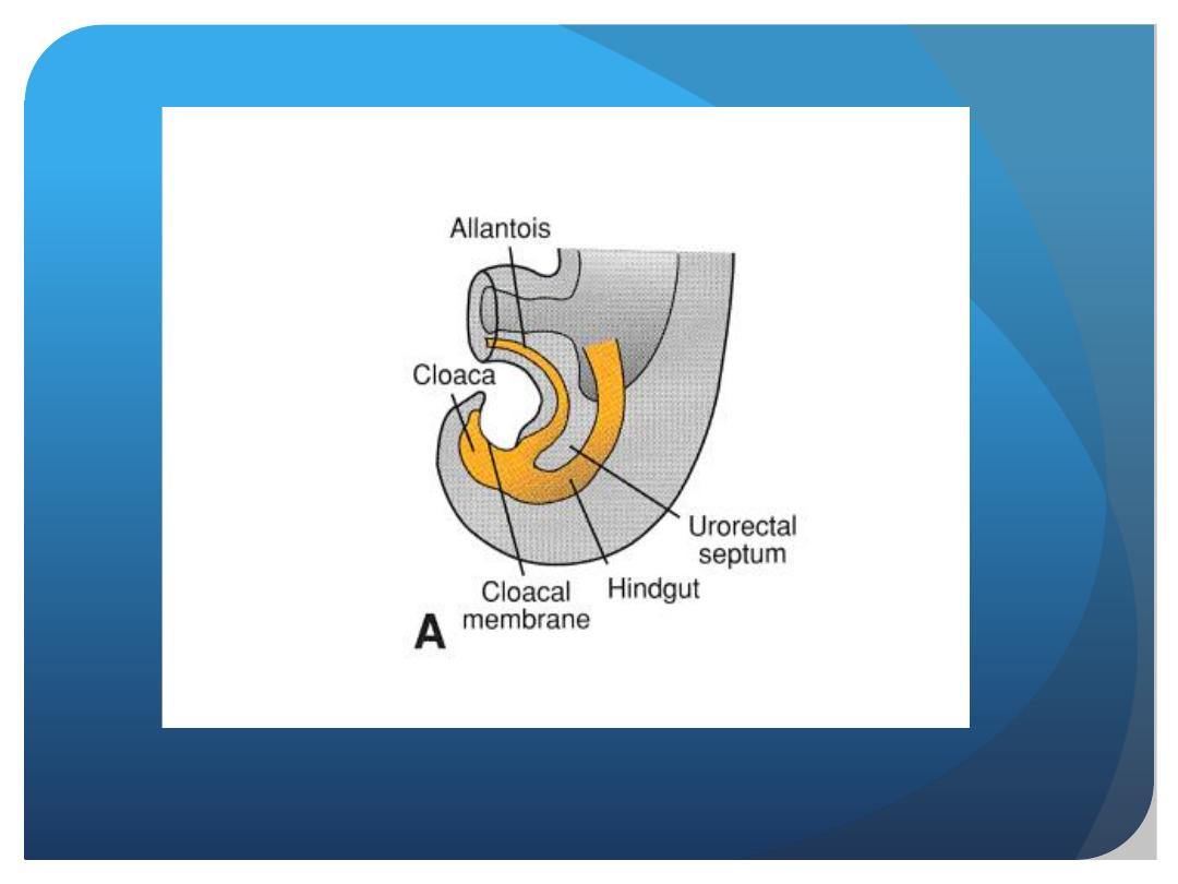
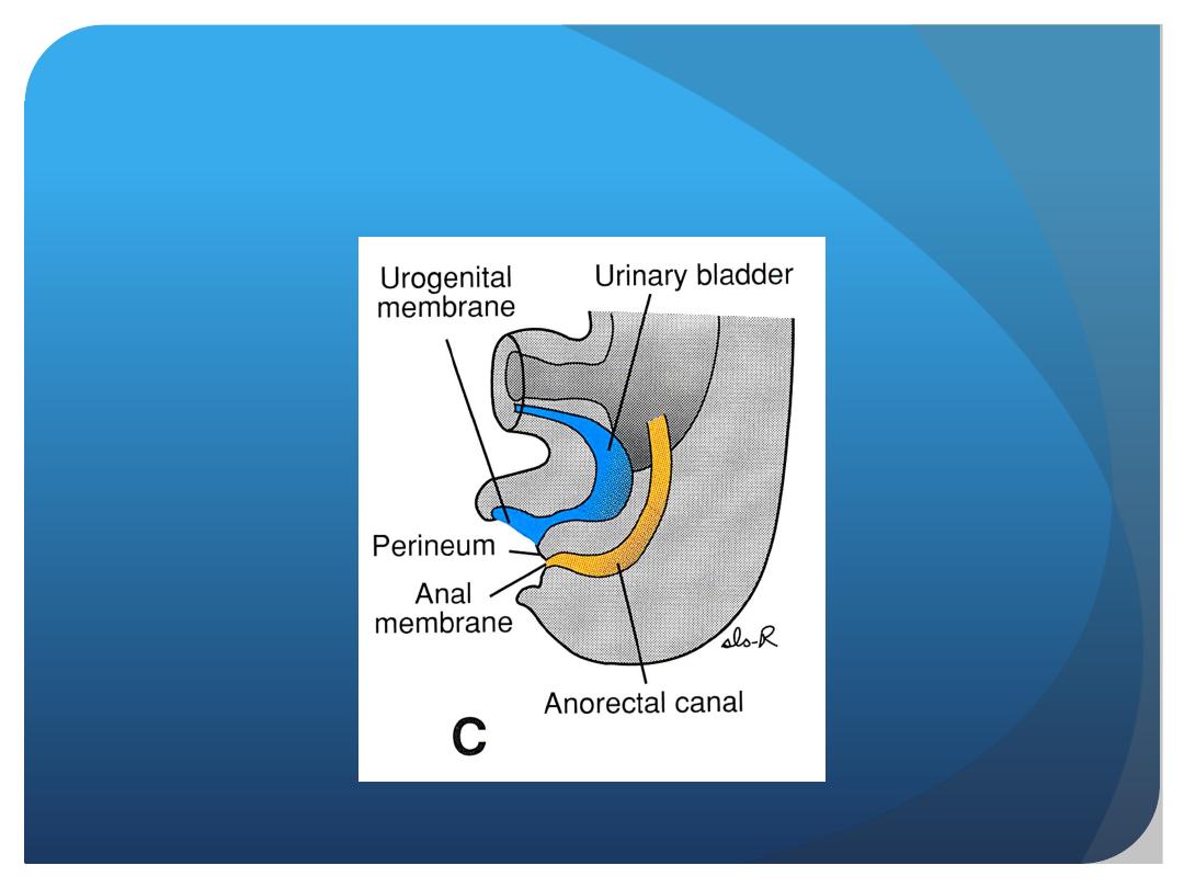
cloaca
urorectal septum
urogenital sinus
Primitive rectum
•Between the two, the tip of the urorectal septum
forms the perineal body.

The phallic
(
The upper and
largest part
urinary bladder
The pelvic
(
Derivatives urogenital sinus

The prostatic and
membranous parts of the
urethra
in male
Urethra in female
The phallic part
The upper and
largest part
The pelvic part
Derivatives of parts of Primitive urogenital
sinus
Penile urthra in male
Vestibule in female
urinary bladder

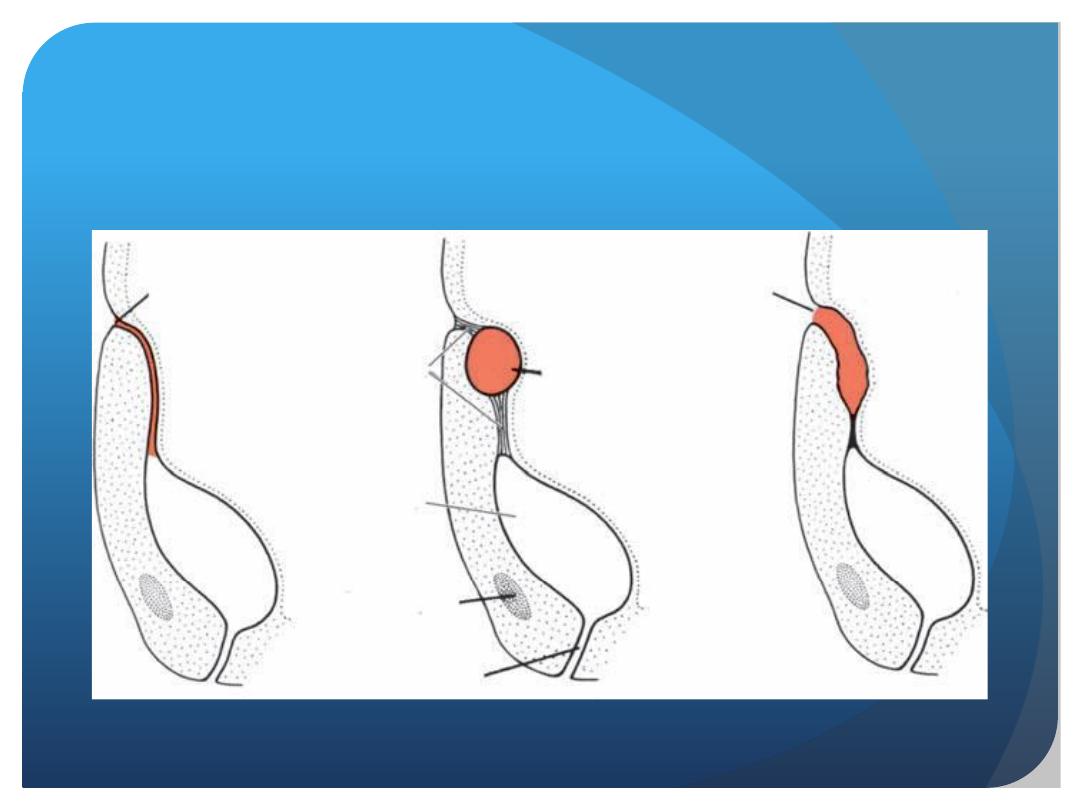
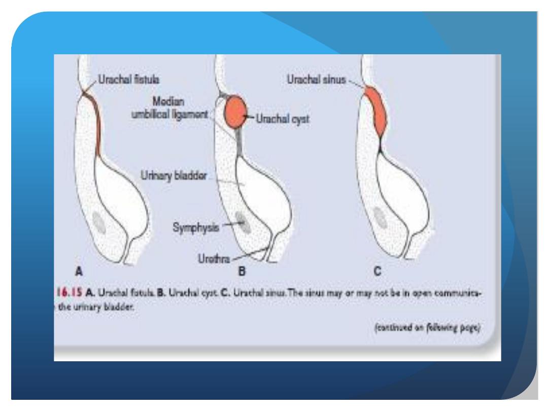
The upper and largest part is the urinary bladder.Initially
the bladder is continuous with the allantois, but when the
lumen of the allantois is obliterated, a thick fibrous cord,
the urachus, remains and
connects the apex of the bladder with the umbilicus. In
the adult, it is known as the median umbilical ligament.
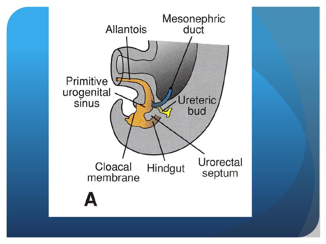

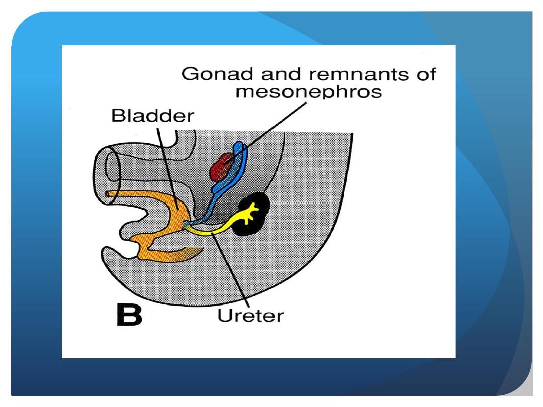
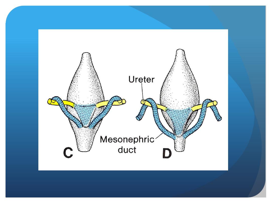
During differentiation of the cloaca, the
caudal portions of the mesonephric
ducts are absorbed into the wall of the
urinary bladder. Consequently, the ureters,
initially outgrowths from the mesonephric
ducts, enter the bladder separately.
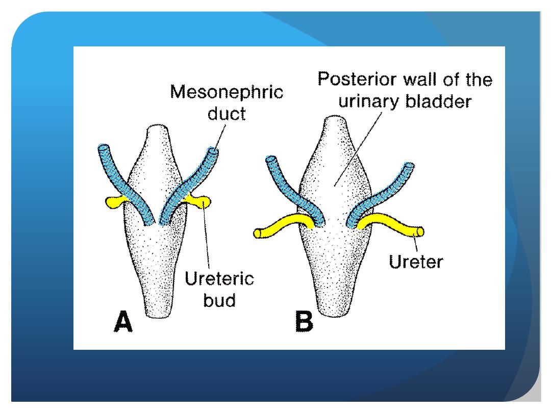

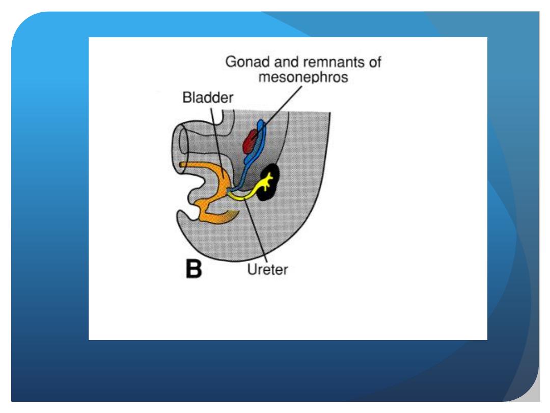
As a result of ascent of the kidneys, the
orifices of the ureters move farther
cranially; those of the mesonephric ducts
move close together to enter the prostatic
urethra and in the male become the
ejaculatory ducts.

• Anomalies of kidneys:
1. Agenesis
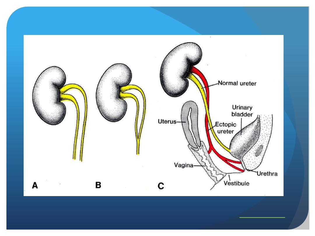
2. Duplication:
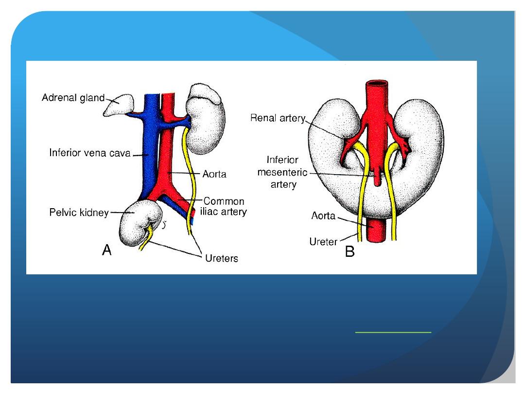
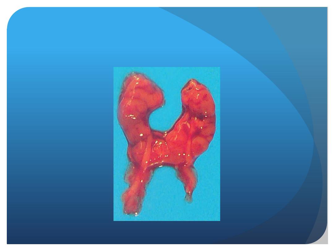
3. Anomalies of shape:Horseshoe kidney
4. Abnormal of position:
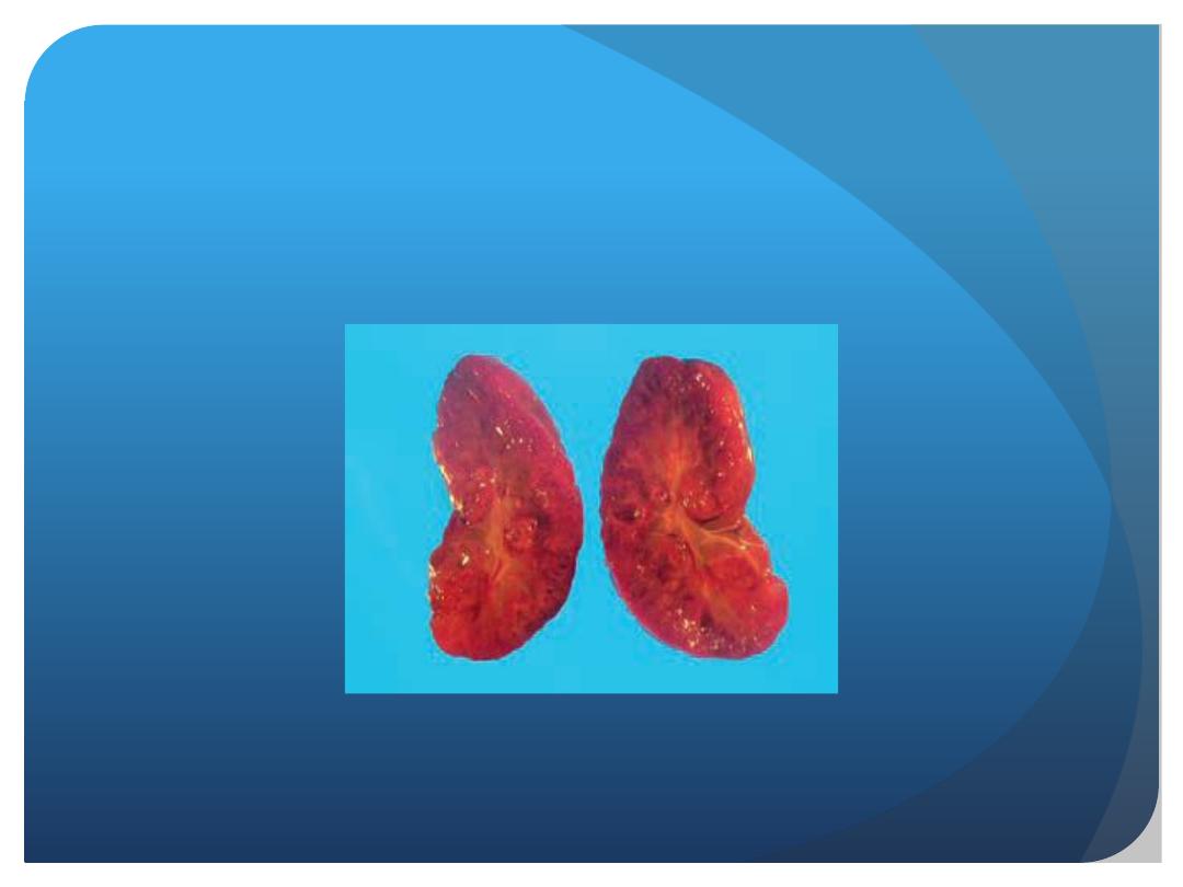
5. Congenital polycystic kindney:

• Congenital polycystic kindney:
• Failure of the excretory tubules of the
to establish contact with the
collecting tubules, leads to the formation
of cysts. Isolated cysts are commonly
seen,but sometimes the whole kidney is a
mass of such cysts.The cysts press upon
normal renal tissue and destroy it.

THANK YOU