بسم الله الرحمن الرحيم
Lecture -1- Medical Physiology
2nd stage Dr. Noor Jawad
The cardiovascular system
Objective:
1. Anatomy and functions of cardiovascular system ?
2. what is the pericardium ?
The cardiovascular system (CVS) consists of
the heart,blood vessels
blood.( approximately 5 liters of blood that the blood vessels transport).
Function
The four major functions of the cardiovascular system are1. To transport nutrients, gases and waste products around the body
2. To protect the body from infection and blood loss
3. To help the body maintain a constant body temperature (‘thermoregulation’)
4. To help maintain fluid balance within the body.
An Overview of the Cardiovascular System
The cardiovascular system perform its impressive work, depending on the heart
together with a network of blood vessels. The network can be subdivided into two circuits:
the pulmonary circuit, which carries carbon dioxide-rich blood from the heart to the gas exchange surfaces of the lungs and returns oxygen-rich blood to the heart
the systemic circuit, which transports oxygen-rich blood from the heart to the rest of the body’s cells, returning carbon dioxide-rich blood back to the heart.
The heart
The heart, is hollow cone-shaped four-chambered muscular pump approximately the size of a fist. The heart rests on the diaphragm between the lungs in the mediastinal space of the intrathoracic cavity in a loose-fitting sac called the pericardium. It is suspended by the great vessels, with its broader side (i.e., base) facing upward and its tip (i.e., apex) pointing downward, forward, and to the left.It is located posterior to the sternum and anterior to the vertebral column. The heart is positioned obliquely, so that the right side of the heart is almost fully in front of the left side of the heart, with only a small portion of the lateral left ventricle on the frontal plane of the heart.
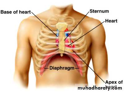
Figure: location of heart.
The heart as a pumpThe heart is separated by septum into right and left pump:
The right pump consist of right atrium and right ventricle, the right atrium receives blood from the systemic circuit, and the right ventricle discharges blood into the pulmonary circuit.
The left pump consist of left atrium and left ventricle, the left atrium collects blood from the pulmonary circuit, and the left ventricle ejects blood into the systemic circuit.
The two are connected in series, when circuits are connected in series, flow must be equal in the two circuits.
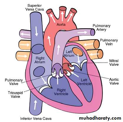
Figure:the chambers of the heart.
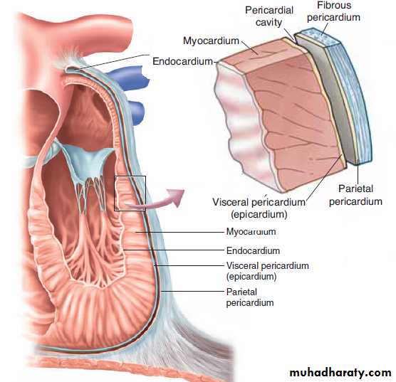
PericardiumThe pericardium forms a fibrous covering around the heart, holding it in a fixed position in the thorax and providing physical protection and a barrier to infection. The pericardium consists of:
A tough outer fibrous layer and a thin inner serous layer. The outer fibrous layer is attached to the great vessels that enter and leave the heart, the sternum, and the diaphragm. The fibrous pericardium is highly resistant to distention; it prevents acute dilatation of the heart chambers and exerts a restraining effect on the left ventricle.
The inner serous layer consists of
a visceral layer, also known as the epicardium, covers the entire heart and great vessels
the parietal layer that lines the fibrous pericardium.
Between the visceral and parietal layers is the pericardial cavity, a potential space that contains 30 to 50 mL of serous fluid. This fluid acts as a lubricant to minimize friction as the heart contracts and relaxes.
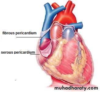
Figure: pericardium.
Clinical points:Pericarditis is inflammation of the pericardium, usually caused by a viral infection. Although this disease can cause sharp, piercing chest pain, it is usually self-limiting and ordinarily does not lead to further problems.
Pericardial effusion is a collection of fluid around the heart in the pericardial sac. If the fluid amount is great enough, it can reduce the heart’s ability to expand and receive blood, reducing its efficiency. This condition is known as cardiac tamponade.
The wall of heart
The wall of heart consist of
1. the inner surface is lined with endocardium, which consists of smooth endothelial cells supported by a thin layer of connective tissue. The endothelial lining of the endocardium is continuous with the lining of the blood vessels that enter and leave the heart.
2.The myocardium, consists largely of cardiac muscle tissue. The muscle fibers of the myocardium are branched and tightly joined to one another. The ventricular muscle is organised into figure of eight bands that squeeze the ventricular chamber forcefully in a way most effective for ejection through the outflow valve. The apex of the heart contracts first and relaxes last to prevent back flow .
3.the epicardium, is the visceral layer of the serous pericardium.