CENTRAL NERVOUS SYSTEM The major regions of the central nervous system (CNS) are the cerebrum, cerebellum, and spinal cord, the CNS is covered by three connective tissue layers, the meninges, but contains very little collagen or fibrous tissue throughout its substance, making it relatively soft and easily damaged by injuries affecting its protective cranium or vertebral bones, Many structural features of CNS tissues can be seen in unstained, freshly dissected specimens. The entire CNS displays organized areas of white matter and gray matter, differences caused by the differential distribution of myelin, the main components of white matter are myelinated axons , often grouped together as tracts, and the myelin-producing oligodendrocytes, White matter contains very few neuronal cell bodies, but astrocytes and microglia are present, gray matter contains abundant neuronal cell bodies, dendrites, the initial unmyelinated portions of axons, astrocytes, and microglial cells. Gray matter is where most synapses occur, and it occupies the thick surface or cortex of both the cerebrum and the cerebellum; most white matter is found in deeper regions. Deep regions of the CNS also have darker aggregates called nuclei consisting of large numbers of neuronal cell bodies and surrounded by white matter.
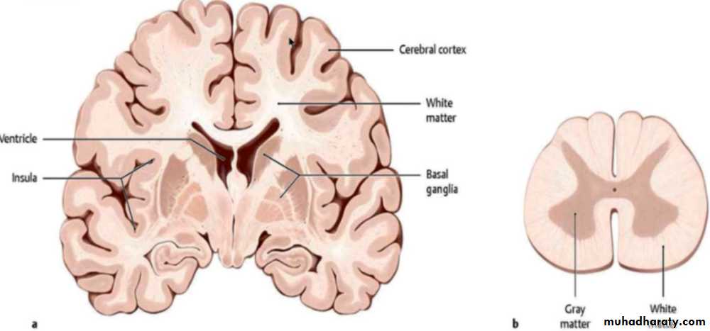
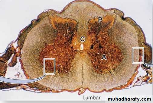
In cross sections of the spinal cord, white matter is peripheral and gray matter is internal and has the general shape of the letter H, in the center is an opening, the central canal which develops from the lumen of the embryonic neural tube. The canal is continuous with the ventricles of the brain, contains CSF, and is lined by ependymal cells. The gray matter forms the anterior horns, which contain motor neurons whose axons make up the ventral roots of spinal nerves, and the posterior horns, which receive sensory fibers from neurons in the spinal (dorsal root) ganglia. Spinal cord neurons are large and multipolar, especially the motor neurons in the anterior horns
Meninges The skull and the vertebral column protect the CNS, but between the bone and nervous tissue are membranes of connective tissue called the meninges. Three meningeal layers are distinguished: the dura, arachnoid, and pia maters.
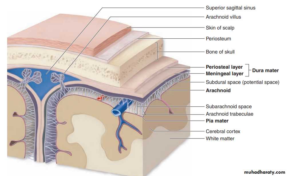
I- Dura Mater: The thick external dura mater (L., dura mater, tough mother) consists of dense, fibro elastic connective tissue that is continuous with the periosteum of the skull, around the spinal cord the dura mater is separated from the periosteum of the vertebrae by the epidural space, which contains a plexus of thin-walled veins and areolar connective tissue. The dura mater is always separated from the arachnoid by the thin subdural space. The internal surface of all dura mater, as well as its external surface in the spinal cord, is covered by simple squamous epithelium of mesenchymal origin.
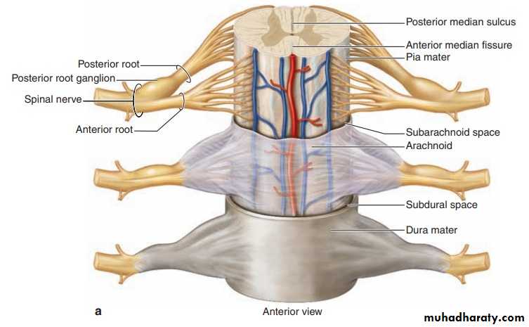
II-Arachnoid The arachnoid (Gr. arachnoeides, spiderweblike) has two components: (1) a sheet of connective tissue in contact with the dura mater and (2) a system of loosely arranged trabeculae composed of collagen and fibroblasts, continuous with the underlying pia mater layer. Surrounding the trabeculae is a large, sponge-like cavity, the subarachnoid space, filled with CSF This fluid-filled space helps cushion and protect the CNS from minor trauma. The subarachnoid space communicates with the ventricles of the brain where the CSF is produced. The connective tissue of the arachnoid is said to be a vascular because it lacks nutritive capillaries, but larger blood vessels run through it. Because the arachnoid has fewer trabeculae in the spinal cord, it can be more clearly distinguished from the pia mater in that area. The arachnoid and the pia mater are intimately associated and are often considered a single membrane called the pia-arachnoid. In some areas, the arachnoid penetrates the dura mater and protrudes into blood-filled venous sinuses located within that layer. These CSF-filled protrusions, which are covered by vascular endothelial cells lining the sinuses, are called arachnoid villi, which function as a site for absorption of CSF into the blood of the venous sinuses.
III- Pia Mater The innermost pia mater (L., pia mater, tender mother) consists of flattened, mesenchymally derived cells closely applied to the entire surface of the CNS tissue. The pia does not directly contact nerve cells or fibers, being separated from the neural elements by the very thin superficial layer of astrocytic processes (the glia limitans), which adheres firmly to the pia mater. Together, the pia mater and the layer of astrocytic end feet form a physical barrier separating CNS tissue from CSF in the subarachnoid space. Blood vessels penetrate the CNS through long perivascular spaces covered by pia mater, although the pia disappears when the blood vessels branch to form the small capillaries. However, these capillaries remain completely covered by the perivascular limiting layer of astrocytic processes