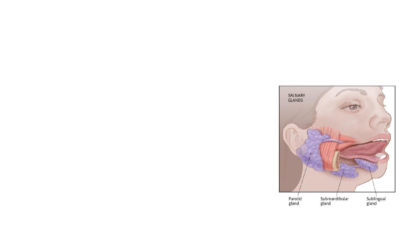
Salivary glands
Firas Al-Hameed
M.B.CH.B C.A.B.S MRCS(ENT)(England)
Thi-Qar Medical School
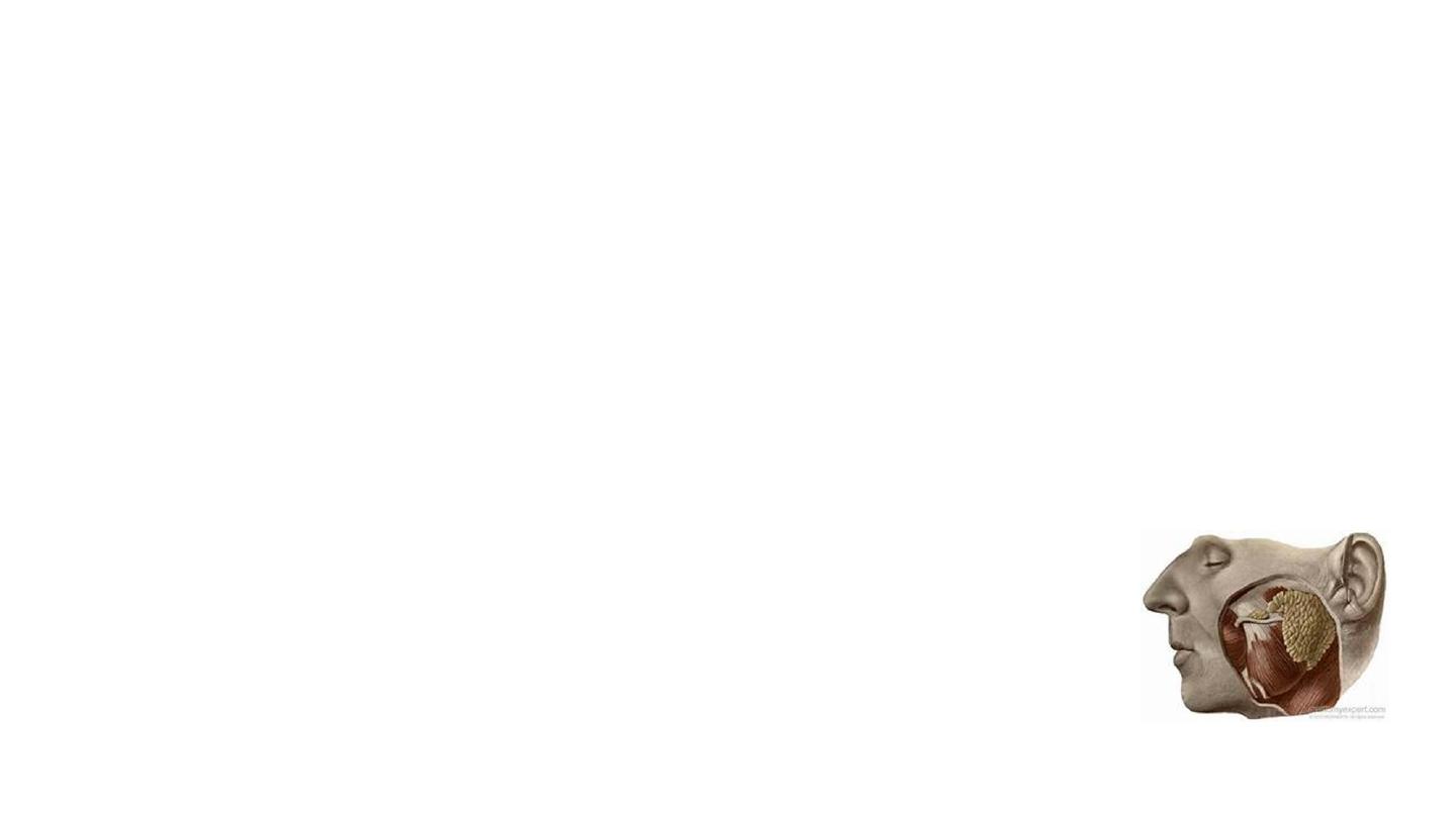
The Parotid Gland
• It produces serous saliva, a watery solution rich in enzymes. This is
then secreted into the oral cavity
• It can be divided into deep and superficial lobes, which are separated
by the facial nerve.
• It lies within a deep hollow, known as the parotid region.
• Superiorly – Zygomatic arch.
• Inferiorly – Inferior border of the mandible.
• Anteriorly – Masseter muscle.
• Posteriorly – External ear and sternocleidomastoid.
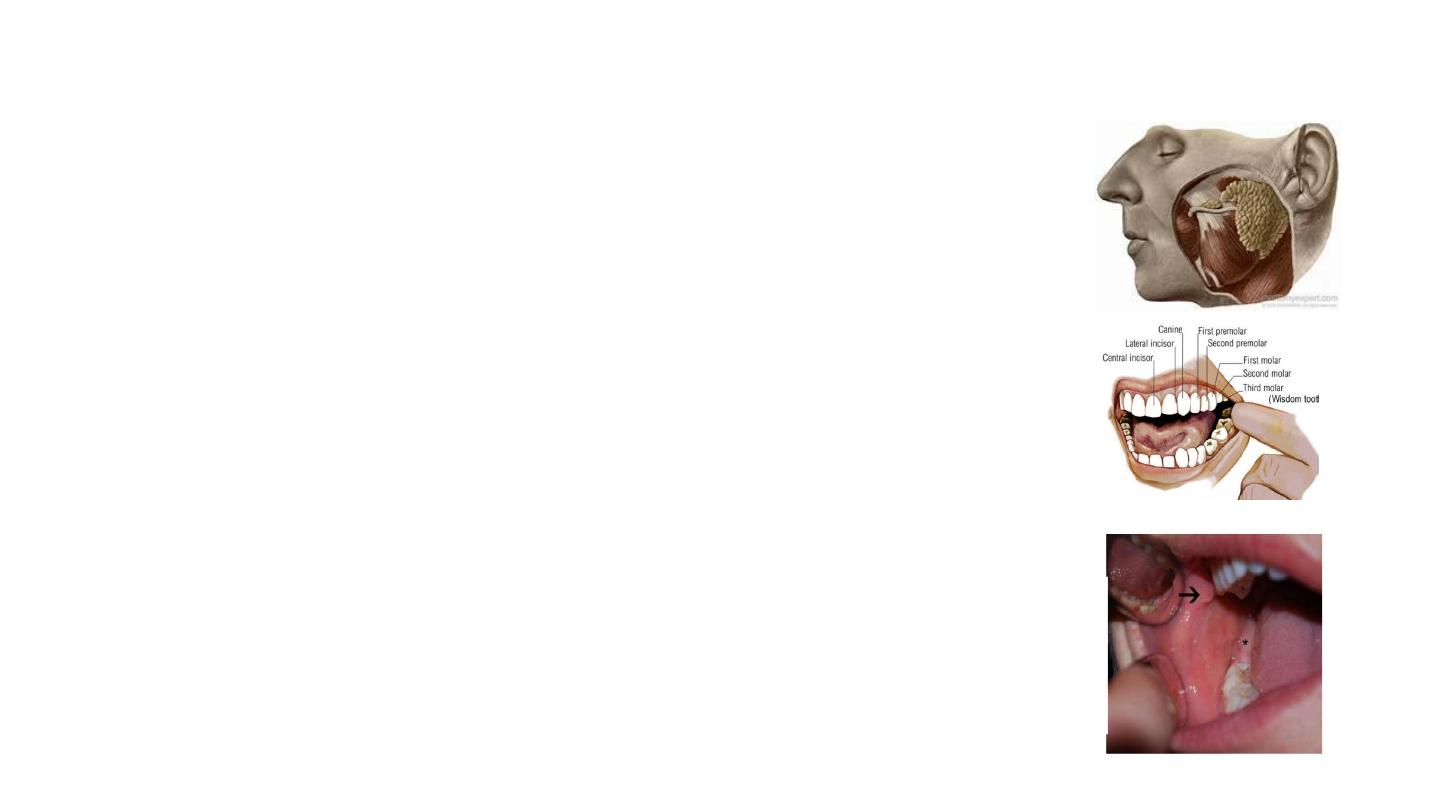
The Stensen duct:
• It arises from the anterior surface of the gland,
traversing the masseter muscle. The duct then
pierces the buccinator, moving medially. It
opens out into the oral cavity near the second
upper molar.
• Length: approximately 5 cm

Anatomical Relationships
• Several important neurovascular structures pass through the gland:
• The facial nerve
(cranial nerve VII), gives rise to five terminal branches
within the parotid gland. These branches innervate the muscles of
facial expression.
• The external carotid artery
(ECA). As it passes through the
posteromedial aspect of the gland, it gives off the maxillary artery, as
well as the superficial temporal artery. Occasionally it also gives off
the posterior auricular artery .
• The retromandibular vein
is formed within the parotid gland by the
union of the superficial temporal and maxillary veins.
• There are numerous lymph nodes distributed throughout and around
the substance of the parotid gland.

Nerve supply
Sensory fibers : auriculotemporal nerve (a branch of CN V) (gland) and
the great auricular nerve (fascia).
Secretomotor fibers
• Parasympathetic
• Begins with the glossopharyngeal nerve (CN IX). This nerve synapses with the
otic ganglion. Then it joins the auriculotemporal nerve .
• The sympathetic
• originates from the superior cervical ganglion. Fibres from this ganglion travel
along the external carotid artery to reach the parotid gland.
Submandibular glands
• Produces 60–67% of saliva secretion
• Mixed serous and mucous salivary secretions
• Located beneath the floor of the mouth, within submandibular
triangle
• It is divided into superficial (large) and deep lobes ( small),
which are separated by the mylohyoid muscle
• The deep lobe lies on the lateral surface of the hyoglossus,
lateral to the root of the tongue.
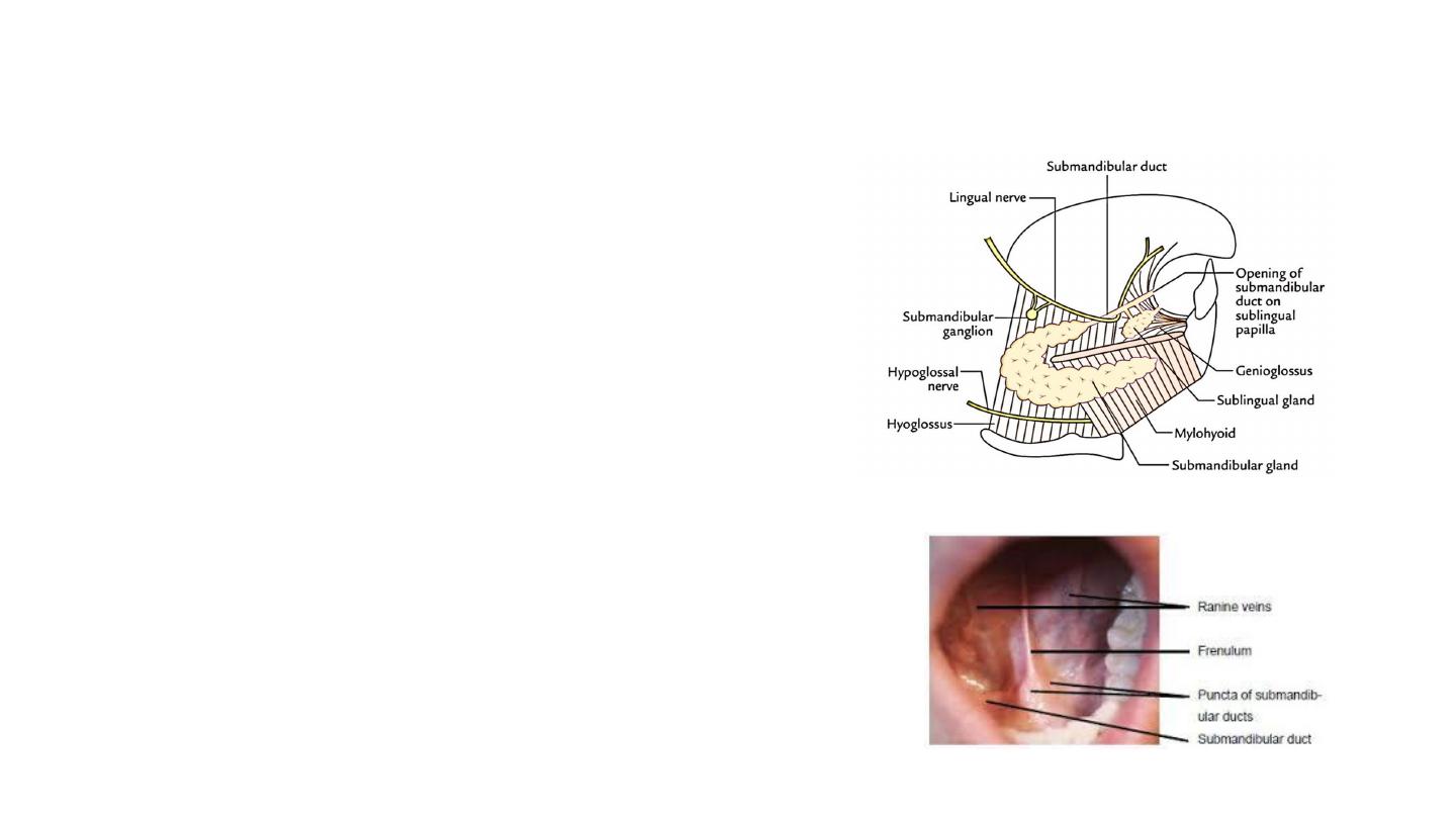
• Submandibular duct (Wharton’s duct)
• Arise from medial portion of the gland
• About 5 cm length
• Crossed by the lingual nerve
• Drains into the sublingual caruncles on either side of the lingual frenulum
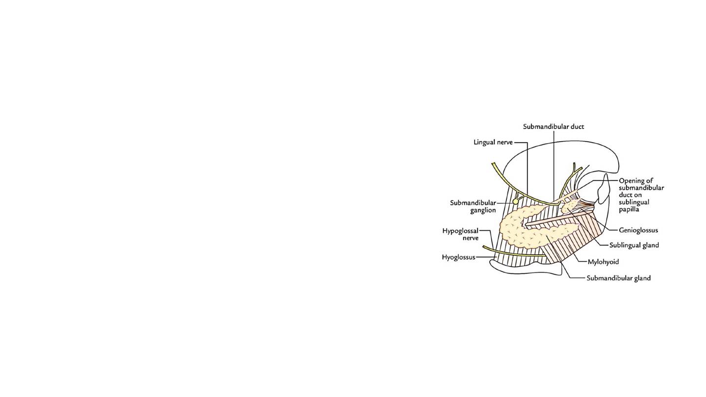
Relationship with Nerves
• Lingual nerve: Beginning lateral to the
submandibular duct, then pass beneath
it.
• Hypoglossal nerve: Lies deep to the
submandibular gland and runs superficial
to hyoglossus and deep to digastric
muscle.
• Facial nerve (marginal mandibular
branch): inferior to the submandibular
gland.

Blood supply:
Sublingual and submental branches of the facial and lingual arteries
Venous drainage:
By common facial and lingual veins
Lymphatics:
Submandibular LN and subsequently into jugulo-digastric lymph nodes
Nerve supply
• Sensory fibers : lingual nerve
• Secretomotor fibers : parasympathetic and sympathetic
• Parasympathetic
• Via the chorda tympani, a branch of the facial nerve, that becomes part of the trigeminal nerve then
synapsing on the submandibular ganglion.
• promotes the secretion of saliva.
• The sympathetic
• Through the arteries that supply it.
• Decreases the volume of secretions, and increase enzymatic secretions.
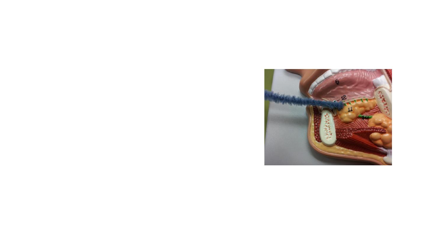
Sublingual gland
• They are the smallest, most diffuse, and the only
unencapsulated major salivary glands. They provide
only 3-5% of the total salivary volume
• They lie anterior to the submandibular gland inferior
to the tongue, as well as beneath the mucous
membrane of the floor of the mouth, bordered
laterally by the mandible and medially by
genioglossus muscle of the tongue.
• Drained by 8-20 excretory ducts called the ducts of
Rivinus
• Innervations, vasculature and lymphatic drainage:
same as submandibular gland

The pharynx
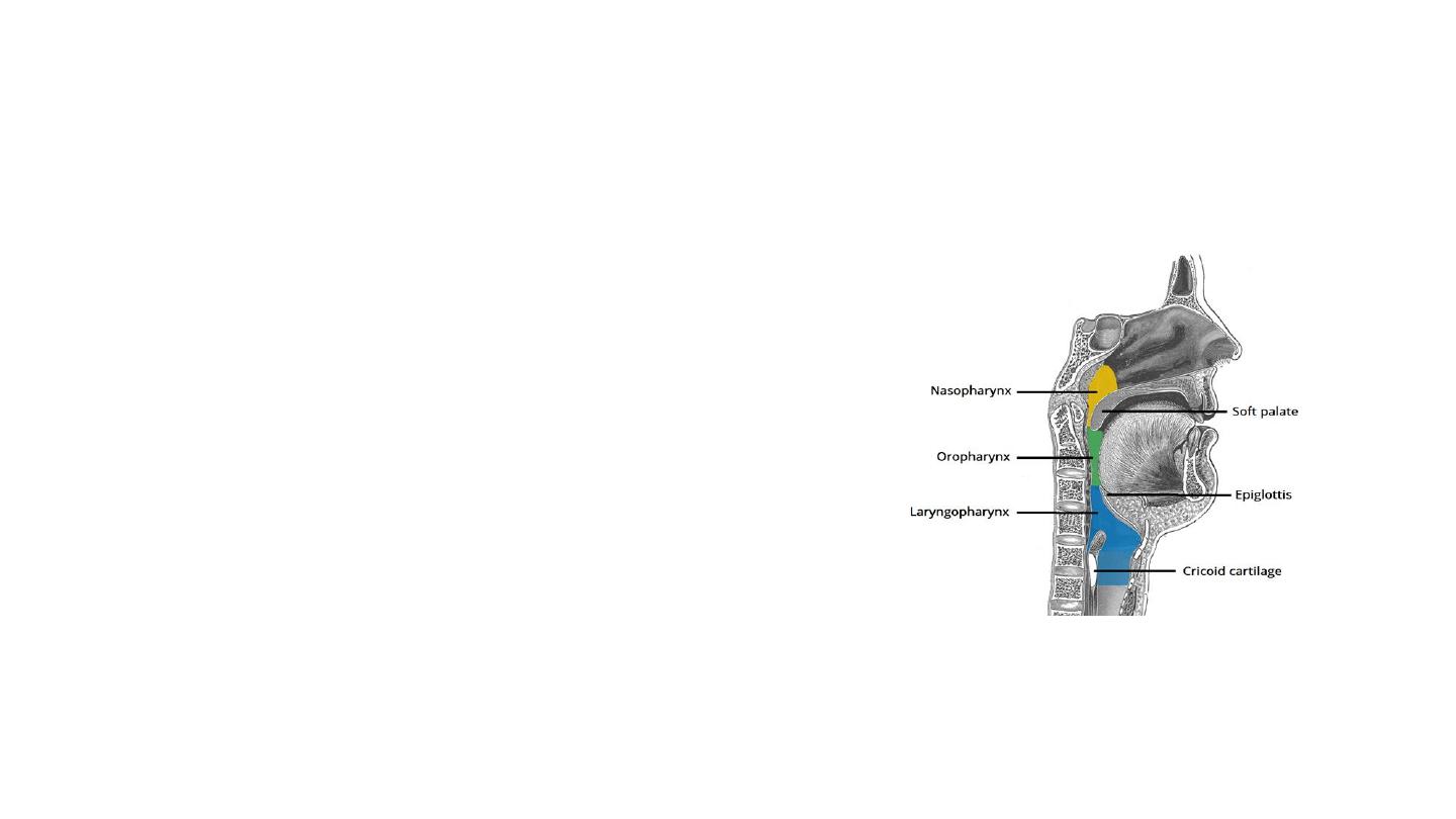
The pharynx
• The pharynx is a muscular tube that connects
the oral and nasal cavity to the larynx and
oesophagus.
• Length: 12-15 cm
• It begins at the base of the skull, and ends at
the inferior border of the cricoid cartilage (C6).
The pharynx is comprised of three parts
(superior to inferior):
• Nasopharynx
• Oropharynx
• Laryngopharynx.
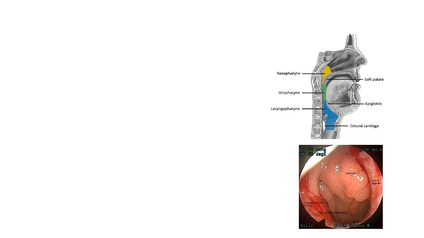
Nasopharynx
• Found between the base of the skull and
the soft palate.
• It is continuous with the nasal cavity.
• Lined with respiratory epithelium
• The posterosuperior nasopharynx contains
the adenoid.
• The openings to the Eustachian tubes,
which lead to the ears.
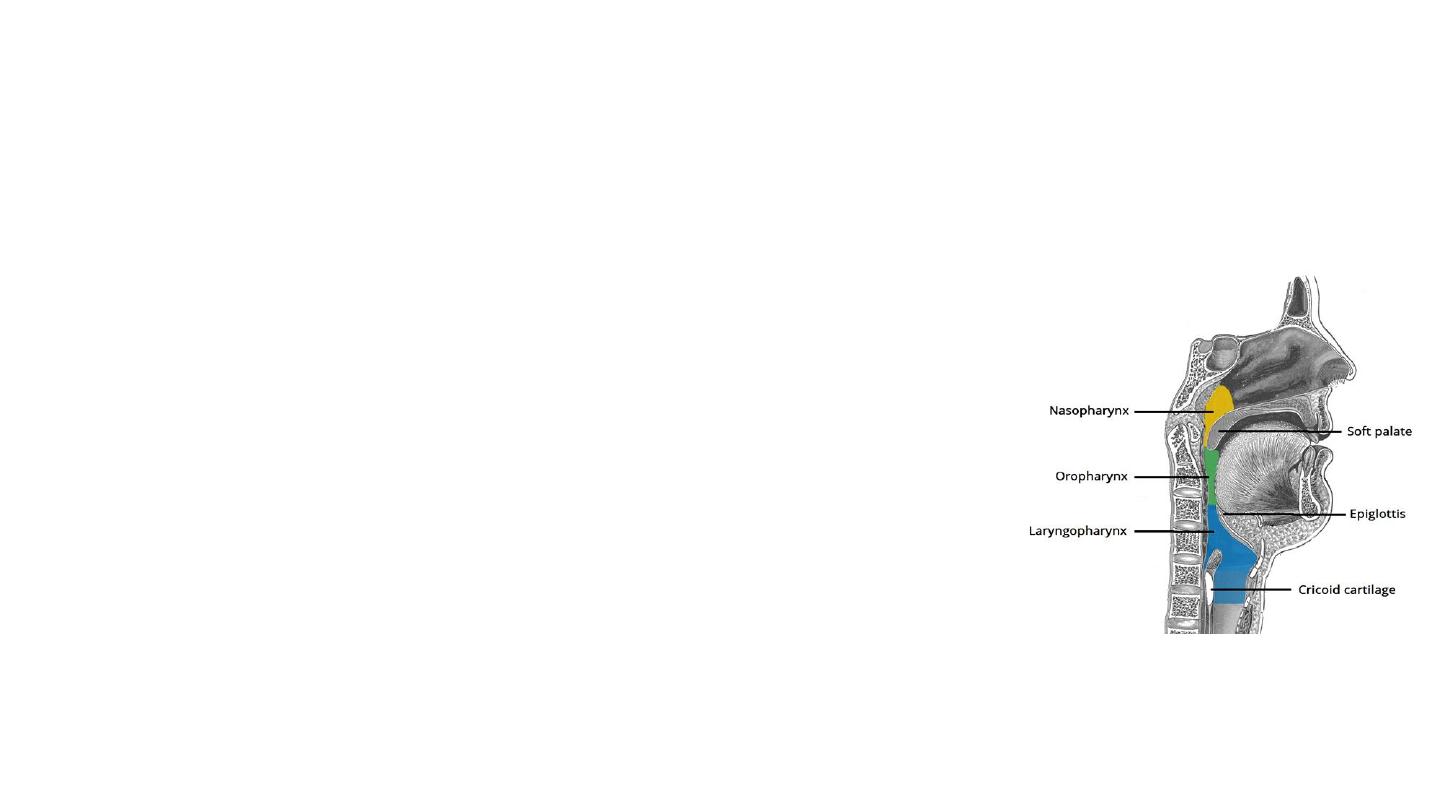
Oropharynx
• The oropharynx is the middle part of the pharynx,
located between the soft palate and the superior
border of the epiglottis.
• It contains the following structures:
• Posterior 1/3 of the tongue.
• Lingual tonsils – lymphoid tissue at the base of the
tongue.
• Palatine tonsils – lymphoid tissue located in the tonsillar
fossa.
• Superior constrictor muscle
• Waldeyer’s ring
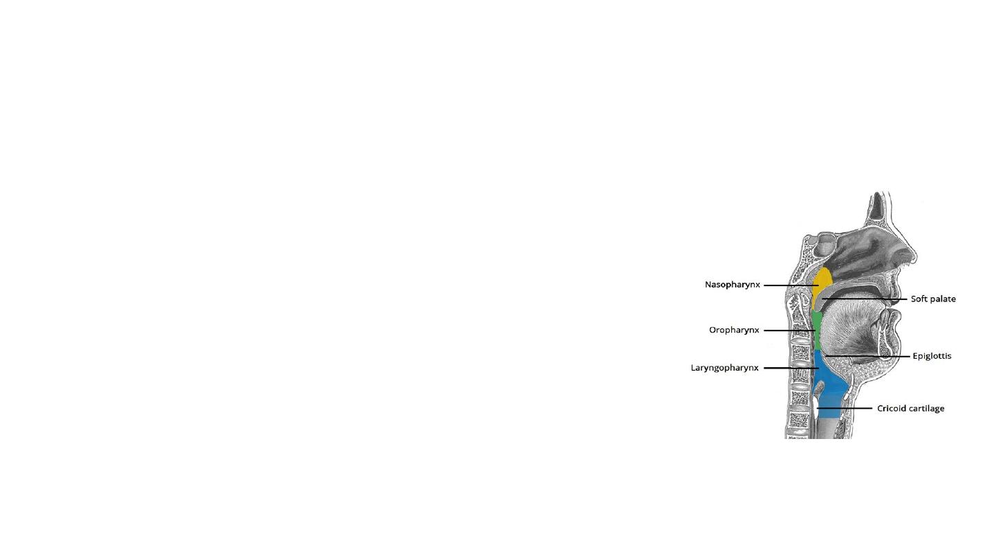
Laryngopharynx
• The most distal part of the pharynx
• Located between the superior border of the
epiglottis and inferior border of the cricoid
cartilage (C6).
• It is continuous inferiorly with the oesophagus.
• It is found posterior to the larynx and
communicates with it via the laryngeal inlet.
• The laryngopharynx contains the middle and
inferior pharyngeal constrictors.
Muscles
• There are two main groups of pharyngeal muscles;
longitudinal and circular.
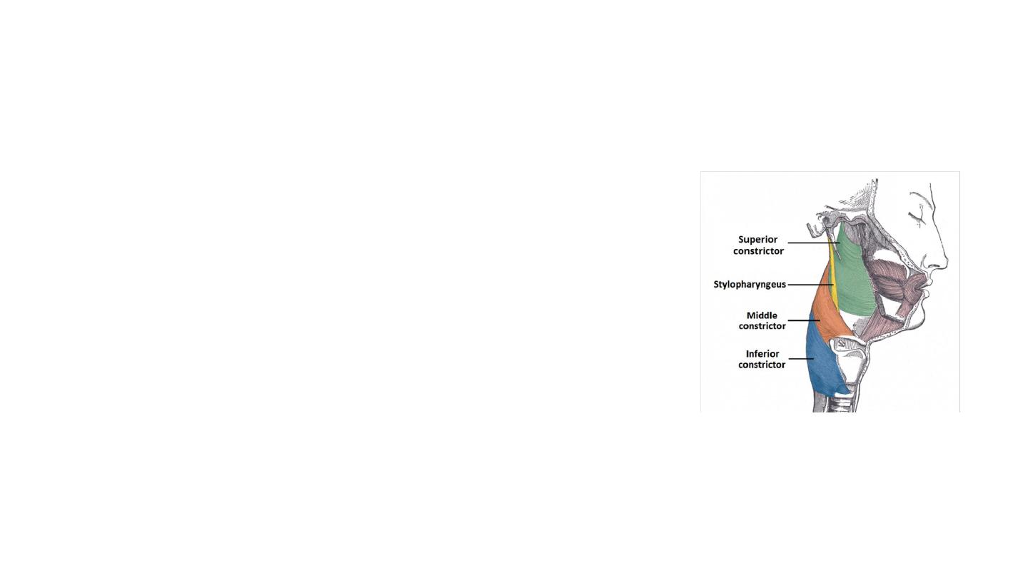
Circular
• There are three circular pharyngeal constrictor muscles; the
superior, middle and inferior pharyngeal constrictors.
• The circular muscles contract sequentially from superior to
inferior to constrict the lumen and propel the bolus of food
inferiorly into the oesophagus.
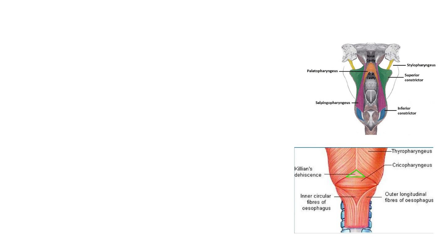
Inferior pharyngeal constrictor – located in the laryngopharynx.
It has two components:
• Superior component (thyropharyngeus) has oblique fibres
that attach to the thyroid cartilage.
• Inferior component (cricopharyngeus) has horizontal fibres
that attach to the cricoid cartilage.
Longitudinal
• They act to shorten and widen the pharynx, and elevate the larynx
during swallowing.
• Stylopharyngeus – arises from the styloid process of the temporal
bone, inserts into the pharynx.
• Palatopharyngeus – arises from hard palate of the oral cavity,
inserts into the pharynx.
• Salpingopharyngeus – arises from the Eustachian tube, inserts
into the pharynx.
• The muscles of the pharynx are mostly innervated by the vagus
nerve – the only exception being the stylopharyngeus
(glossopharyngeal nerve).

Innervation
Sensory:
• Pharyngeal branches from the glossopharyngeal nerve (CN IX) (main)
• Parts of the nasopharynx is innervated by the trigeminal nerve (CN V)
• The inferior aspect of the laryngopharynx (surrounding the beginning of the
larynx) is innervated by the internal branch of the vagus nerve (CN X)
• Motor
• All the muscles of the pharynx are innervated by the vagus nerve (CN X),
except for the stylopharyngeus, which is innervated by the glossopharyngeal
nerve (CN IX).

Vasculature
• Arterial supply to the pharynx : via branches of the external carotid
artery:
• Ascending pharyngeal artery
• Branches of the facial artery
• Branches of the lingual and maxillary arteries.
• Venous drainage is achieved by the pharyngeal venous plexus, which
drains into the internal jugular vein.

Thank you
Nottingham 2020