Skull and brain imaging
ByDr. Firas Abdullah
Aims of our lectures:
To know the normal appearance of skull on X-rayTo learn the normal CT and MRI of brain and skull
To discuss some cerebral pathologies and see some cases of traumaTo know about pathology of sinus, orbit, and neck
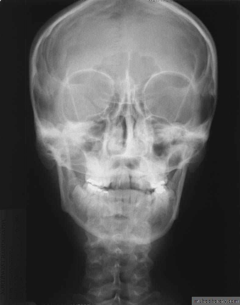
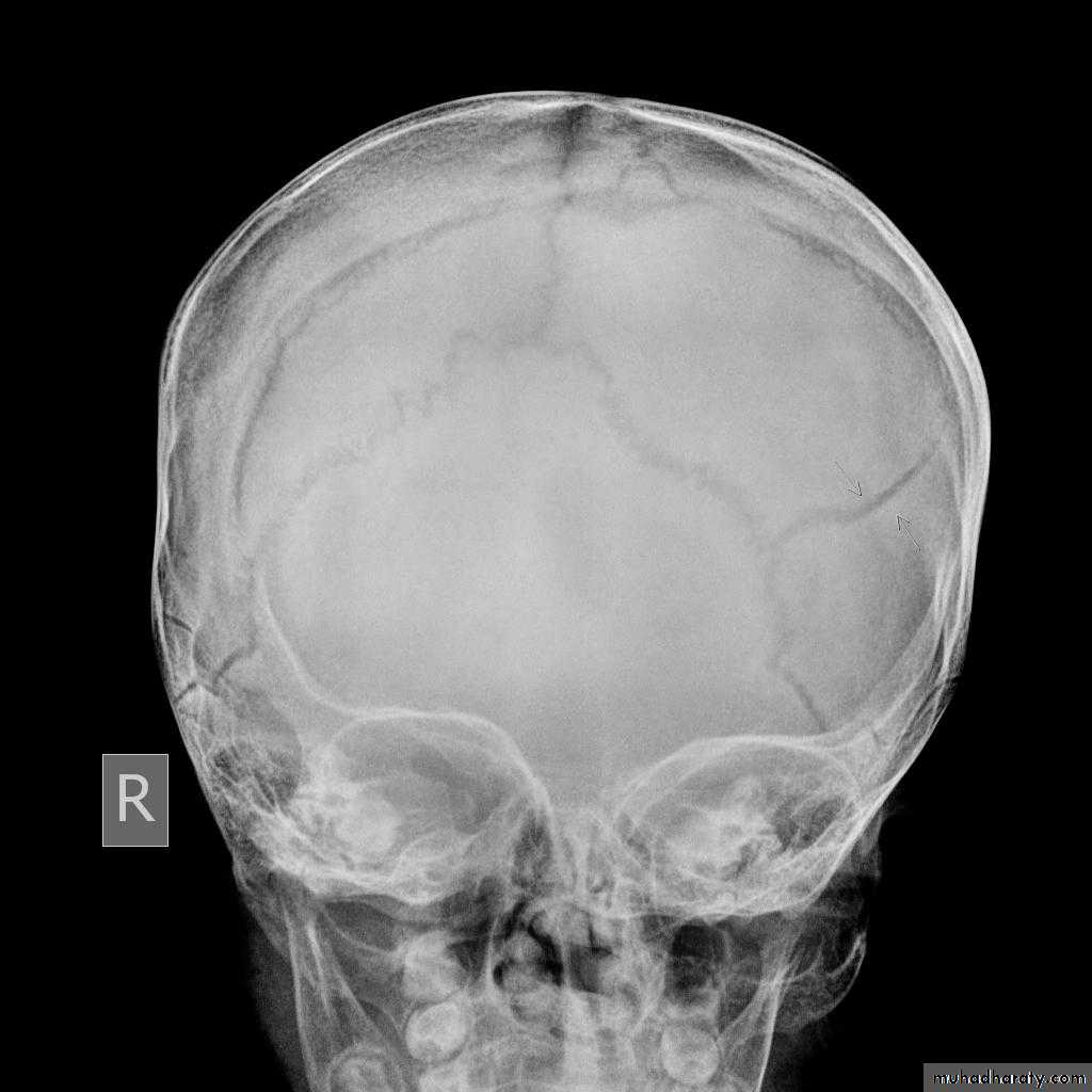
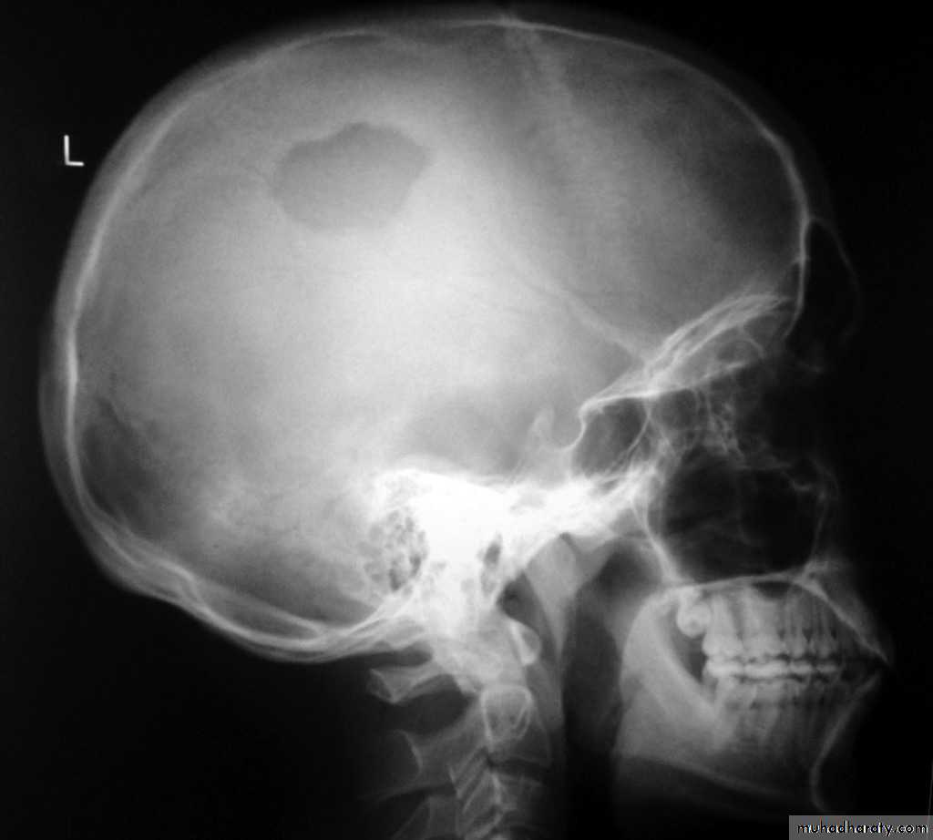
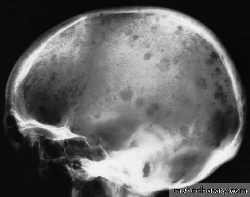
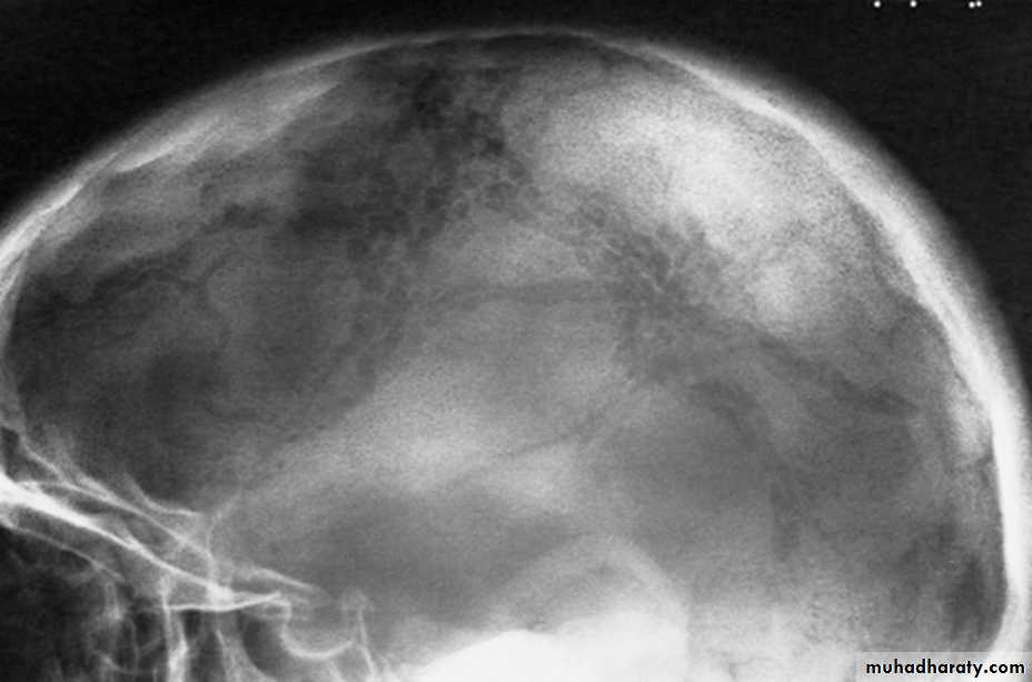
Skull X-rayBony configuration and shape
Bone densityAny Lytic lesion
Any fracture
Any calcification
Diploë, pituitary fossa, paranasal sinuses, orbits
The sutures
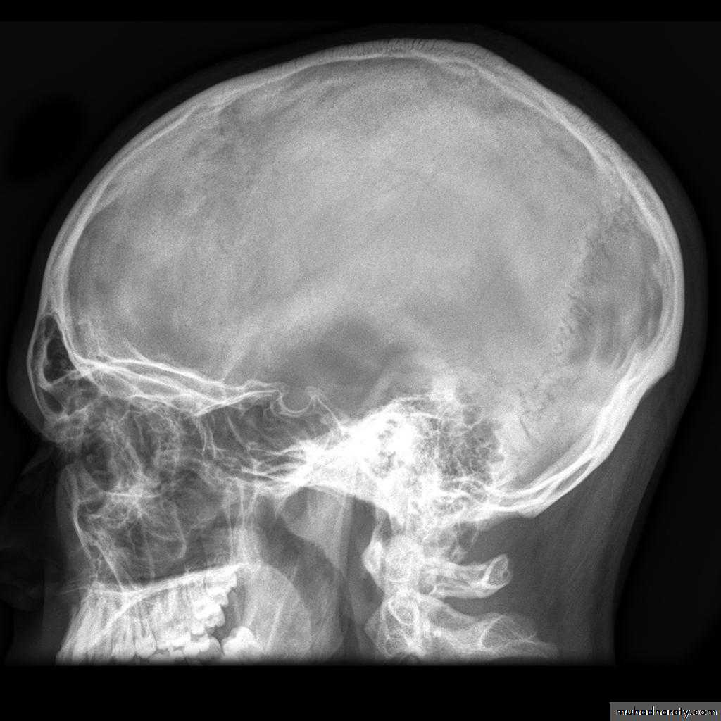
The normal pituitary fossa as shown in a lateral skull film can vary considerably in size. Normal figures are (length of 11-16 mm and a depth of 8-12 mm)
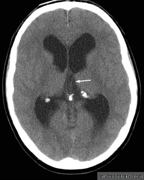
• Pineal
• Choroid plexuses• Dura (falx; tentorium; over vault)
• Basal ganglia and dentate nuclei
• Pituitary gland
• Lens
Normal intracranial calcification
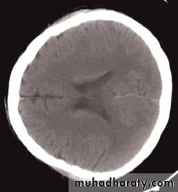
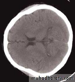
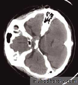
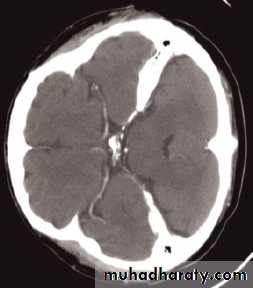
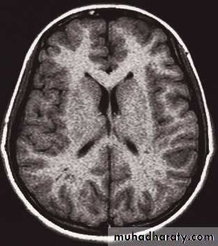
Computed tomography of the brain
• A routine CT examination of the brain involves making 20–30 axial sections.• The axial plane is the routine projection but computer reconstructions can be made from the axial sections, which then provide images in the coronal or sagittal planes
• The window settings are selected for the brain and are also altered to show the bones
• I.V. contrast indication
• IV contrast tends not to be used in patients who are known to have a very recent cerebral haemorrhage or infarct. Areas of calcification may be obscured on post contrast scans.
Computed tomography of the brain
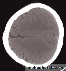
The cardinal signs of an abnormality on a CT scan are:
• Abnormal tissue density• Mass effect
• Enlargement of the ventricles.
Abnormal tissue density
• Abnormal tissue may be of higher or lower density than the normal surrounding brain.• High density is seen with recent haemorrhage, calcified lesions, and areas of contrast enhancement
• Low density is usually due to neoplasms or infarcts, or to oedema, which commonly surrounds neoplasms, infarcts, haemorrhages and areas of inflammation.
Abnormal tissue density
• Oedema characteristically shows finger-like projections and does not enhance with intravenous contrast medium.
• As a rule, it is not possible to diagnose the nature of a mass based on attenuation values alone; an exception is lipoma which, because it contains fat, has a value of approximately (- 100 Hounsfield units).
Mass effect
• The lateral ventricles should be examined to see if they are displaced or compressed.• Shift of midline structures, such as the septum pellucidum , the third ventricle, or the pineal, is a common finding with intracranial masses.
Enlargement of ventricles
There are two basic mechanisms which cause the cerebral ventricles to enlarge:• Obstruction to the CSF pathway, either within the ventricular system (non-communicating hydrocephalus) or over the surface of the brain (communicating hydrocephalus)
• Secondary to atrophy of brain tissue
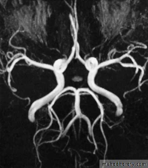
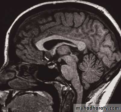
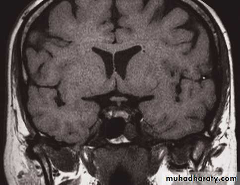
MRI of the brain
• Axial, coronal and sagittal projections are all considered standard• T1-weighted and T2-weighted images.
• It is possible to recognize flowing blood and, therefore, the larger arteries and veins stand out clearly without the need for contrast medium.
• The characteristics of grey and white matter are different, and both are clearly different from the CSF in the ventricular system and subarachnoid space.
MRI of the brain
• The disadvantages of MRI compared with CT are the inability to show calcification, lack of bone detail, the relative expense of the technique, and the difficulty of monitoring seriously ill patients
• IV contrast in T1 WI
• MRA and MRV, Recent advances of MRI: perfusion, diffusion, spectroscopy, functional MRI, and tractography
MRI of the brain
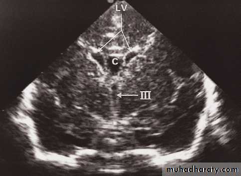
Neurosonography
• Simple to scan the heads of neonates and young babies to obtain images of the ventricular system and the adjacent brain.• Scanning is best done through an open fontanelle where there is no bone to impede the transmission of ultrasound.
• Little discomfort is caused to the baby and the procedure is readily carried out even on ill babies in intensive care units.
• Neurosonography has proved particularly useful in detecting intracerebral haemorrhage and the ventricular dilatation that may follow. It has also been used to demonstrate the presence and cause of other forms of hydrocephalus and congenital abnormalities of the brain.