APPROACH TO RENAL DISEASE
بسم الله الرحمن الرحيم
Renal function
include• the clearance of nitrogenous waste products,
• regulation of electrolytes and pH,
• maintenance of blood pressure,
• regulation of volume,
• synthesis of active forms of vitamin D,
• synthesis of erythropoietin.
• Synthesis of Renin
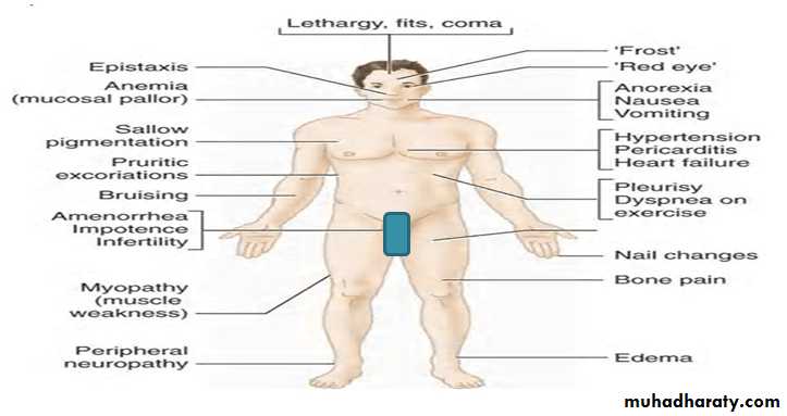
Signs & symptoms of uremia
General approach
Most of the diagnoses of renal disease can be made with a CAREFUL HISTORY and PHYSICAL EXAMINATION supplemented by review of BASIC LABORATORY TESTS, especially the urinary sediment.The specificity of the diagnosis can be improved by the use of serologic analysis, imaging,
occasionally, invasive procedures such as angiography and renal biopsy.
History
The history include
• Medication use .
• Family history of renal disease .
• The time of onset of symptoms of renal dysfunction .
• Changes in bladder function, including nocturia, polyuria, and hesitancy.
• Fatigue and weakness .
• Dyspnea on exertion, a manifestation of fluid overload or acidosis .
History
skin→→ including petechial rash, purpura, digital gangrene, and splinter hemorrhages.Otitis, sinusitis, epistaxis, hemoptysis, and nasal septal ulcers →→ Wegener's granulomatosis.
Pulmonary hemorrhage →→ Goodpasture's syndrome or (anti-GBM) disease.
Abdominal distention may be seen in nephrotic syndrome with ascites, as well as ADPCKD.
Abdominal pain and tenderness →→ Henoch-Schönlein purpura and classic polyarteritis nodosa.
Neurologic symptoms may be a manifestation of vasculitis, such as microscopic polyangiitis
and cryoglobulinemia.
PHYSICAL EXAMINATION
The vital signs are crucial. A patient with a “normal blood pressure” may be relatively hypotensive in the setting of renovascular disease.Pulsus paradoxus may reflect cardiac tamponade.
The eyes may exhibit conjunctivitis, episcleritis, or uveitis.
In the abdomen, ascites may be seen in cirrhosis, nephrosis, and congestive heart failure.
Hepatomegaly is seen in passive congestion and amyloidosis.
Splenomegaly may be seen in amyloidosis, endocarditis, and lymphoma.
Kidney and liver enlargement may be seen in ADPCKD autosomal dominant polycystic kidney disease.
Lower extremity edema can be seen in cirrhosis, nephrotic syndrome, and congestive heart failure.
Splinter hemorrhages as well as Osler's nodes and Janeway lesions may represent bacterial endocarditis.
Rashes can be seen in many of the vasculitides.
PHYSICAL EXAMINATION
jugular venous pressure volume status , should be assessed daily.The presence of a pericardial friction rub can be observed in the serositis (SLE) or the pericarditis associated with uremia.
Infiltrative diseases, such as amyloidosis and sarcoidosis,→→ restrictive cardiomyopathy with associated congestive heart failure.
The presence of (S4) →→ cardiac hypertrophy, and S3 may be a sign of congestive heart failure.
Vascular bruits reflect generalized atherosclerosis, and the presence of an abdominal bruit may be an important clue to the presence of renovascular disease.
Peripheral neuropathy may be seen in vasculitis with involvement of the nerves as mononeuritis multiplex.
Frank cerebrovascular accidents may be seen in SLE and in the antiphospholipid antibody syndrome.
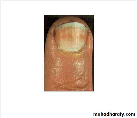
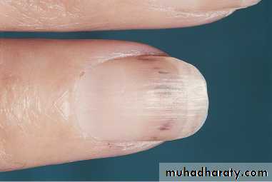
HALF AND HALF NAIL
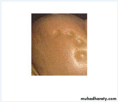
PITTING ODEMA
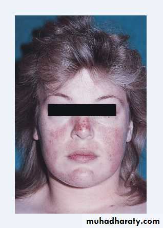
BUTERFLY RASH
SPLINTER HEMORRAGE
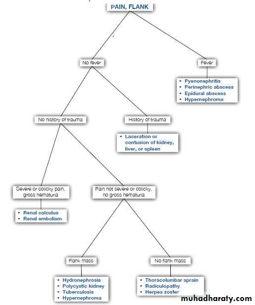
Investigation
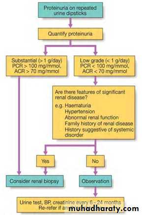
Twenty-four Hour Urine Collection for Protein ExcretionProteinuria ≥ 3.5 g in 24 hours indicates glomerular disease
NEPHROTIC SYNDROM ) .)
electrophoresis gives valuable insight into the composition of the proteinuria.
Occasionally, overflow proteinuria such as light chains in Bence Jones proteinuria, can be ≥ 3.5 g/day without any of the manifestations of nephrotic syndrome.
A urine protein electrophoresis study is important in making the distinction.
Collection way ( tachnique).
Protein-to-Creatinine Ratio
The 24-hour urine collection for protein excretion is cumbersome and subject to inaccuracies.A spot urine sample for protein and creatinine can be used to estimate the amount of protein excreted.
A protein-to-creatinine ratio of 3 estimates that the 24-hour protein excretion is about 3 g.
The ratio may be inaccurate in patients with orthostatic proteinuria.
Urine for Microalbumin
microalbuminuria→→The excretion of abnormal quantities of albumin below the level detectable by the urine dipstick .
Normal albumin excretion is ≤ 30 mg/day.
This is detected by radioimmunoassay or enzyme immunoassay.
Microalbuminuria is the earliest clinically detectable stage of diabetic nephropathy.
Fractional Excretion of Sodium
The excretion of sodium in the setting of oliguria and acute renal failure often gives insight into the appropriateness of tubular function.In the setting of oliguria, an FeNa ≤ 1% often denotes prerenal azotemia, whereas an FeNa of greater than ≥1% denotes intrinsic renal failure .
Fractional Excretion of Urea
If diuretics are being used, the FeNa is unreliable,the fractional excretion of urea can be used as a surrogate for the assessment of volume status.
Urea reabsorption varies with the volume status, increasing in the setting of volume depletion.
A fractional excretion ≤ 30% state of decreased effective circulating volume .
Urine Potassium Excretion
Potassium excretion depends on adequate distal sodium delivery and reabsorption to provide a sufficient electrochemical gradient for tubular potassium excretion.Failure to excrete potassium is seen in volume depletion.
The excretion ≤ 15 mmol/day of potassium in the face of hyperkalemia suggests an inadequate renal response.
Twenty-four Hour Urine Collection for Calcium, Uric Acid, Oxalate, Citrate, Sodium, and Creatinine
These studies are performed in the evaluation of the patient with recurrent kidney stones
GUE ---- Specific Gravity
The specific gravity of the urine can be raised by the presence of an increased number of solutes or by molecules with a high molecular weight, such as glucose or contrast dye.There is a linear relationship between specific gravity and osmolality, (unless there is glycosuria or excretion of contrast media, in which case the specific gravity is higher).
A fixed specific gravity of 1.010 is characteristic of chronic kidney disease.
GUE ---- Glucose
Detected by an assay using dipsticks impregnated with the enzyme glucose oxidase.Glycosuria is seen in
• diabetes mellitus,
• pregnancy
• pan-proximal tubular dysfunction ( glycosuria, aminoaciduria, phosphaturia) -- Fanconi's syndrome .
• Alimentary glycosurea (lag storage disease)
GUE ---- Protien
The dipstick test is a sensitive assayIt is most sensitive to the presence of albumin
and is much less sensitive to other proteins, such as the light chains.
The presence of 1+ protein correlates with about 30 mg/dL of albuminuria, and 3+ protein correlates with greater than 500 ??????? mg/dL of proteinuria.
PROTIENUREA --- ORTHOSTATIC
--- TRANSIENT
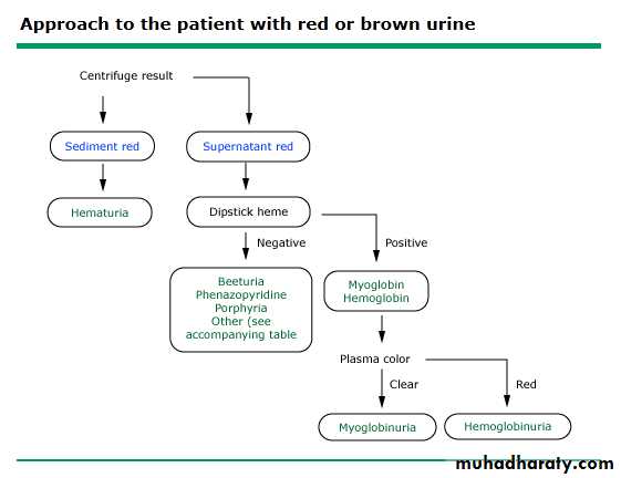
GUE ---- HEME
The dipstick uses the peroxidase-like activity of the hemoglobin and myoglobin molecules to detect the presence of heme pigment.
+VE in the prescence of RBC , hemoglobin, Myoglobin.
GUE ---- Leukocytes
The detection of leukocytes depends on the presence of leukocyte esterase in leukocytes.They usually are present in infections and in inflammatory conditions.
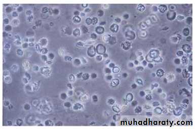
GUE --- CELLS
Dysmorphic RBCs - Phase contrast microscopy aids in the identification of dysmorphic RBCs. The presence of RBC casts is often conclusive evidence for the presence of glomerulonephritis.WBCs are seen most commonly in urinary tract infections.& interstitial nephritis.
Mononuclear cells often appear with transplant rejection.
Tubular cells are seen in many conditions involving tubulointerstitial diseases. They also are seen in ischemic and nephrotoxic injury,
Eosinophils Classically associated with allergic interstitial nephritis,
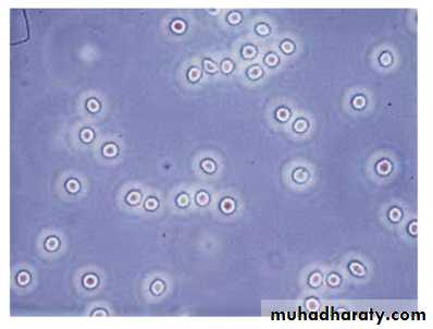
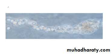
RBC ISOMORPHIC DYSMORPHIC
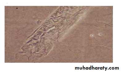
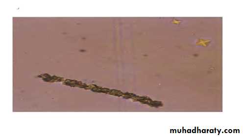
CASTS
Hyaline casts (Tamm-Horsfall proteins) that are formed normallyGranular casts are seen in the setting of tubular injury .
Pigmented granular ?????? casts are seen in rhabdomyolysis with myoglobinuria or, rarely, hemoglobinuria.
RBC casts are diagnostic of
glomerulonephritis.
WBC casts are seen commonly in pyelonephritis and interstitial nephritis
Tubular cell casts are seen with any acute tubular injury and are the dominant cellular casts in ischemic acute tubular necrosis
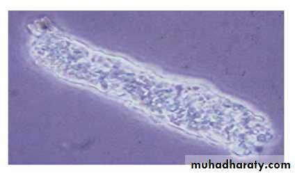
HYALINE CASTS
GRANULAR CASTS
RBC CASTS
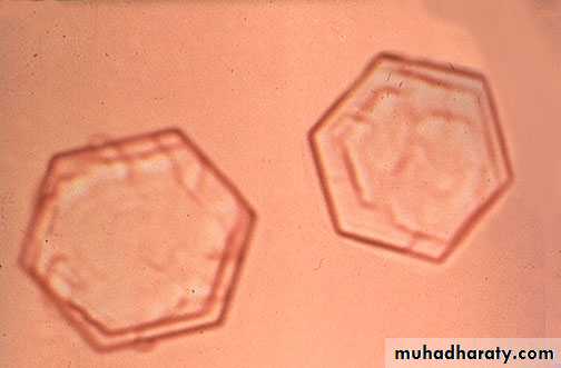
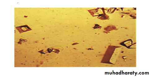
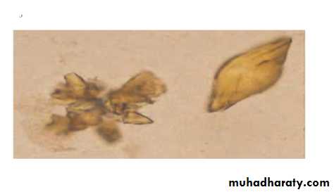
CRYSTALS
Crystals often can be a normal finding in the urine or serve as clues to pathophysiologic processes.hexagonal crystals seen with cystinuria, are always abnormal .
calcium oxalate crystals , may be a normal finding or may be evidence for ethylene glycol intoxication in a patient with anion gap metabolic acidosis, acute renal failure.
Triple phosphate crystals are composed of ammonium magnesium phosphate and are coffin shaped. These are seen in urinary tract infections with urea splitting organisms.
Uric acid crystals, sodium urate crystals and calcium phosphate amorphous crystals all are common and do not denote any pathologic significance.
CYSTIN CRYSTAL
OXALATE CRYSTALS
TRIPLE PHOSPHATE CRYSTALS
URATE CRYSTALS
RENAL FUNCTIOON TESTS
• 1- serum creatinine.• 2- blood urea nitrogen (BUN), which is a product of protein catabolism,
is about 10-fold higher than creatinine concentration, and the BUN-to-creatinine ratio commonly is used as a marker of volume status.
BUN may be inappropriately high, such as with gastrointestinal bleeding or the use of steroids or tetracyclines.
The BUN may be low if there is poor dietary intake of protein and in liver disease.
creatinine clearance
can be estimated from the serum creatinine concentration by the Cockcroft-Gault formula:crc =(140-age) x wt(kg)/72x serum creatinine x( 0.85 if female )
The Modification of Diet in Renal Disease (MDRD) study equation.
Inulin clearance
Inulin,a 5200-D uncharged polymer of fructose, is an ideal marker for the measurement of GFR because it is not reabsorbed, secreted, synthesized, or metabolized. It is not available for routine clinical assessment .
Other tests
Complement— c3, c4, ch50Serum immune electrophoresis
Bence jones protiens
Antineutrophil cytoplasmic antibody (c-ANCA)(p-ANCA)
Anti-Glomerular Basement Membrane Antibody
THANK YOU
QUIZ