The immune system
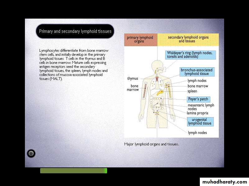
It is a group of structures (cells, tissues and organs) that is collectively referred to the lymphoid system. The major lymphoid organs and tissues are classified into either:Primary (central): where the cells of immune system are produce &/or developed, like the thymus and bone marrow.
Secondary (peripheral): where the cells of immune system are functioned. like the spleen (when we speak about blood born invader), lymph nodes (when we speak about skin invader), mucosa associated lymphoid tissue -MALT-, tonsils & Peyer’s Patches (when we speak about mucus membrane invader).
Primary (central): where the cells of immune system are produce &/or developed, like the thymus and bone marrow.
Secondary (peripheral): where the cells of immune system are functioned. like the spleen (when we speak about blood born invader), lymph nodes (when we speak about skin invader), mucosa associated lymphoid tissue -MALT-, tonsils & Peyer’s Patches (when we speak about mucus membrane invader).
The cell of immune system:
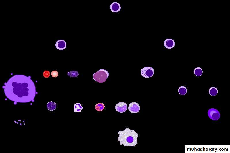
Hematopoietic stem cells consist of myeloid and lymphoid lineages which are both involved in dendritic cell formation. Myeloid cells include monocytes, macrophages, neutrophils, basophils, eosinophils, erythrocytes, and megakaryocytes to platelets. Lymphoid cells include T cells, B cells, and natural killer cells. The peripheral blood contains two large populations of cells: the red cells (RBCs), whose main physiological role is to carry oxygen to tissues, and the white blood cells (WBCs), which have as their main physiological role the elimination of potentially harmful organisms or compounds.INCLUDEPICTURE "https://teachmephysiology.com/wp-content/uploads/2018/09/haematopoeisis.png" \* MERGEFORMATINET
Phagocytic cells
Phagocytic cells, such as monocytes, macrophages, and granulocytes, play significant roles as effectors of the immune response. One of their main functions is to eliminate antigens that have elicited an immune response. This is achieved by means of antibodies and complement. However, if the antigen is located on the surface of a cell, antibody induces the attachment of cytotoxic cells that cause the death of the antibody-coated cell [antibody-dependent cellular cytotoxicity (ADCC)]. phagocyte which internalize antigens and pathogenic microorganisms and degrade them. They fall into two categories:Mononuclear phagocytes: It is long-lived phagocytic cells. Many organs contain phagocytic cells derived from blood monocytes which are manufactured in the bone marrow. Monocyte cells pass out of the blood vessel and become macrophages in the tissue. Resident phagocytic cells of different tissues were previously referred to as reticuloendothelial system (RES) like the microglial cell (brain), alveolar macrophages (lung), Kupffer cell (liver), mesangial phagocytes (kidney), synovial A-cells (joints) and the monocyte in the blood.
Polymorphonuclear Neutrophils: short-lived phagocytic cells, it constitutes the majority of the blood leucocytes. They migrate into tissue, particularly at site of inflammation, in one-way trap. Since, it engulfs material, destroy it and then die.
Lymphocytes:
A lymphocyte is one of the subtypes of white blood cells in a vertebrate's immune system. It is an important cell because of its central role in the immune response. The adaptive immune system is coordinated by lymphocytes (a class of leukocyte) and results in the production of antibodies. They are responsible for the specific immune recognition of the pathogens. There are several subpopulations of lymphocytes have been defined, including natural killer cells (which function in cell-mediated, cytotoxic innate immunity), T cells (for cell-mediated, cytotoxic adaptive immunity), and B cells (for humoral, antibody-driven adaptive immunity).B lymphocytes: represent a precursor of antibody-producing cells, known as plasma cells. They are genetically programmed to encode a surface receptor specific for a particular Ag. Having recognized its specific Ag they multiply and differentiated into plasma cells, which produce large number of antibodies.
T lymphocytes: Several T- lymphocytes subpopulations have been observed.
Helper T lymphocytes (Th), which play a very significant amplification role in the immune responses. Two functionally distinct subpopulations of Th lymphocytes emerging from a precursor population (Th0) have been defined. The Th1 population assists the differentiation of cytotoxic cells and also activates macrophages (One group interacts with MNP and helps them destroy intracellular pathogens). Activated macrophages, in turn, play a role as effectors of the immune response. The Th2 lymphocytes are mainly involved in the amplification of B lymphocyte responses (these cells interact with B-cells and help them to divide, differentiate and make Antibody). The amplifying effects of Th lymphocytes are mediated in part by soluble mediators—cytokines— and in part by signals delivered as a consequence of cell–cell interactions.Cytotoxic T lymphocytes, which are the main immunologic effector mechanism involved in the elimination of non-self or infected cells. This kind of cells is responsible for the destruction of host cells which have become infected by viruses or another intracellular pathogen. These T-cells generate their effects, either by releasing soluble proteins (cytokines) or by direct cell-cell interactions.
Immunoregulatory T lymphocytes, which have the ability to downregulate the immune response through the release of cytokines such as interleukin-10 (IL-10) and through the expression of membrane molecules such as CTLA4, whose interaction with the corresponding receptors delivers a downregulatory signal.
Other T-cells can suppress the immune response, and this might operate through direct killing of APCs or through suppressive cytokines like TGFb or via a negative regulation of signal transduction (CTLA-4) or via idiotype network).
Natural killer cells: they play a dual role in the elimination of infected and malignant cells. These cells are unique in that they have two different mechanisms of recognition: they can identify malignant or viral-infected cells by their decreased expression of histocompatibility antigens (HLA), and they can recognize antibody-coated cells and mediate ADCC.
INCLUDEPICTURE "https://upload.wikimedia.org/wikipedia/commons/thumb/0/0d/Immune_Response.svg/1200px-Immune_Response.svg.png" \* MERGEFORMATINET
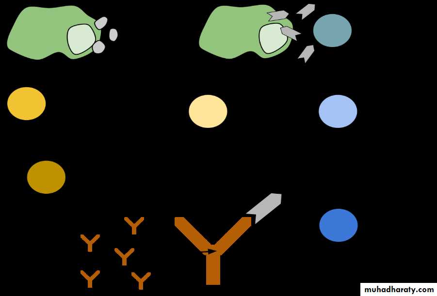
Cytotoxic cells: They have the ability to recognize and destroy other cells. They can be divided into:
Large granular lymphocytes which recognize the surface changes that occur on a variety of tumor cells and virally infected cells. They recognize cells which lack or have lost their MHC-molecules. NK-cells- or like (macrophage, neutrophil & NK) large granulocyte lymphocytes recognize and destroy some target cells which have become coated with specific Ab in what is called Ab-dependent cell-mediated cytotoxicity (ADCC).
Eosinophil PMN: they comprise 2-5% of leukocytes and their cytoplasmic granules stain acidic dye. It plays a role to engage and damage large extracellular parasites, they damage their different targets by releasing the contents of their intracellular granules close to them. Eosinophils also release histamine and aryle-sulphatase which inactivate histamine and some of the leukotrienes (SRS-A).
Auxiliary cells: this kind of cells can mediate inflammation. For example
Basophils and mast cells, they have granules containing a variety of mediators that produce inflammation in surrounding tissues. They can also synthesize and secrete a number of mediators which control the development of immune reaction. Mast cells lie close to blood vessels in all tissue and basophiles are mobile, circulating cells. They represent less than 0.2% of leukocytes, the cytoplasmic granules that stain with basic dyes.platelets: They also release inflammatory mediators when activated during thrombogenesis or by mean of Ab-Ag complexes.
Ag-presenting cells (APCs) are a heterogeneous population of leukocytes such as macrophages, macrophage-related cells, and dendritic cells with very efficient immunostimulatory capacity. These cells play a very significant role in the induction stages of the immune response by trapping and presenting both native antigens and antigen fragments in a most favorable way for the recognition by lymphocytes. In addition, these cells also deliver activating signals to lymphocytes engaged in antigen recognition, both in the form of soluble mediators (interleukins, such as IL-1, IL-12, and IL-18) and in the form of signals delivered by cell–cell contact. APCs are the interface between the innate and adaptive immune systems. They are found primarily in the skin, lymph node, spleen, within or underneath most mucosal epithelia and in thymus. Langerhans cells in the skin and interdigitating cells (IDCs) which is a migrating cell provides an efficient mechanism for carrying Ag from the skin and mucosa to the Th-cells located in the lymph node. These APCs are rich in class II MHC-molecules, which are important for presenting Ag to Th-cells. They bind Ag via complement receptor (CD21, CD35) and Fc-gamma Receptor. Macrophage and classical B-cells are rich in MHC-II, thus able to present Ag to T-cells and these cells also called professional APCs. Somatic cells do not normally express MHC-II but cytokines such as IFN-gamma and TNF-alpha can induce the expression of MHC-II on some of these cells and become able to present Ag like the skin and thyroid epithelium and endothelia. These cells known as non-professional APCs.
Soluble mediators of the immune system:
A wide variety of molecules are involved in the development of the immune response. These includes Abs, complements and cytokines.A-complement proteins; about 30 serum proteins, present in an inactive state, are activated by different mechanisms to mediate variable functions in the immune system like; opsonization (enhanced phagocytosis), chemotaxis (unidirectional migration of the inflammatory cells), anaphylaxis (mast cells degranulation and release of further inflammatory mediator) and lysis of targeted cells.
B- Antibodies: they are also called immunoglobulin (Igs), they are group of serum molecules produced by B-cells, it’s a soluble form of B-cells surface Ag-receptors. In general, each Abs can bind specifically to just one Ag. The part of an AB molecule that bind to Ag called Fab-portion and the other part called constant portion (Fc) which interact with other cells of immune system and act as opsonin which enhance phagocytosis
C. Cytokines are large group of molecules involved in signaling between cells. The principle sets of cytokines are:
Interferons it is important in limiting the spread of certain viral infection like IFN alpha and beta since these IFNs induce a state of antiviral resistance in uninfected tissue cells. IFN gamma which released by activated T-cells and its very important in cell-mediated immunity.
Interleukines (Ils) these are a large group of cytokines IL1-IL22 produced by T-cells, mononuclear phagocytes, most of them are involved in directing other cells to divide and differentiate.
Colony stimulating factors (CSFs) they are involved in directing the division and differentiation of bone marrow stem cells and the precursors of blood outside the bone marrow.
Chemokines; they direct movement of cells around the body, from the blood to the appropriate location. Some of the chemokines also activate cells to carry out particular functions.
Others; like TNF alpha & beta and transforming growth factors-beta.
Antigen:
Antigen (Ag) is a substance that reacts with the products of a specific immune response. It is usually exogenous substances (cells, proteins, and polysaccharides), which are recognized by receptors on lymphocytes, thereby eliciting the immune response. The receptor molecules located on the membrane of lymphocytes interact with small portions of those foreign cells or proteins that are designated as antigenic determinants or epitopes. An adult human being has the capability to recognize millions of different antigens, producing antibodies.Immunogen is a substance that induces a specific immune response. A stimulus that produces a humoral or cell-mediated mediated immune response humoral immune response by antibody and Cell mediated immune response by T cells. It can be classified into:
T-dependent antigens are antigens that do not directly stimulate the production of antibody without the help of T cells.
Mostly proteins
require macrophages or another APCRequire T-helper cells for formation of Abs
Require major histocompatibility antigensMemory
T-independent antigens are antigens which can directly stimulate the B cells to produce antibody without the requirement for T cell help.Complex carbohydrates
The antigen stimulates antibody production without T-cell help (Do not require processing)
The immune response to these Ag usually by IgM
Can directly interact with B cellsLittle or no memory
By definition All immunogens are antigens, but not all antigens are immunogens.Epitope (antigenic determinant): it is an active region of an immunogen (or antigen) that binds to antigen-specific receptors on lymphocytes or to secreted antibodies.
Superantigen: A class of antigens that cause non-specific activation of T-cells, resulting in polyclonal T-cell activation and massive cytokine release.
Exogenous and Endogenous antigens:
Exogenous antigens are antigens that have entered the body from the outside, for example, by inhalation, ingestion or injection. The immune system's response to exogenous antigens is often subclinical. By endocytosis or phagocytosis, exogenous antigens are taken into the antigen-presenting cells (APCs) and processed into fragments. APCs then present the fragments to T helper cells (CD4+) by the use of class II MHC molecules on their surface. Some antigens start out as exogenous, and later become endogenous (for example, intracellular viruses). Intracellular antigens can be returned to circulation upon the destruction of the infected cell.Endogenous antigens, by definition, are part of self, and the immune system is usually tolerant to them. The response to self-antigens may have an important role in normal catabolic processes. The loss of tolerance to self-antigens, however, can also have pathogenic implications (autoimmune diseases).
They are generated within normal cells as a result of normal cell metabolism, or because of viral or intracellular bacterial infection. The fragments are then presented on the cell surface in the complex with MHC class I molecules. If activated cytotoxic CD8+ T cells recognize them, the T cells secrete various toxins that cause the lysis or apoptosis of the infected cell. In order to keep the cytotoxic cells from killing cells just for presenting self-proteins, the cytotoxic cells (self-reactive T cells) are deleted as a result of tolerance (negative selection).
Antigenicity and Immunogenicity
Antigenicity is defined as the property of a substance (antigen) that allows it to react with the products of a specific immune response (antibody or T cell receptor). On the other hand, immunogenicity is defined as the property of a substance (immunogen) that endows it with the capacity to provoke a specific immune response (The ability to induce a humoral or cell mediated immune response).Hapten is a substance that fails to induce immune responses in their native form due to low molecular weight (MW) and / or their chemical simplicity. Hapten is term used to describe any antigen that is unable to induce an immune response. It has the property of antigenicity but not immunogenicity. It can react with the products of a specific immune response with no antibodies formation. This non antigenic substance can be immunogenic if coupled to a larger molecule referred to as a carrier molecule. For example, small molecules with a low molecular weight (Less than 10,000) that could never induce an immune response when administered by themselves unless it coupled to a carrier molecule.
Factors Influencing Immunogenicity: Many different substances can induce immune responses. The following characteristics influence the ability for a substance to behave as an immunogen:
Foreignness: Only substances recognized as “non-self” will trigger the immune response. Microbial products and exogenous molecules are obviously “non-self” and may be strongly immunogenic. (More foreign substance can give more immunogenic).
Molecular size: The most potent immunogens are macromolecular proteins (MW.100,000 Daltons). Molecules smaller than 10,000 Daltons are often only weakly immunogenic, unless coupled to an immunogenic carrier protein.
Chemical structure: Protein represents the best immunogenic molecules, followed by CHO, whereas lipid and nucleic acids are weak immunogic.
Chemical complexity: There appears to be a direct relationship between antigenicity and chemical complexity—aggregated or chemically polymerized proteins are much stronger immunogens than their soluble monomeric counterparts.
Degradability: Peptides composed of D-amino acids, which are resistant to enzymatic degradation, are not immunogenic, while L-isomers are susceptible to enzymes and are thus immunogenic whereas, CHO are not processed or presented and are thus unable to activate T cells, although they can activate B cells.
Other factors play a role in whether or not a substance is immunogenic
Genetic make-up
Some substances are immunogenic in one species but not in another.
Immune responsiveness controlled by genes mapping within the MHCAge: it can also influence immunogenicity. Usually the very young and the very old have a diminished ability to mount an immune response in response to an immunogen.
Dosage and route of administration
Threshold amount of AgInsufficient doses tend to render the responding cells unresponsive – tolerance
Number of doses administeredRepeated doses stimulate a stronger response
Route of AdministrationDetermines which organs and cell populations respond, subcutaneous (S.C.) and intramuscular better than oral or intravenous injection.
Immunologic Adjuvants: is substances that can enhance the immune response to an immunogen. The use of adjuvants, however, is often hampered by undesirable side effects such as fever and inflammation.
It is not immunogenic by itself.
Cannot evoke immune response by itself.There are two types of adjuvants
Repository: like the Al- & Ca- salts, they act by slowing the release of immunogen when they form insoluble complex with it.Emulsifying agent: like the Freund’s adjuvant which composed of water, oil and killed mycobacterium, the different size droplets are degraded at different rates (prolonged presence) and the induce granuloma reaction act as physical barrier, delayed it release.