1
Ophthalmology
Dr. Muataz Hasan
THE LACRIMAL SYSTEM
Anatomy of the lacrimal system
1. Secretory system
2. Drainage system
The lacrimal secretory system
The lacrimal secretory system is formed of
1. The main lacrimal gland.
2. The accessory lacrimal glands.
3. Conjunctival goblet cells.
1. The main lacrimal gland: Almond in shape and is formed of 2 parts:
i. Orbital portion: It is the main part of the gland, situated in a shallow bony
fossa in the anterolateral part of the roof of the orbit.
ii. Palpebral portion (1/4 of the whole gland): It is continuous with the orbital
portion posteriorly
iii. The lacrimal ducts: 10-12 ducts arise from the orbital portion of the gland
to pass through the palpebral portion and then open in the lateral part of
superior fornix.
2. Accessory lacrimal glands:
They are microscopic in size and open into conjunctival sac by fine ductules:
i. The glands of Krause: In the conjunctival fornices (40 in the upper fornix and
10 in the lower fornix)
ii. The glands of wolfring: Located in the palpebral conjunctiva opposite the
mid- tarsus.
3. Goblet cells of the conjunctiva: They are unicellular mucionous glands.

2
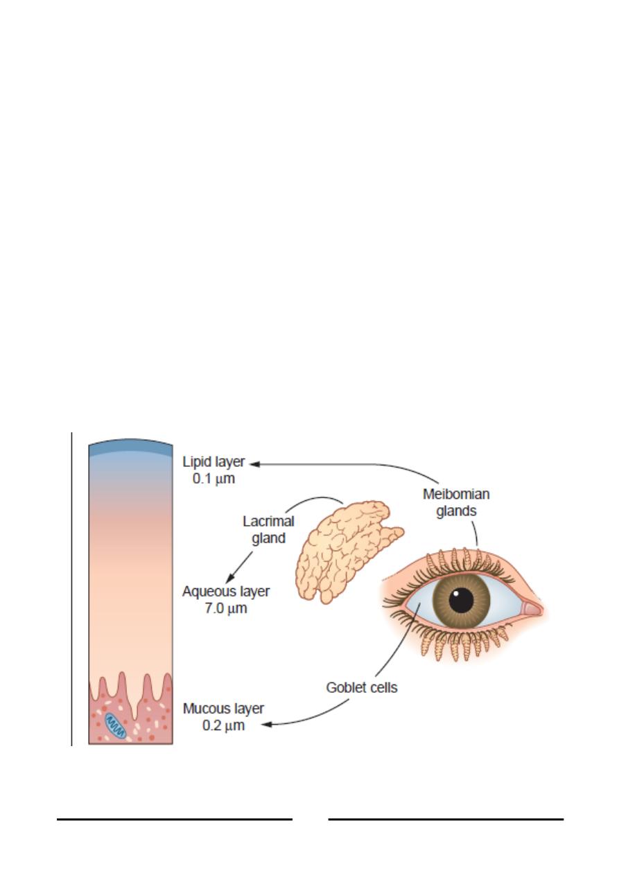
Precorneal tear film:
It is formed of 3 layers.
1. Outer lipid layer: secreted by the meibomian glands.
Function:
Prevent rapid evaporation of tears.
Lubricates the eyelids over the globe.
2. Middle aqueous layer: Secreted by the lacrimal gland.
Function:
Supplies oxygen to the corneal epithelium.
Antibacterial as it contains lysozymes.
3. Inner mucinous layer: Secreted by the goblet cells.
Function:
Makes the corneal epithelium hydrophilic.

3
The Lacrimal Drainage system
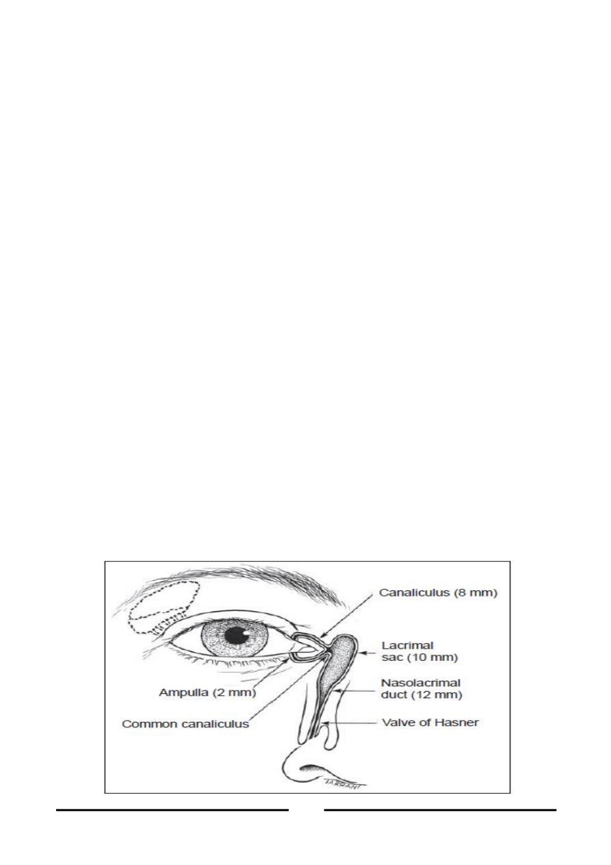
The lacrimal drainage system is formed of 2 canaliculi, the lacrimal sac, and the
nasolacrimal duct ending in the inferior meatus of the nose.
1. Two puncti:
Located at the posterior edge of the lid margin, not seen except when the lid is
everted. Each punctum lies 6 mm. from medial canthus on a slightly elevated
portion called the papilla.
2. Two canaliculi:
Are fine tubes which carry tears from the puncti to the lacrimal sac. Each
canaliculus is made of 2 portions:
a. Vertical part: 2mm
b. Horizontal part: 8mm
Before entering the lacrimal sac, they unite into a common canal, 1-2 mm long
that opens at the junction between the upper 1/3 and the lower 2/3 of the sac.
3. Lacrimal Sac:
Site: the lacrimal sac lies in the lacrimal fossa in the medial wall of the orbit.
Size: 8x12mm (when distended).
The lacrimal sac is formed of:
a. Body: This forms the main part.
b. Fundus: Blind upper portion, it lies above the medial palpebral ligament.
c. Neck: The neck is narrow and continuous with the nasolacrimal duct.

4
4. Nasolacrimal duct:
The nasolacrimal duct is 12-24 mm long. It passes from the end of the sac to
open in the inferior meatus of the nose. The direction of the duct is:
downwards, slightly backwards and laterally.
5. Nose: the opening of nasolacrimal duct into the meatus of the nose is guarded
Hasner's valve.
Tear Drainage
1. Evaporation: 25% of tears.
2. Excretion:
a. Passive: Gravity and capillarity.
b. Active: lacrimal pump through the action of the lacrimal portion of
orbicularis muscle (Horner’s muscle).
WATERY EYE
1. Lacrimation
Lacrimation is over secretion of tears
Etiology:
a. Emotional conditions
b. Reflex lacrimation from foreign body or inflammation.
2. Epiphora:
Epiphora is overflow of tears onto the cheek due to inadequate drainage, which
may be due to lacrimal pump failure or obstruction of the lacrimal passages.
CLINICAL EVALUATION AND INVESTIGATION OF EPIPHORA
History:
Exclude lacrimation as a cause
1. Bilateral watering is usually due to lacrimation.
2. Unilateral watering: is usually due to epiphora.

5
Examination:
1. Eyelid: exclude the presence of ectropion and trichiasis.
2. Lacrimal sac swelling and dacryocystitis.
3. Nose: polypi, deviated septum.
Investigations:
1. Regurgitation test: press on the lacrimal sac against the bone.
a. A + ve regurge = reflux of pus or tears from the puncti in case of
obstruction of the nasolacrimal duct. This is a definite proof of obstruction.
b. A – ve regurge = No reflux with patent lacrimal passages.
2. Jones test:
Type I test: instill a drop of fluorescein in the conjunctival sac and a cotton
pellet soaked in xylocaine spray (local anesthetic) under the inferior turbinate
of the nose.
i.
If the cotton is stained with fluorescein the lacrimal passages are patent.
ii.
If no fluorescein is recovered, proceed to type II jones test.
Type II test: the lacrimal passage is irrigated with saline.
i.
If fluorescein is recovered, there is partial or functional obstruction.
ii.
If fluorescein is not recovered and saline does not reach the nose, there is
complete block.
3. Dacryocystography: a radio contrast medium is injected and X – ray is done
at intervals to detect filling
4. Plain X- ray: for diagnosis of tumors or fractures.
Treatment of Epiphora
1. Treatment of the cause: e.g Ectropion and nasal causes of epiphora.
2. Stenosis of puncti and canaliculi:
Dilatation and probing

6
One snip ampullotomy: vertical snip is made in posterior wall of canal.
Laser punctoplasty.
3. Obstruction of puncti and canaliculi:
Three snip operation: a triangle is removed from posterior wall of the
canaliculus.
4. Obstruction of Nasolacrimal duct:
a. Congenital obstruction:
Hydrostatic massage.
Dilatation and probing
Dacryocystorhinostomy.
b. Acquired obstruction:
Dilatation and probing usually fails.
Dacryocystorhinostomy.
Dacryocystectomy.
LACRIMAL SAC DISORDERS
Acute dacryocystitis
Definition: Acute suppurative inflammation of the lacrimal sac.
Etiology:
Predisposing factor: nasolacrimal duct obstruction.
Causative agent: pneumococci, staphylococci and streptococci.
Clinical picture:
Symptoms:
Severe pain.
Fever

7
Signs:
1. Marked edema and redness of skin over the sac.
2. Regurgitation test: excessive reflex of pus.
3. Tender swelling of lacrimal sac.
4. Abscess formation with fluctuation.
Complications:
1. Lacrimal fistula: the sac may burst anteriorly through the skin.
2. Pyocele: canaliculi may become obstructed.
3. Orbital cellulitis and cavernous sinus thrombosis.
4. Chronic dacryocystitis.
Treatment:
1. During the acute phase.
a. Antibiotics: systemic and topical.
b. Hot fomentations.
c. Lotions: to clean the pus.
d. Incision and drainage if an abscess forms.
2. After the acute attack subsides: dacryocystorhinostomy with fistulectomy if
needed.
CHRONIC DACRYOCYSTITIS
Definition:
A chronic inflammation of lacrimal sac secondary to obstruction of the naso-lacrimal
duct. It is the commonest lacrimal sac disorder.
Etiology:
Predisposing factor: Nasolacrimal duct obstruction.
Causative agent:
i. Pneumococci in 80%
ii. Staphylococci, streptococcus, trachoma, and fungi

8
Clinical picture:
Symptoms:
1. Watery eye.
2. Discharge.
Signs:
The inner canthus is red and hyperemic.
Swelling of lacrimal sac below the medial palpebral ligament.
Regurgitation test +ve : pressure on the swelling causes regurge of mucous or
pus.
Treatment:
The aim of treatment is:
1. To restore communication between the lacrimal sac and the nose.
2. To treat infection
Treatment of congenital dacryocystitis:
1. Antibiotics: systemic and topical (drops and ointment)
2. Hydrostatic massage: the mother is instructed to press on the lacrimal sac in a
downward direction. This may help to remove any remnants of epithelium or
to open hasner’s valve. This is tried for a long period up to 1 year.
3. Probing: is successful if done carefully as the lacrimal passages are still elastic
and can be stretched on the probe.
4. Irrigation: repeated syringing with saline may cure the condition.
5. Dacryocystorhinostomy
Treatment of acquired dacryocystitis:
1. Treatment of the cause of obstruction: e.g relieves congestion, removal of a
nasal polyp.
2. Dacryocystorhinostomy: operation of choice.
3. Dacryocystectomy: in neglected cases.

9
DACRYOCYSTORHINOSTOMY (DCR)
Principle: is to create a surgical opening between the lacrimal sac and the nasal
mucosa of the middle meatus, allowing drainage of tears directly into the nose
bypassing the obstructed naso – lacrimal duct.
Indications:
1. Chronic dacryocystits.
2. Mucocele of lacrimal sac.
3. Lacrimal fistula (DCR and fistulectomy)
DACROCYSTECTOMY
Removal of the lacrimal sac.
Indications: indicated in cases where DCR cannot be done, and the lacrimal sac is
fibrosed.
DRY EYE
Etiology:
1. Old age due to decreased amount of tears.
2. Congenital absence of the lacrimal gland.
3. Inflammation of lacrimal gland e.g sarcoidsis.
4. Tumors of lacrimal gland: e.g mixed lacrimal gland tumor.
5. Keratoconjunctivits sicca: Autoimmune disease leading to atrophy and fibrosis
of the lacrimal gland, it occurs usually in females and may be associated with
arthritis and dry mouth (sjogren's syndrome)
6. Conjunctival scarring: Due to Tachoma, chemical burns, stevens- Johnson
syndrome and ocular cicatricial pemphigoid.
7. Drugs as antiglaucoma therapy.
8. Vitamin A difficiency.
Clinical picture:
Symptoms: irritation and foreign body sensation.

10
Signs:
1. Deficient precorneal tear film and loss of corneal luster.
2. Punctuate epithelial erosion of the cornea.
Investigations:
1. Tear film break – up time (BUT) is diminished (normally it is 15 second).
2. Schirmer's test: A normal person wets 10-30 mm. of a whatman number 41
filter paper strip (5mm. wide x 30 mm. long) in 5 minutes. Values less than
5mm. indicate hyposecretion.
3. Rose Bengal staining of devitalized epithelial cells.
Treatment:
1. Protective glasses and contact lenses.
2. Artificial tears eye drops.
3. Occlusion of the puncti to reduce tear drainage.
4. Systemic steroids.
