
Professional plaque control
UNIVERSITY OF MOSULCOLLEGE OF DENTISTRY
By: Dr. Huda A. Salim
B.D.S, M.Sc. In Oral and Maxillofacial Surgery
Mosul University/College of Dentistry/Oral and Maxillofacial Department.
2020-2021
Department of:
Oral and Maxillofacial Surgery.
UNIVERSITY OF MOSUL
COLLEGE OF DENTISTRYScaling
Removal ofplaque retentive
factors
Root Planing
Professional Plaque ControlUNIVERSITY OF MOSUL
COLLEGE OF DENTISTRY The periodontal pocket, which is defined as a pathologically deepened gingival sulcus.
is one of the most important clinical features of periodontal disease. Deepening of the gingival sulcus may occur as a result of coronal movement of the gingival margin, apical displacement of the gingival attachment, or a combination of the two processes.
UNIVERSITY OF MOSUL
COLLEGE OF DENTISTRY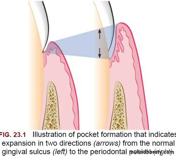
UNIVERSITY OF MOSUL
COLLEGE OF DENTISTRYClassification:
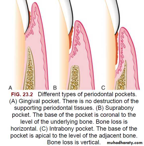
Pockets can be classified as follow:1-Gingival pocket (also called “pseudo-pocket”) is
formed by gingival enlargement without destruction of the underlying periodontal tissues. The sulcus is deepened because of the increased bulk of the gingiva.
UNIVERSITY OF MOSUL
COLLEGE OF DENTISTRY020-2021
2-Periodontal pocket: produces destruction of the supporting periodontal tissues, leading to the loosening and exfoliation of the teeth.
Based on the location of the base of the pocket in relation to the underlying bone, periodontal pockets can be classified into the following types:
A- Suprabony (supracrestal or supraalveolar): occurs when the bottom of the pocket is coronal to the underlying alveolar bone.
B- Intrabony (infrabony, subcrestal, or intraalveolar): occurs when the bottom of the pocket is apical to the level of the adjacent alveolar bone. With this second type, the lateral pocket wall lies between the tooth surface and the alveolar bone.
UNIVERSITY OF MOSUL
COLLEGE OF DENTISTRY020-2021
UNIVERSITY OF MOSUL
COLLEGE OF DENTISTRY020-2021
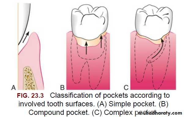
• Pockets can involve one, two, or more tooth surfaces, and they can be of different depths and types on different surfaces of the same tooth and on a proximal surfaces of the same interdental space.
• Pockets can also be spiral (i.e., originating on one tooth surface and twisting around the tooth to involve one or more additional surfaces). These types of pockets are most common in furcation areas.
UNIVERSITY OF MOSUL
COLLEGE OF DENTISTRYClinical Features:
Bluish red thickened marginal gingiva. Bluish red vertical zone from the gingival margin to the alveolar mucosa
Gingival bleeding and suppuration.
Tooth mobility.
Diastema Formation.
• Symptoms such as localized pain or pain “deep in the bone.”
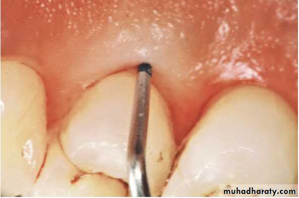
• The only reliable method of locating periodontal
pockets and determining their extent is careful
probing of the gingival margin along each tooth
surface.
UNIVERSITY OF MOSUL
COLLEGE OF DENTISTRYPocket contents:
Periodontal pockets contain:
Debris that consists principally of microorganisms and their products (enzymes, endotoxins, and other metabolic products).
Gingival fluid.
Food remnants.
Salivary mucin.
Desquamated epithelial cells, and leukocytes.
Pus is a common feature of periodontal disease, but it is only a secondary sign.
(GCF) is an ultra filtrate of blood, present in the gingival sulcus, space that contains several molecular components like bacterial degradation products, host tissue degradation products, and inflammatory mediators
UNIVERSITY OF MOSUL
COLLEGE OF DENTISTRYClassification of Periodontal Instruments:
Periodontal instruments are designed for specific purposes, such as calculus removal, biofilm removal, and root planing.Periodontal instruments are classified according to the purposes they serve, as follows:
1. Periodontal probes.(locate, measure, and mark p.)
2. Explorers.
3. Scaling, root-planing, and curettage instruments.
4. Periodontal endoscopes.
5. Cleansing and polishing instruments.
UNIVERSITY OF MOSUL
COLLEGE OF DENTISTRYDepartment of:
HERE
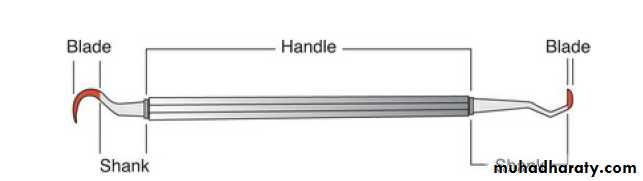
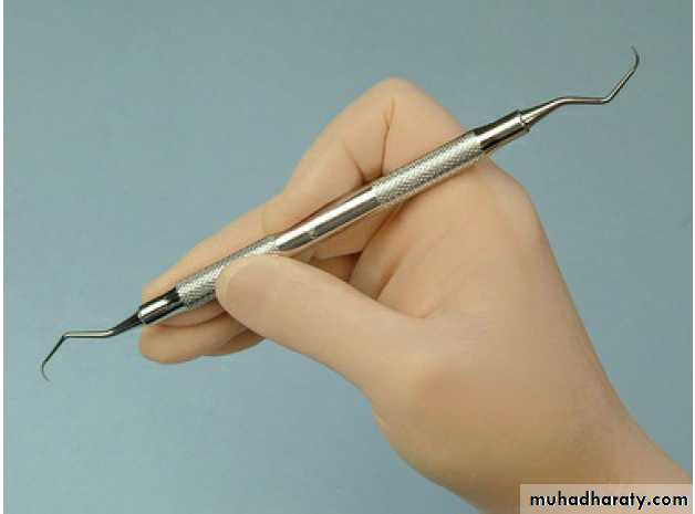
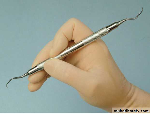
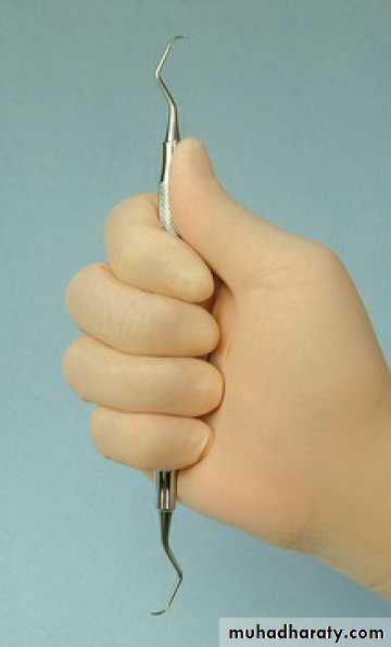
The parts of each instrument are referred to as the working end, shank, and handle
UNIVERSITY OF MOSUL
COLLEGE OF DENTISTRYDepartment of:
HERE
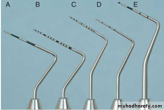
Periodontal Probes: Periodontal probes are used to measure the depth of pockets and to determine their configuration. The typical probe is a tapered,
rod like instrument calibrated in millimeters, with a blunt, rounded
tip
UNIVERSITY OF MOSUL
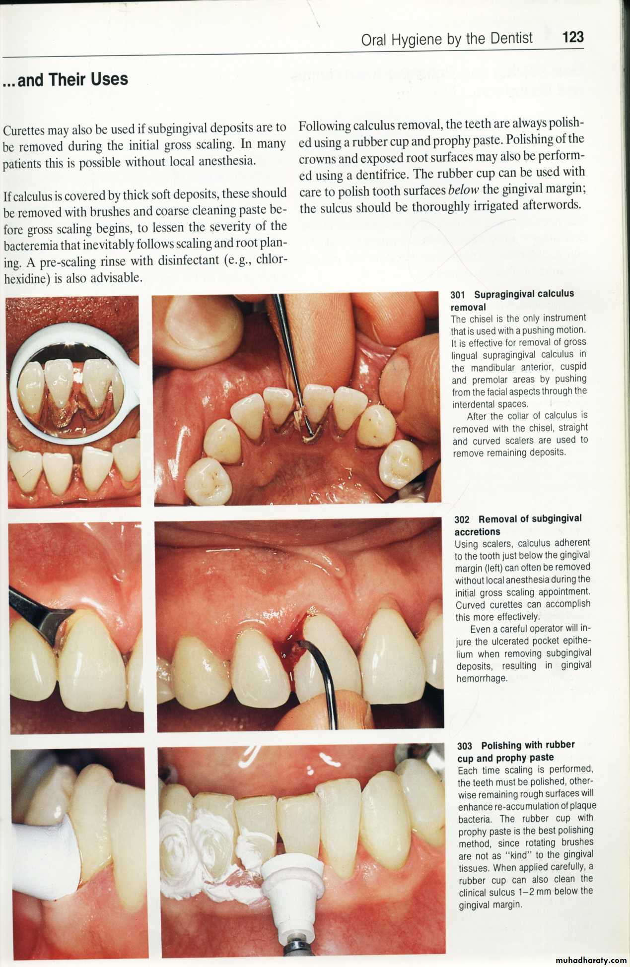
COLLEGE OF DENTISTRYSCALING:
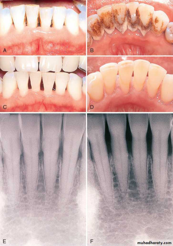
Scaling is the process by which biofilm and calculus are removed from both supragingival and subgingival tooth surfaces. Root planing is the process by which residual embedded calculus and portions of Cementum are removed from the roots to produce a smooth, hard, clean surface.
Scaling and root planing are not separate procedures; all the principles of scaling apply equally to root planing.
The primary objective of scaling and root planing is to restore
gingival health by completely removing elements that provoke
gingival inflammation (i.e., biofilm, calculus, and endotoxin) from
the tooth surface
UNIVERSITY OF MOSUL
COLLEGE OF DENTISTRY
HERE
UNIVERSITY OF MOSUL
COLLEGE OF DENTISTRY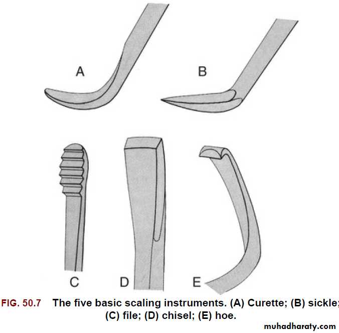
UNIVERSITY OF MOSUL
COLLEGE OF DENTISTRY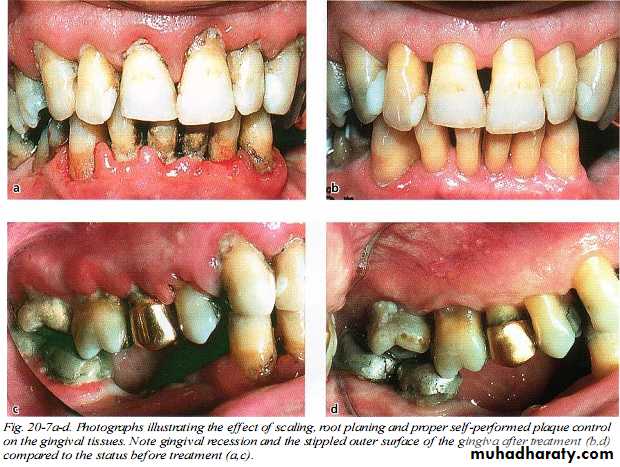
UNIVERSITY OF MOSUL
COLLEGE OF DENTISTRYDepartment of:
HERE
• When biofilm and calculus form on enamel, the deposits are usually superficially attached to the surface and are not locked into irregularities.
• Scaling alone is sufficient to completely remove biofilm and calculus from enamel, leaving a smooth, clean surface.
• Root surfaces exposed to biofilm and calculus pose a different problem. Deposits of calculus on root surfaces are frequently embedded in cemental irregularities.
• Subgingival calculus is porous and harbors bacteria and endotoxin and therefore should be removed completely
UNIVERSITY OF MOSUL
COLLEGE OF DENTISTRYDepartment of:
HERE
Scaling and root planing should not be viewed as separate procedures unrelated to the rest of the treatment plan.
Sickles, curettes, and ultrasonic and sonic instruments are most often used for the removal of supra gingival calculus; hoes and chisels are less frequently used.
Sub gingival scaling and root planing are much more complex and difficult to perform than supra gingival scaling
v
UNIVERSITY OF MOSUL
COLLEGE OF DENTISTRY020-2021
A
B
C
UNIVERSITY OF MOSUL
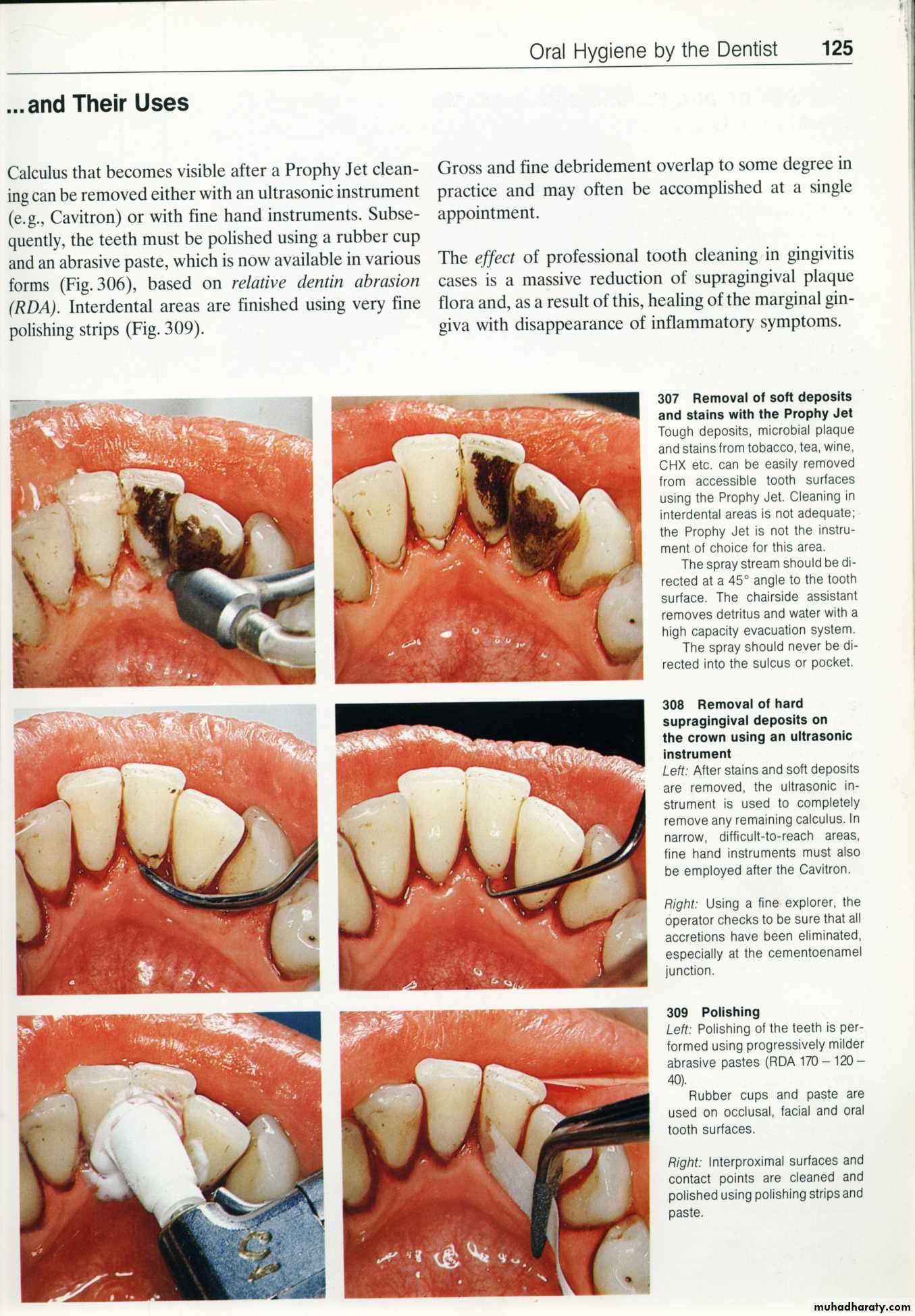
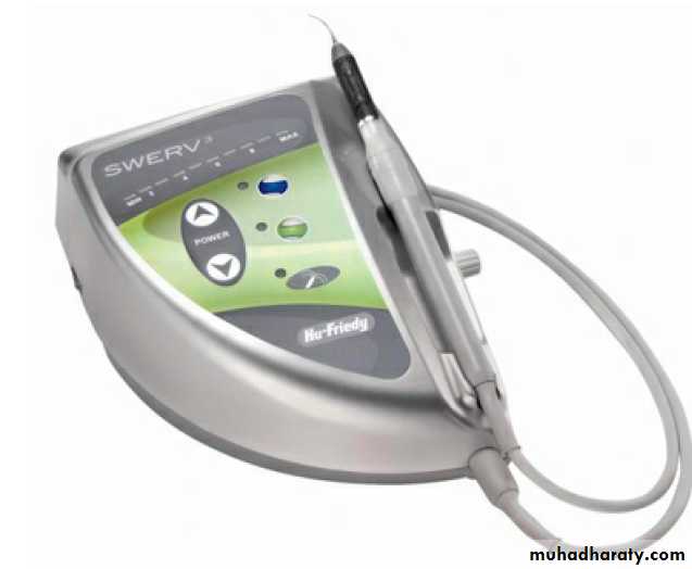
COLLEGE OF DENTISTRYUltrasonic and Sonic Scaling:
Ultrasonic scalers may be used for removing plaque and stain, scaling, root planing, curetting, and surgical debridement.
Ultrasonic scaler used for
• S & RP.• Curetting.
• debridement during p.d. surgery.
• removal of stain.
• overhanging restoration.
Rough sur. left after use large tip
But such roughness not interfere with healing.
The vibrational energy produced by the ultrasonic instrument makes it useful for removing heavy, tenacious deposits of calculus and stain.
UNIVERSITY OF MOSUL
COLLEGE OF DENTISTRY
Department of:
HERE
2 type of ultrasonic s.:
Magnetostrictive:
vibration of tip is elliptical (all sides of tip are active). frequency range of 18,000 to 50,000 cycles per second.
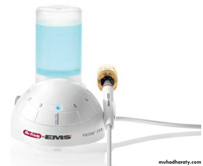
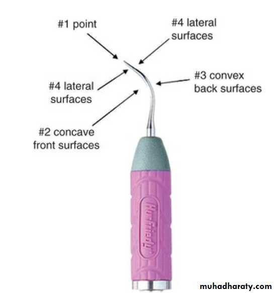
Piezoelectric; vibration of tip is linear meaning, or back and forth, the 2 sides of tip are most active. frequency range of 18,000 to 50,000 cycles per second. Piezoelectric tips move, primarily in a linear pattern, giving the tip two active surfaces.
• Magnetostrictive ultrasonic inserts generate heat and require water for cooling.
• Sonic and piezoelectric units do not generate heat but still use water forcooling of frictional heat and for flushing away debris.
UNIVERSITY OF MOSUL
COLLEGE OF DENTISTRY020-2021
A
B
C
D
UNIVERSITY OF MOSUL
COLLEGE OF DENTISTRY020-2021
Ultrasonic s.:
• Patient with artificial pacemaker2- patient with communicable diseases that can be transmitted by aerosols should not be treated with ultrasonic or sonic scaling devices.
3-for implant ,porcelain ,bonded restoration which can be fractured or removed
UNIVERSITY OF MOSUL
COLLEGE OF DENTISTRY020-2021
Advantages:
Less chair side time & less trauma so less postoperative discomfort Stain removal easier.
Superior to hand instrument in cleaning furcation area.UNIVERSITY OF MOSUL
COLLEGE OF DENTISTRY
020-2021
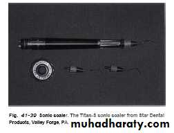
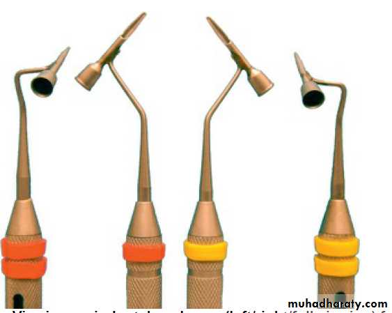
Sonic scaler:
Sonic units work at a frequency of 2000 to 6500 cycles per second and use a high- or low-speed air source from the dental unit.
Sonic units consist of a hand piece that attaches to a compressed air line and uses a variety of specially designed tips.
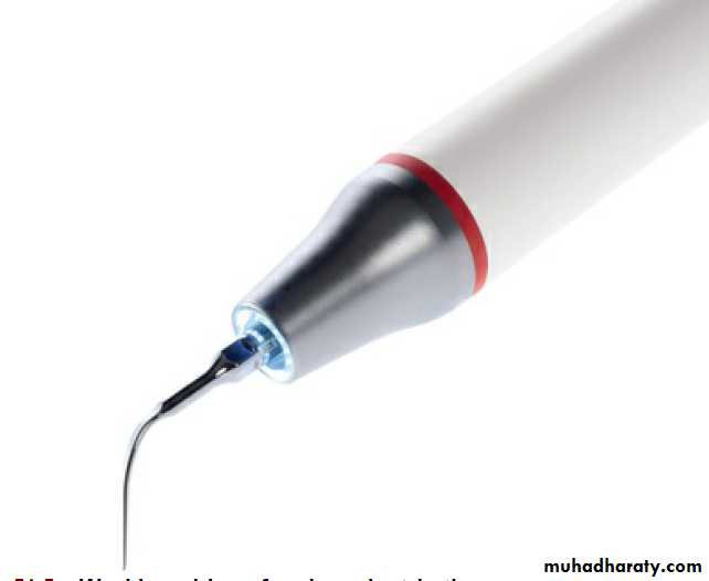
A sonic scaler tip travels in an elliptical or orbital stroke pattern. This stroke pattern allows the instrument to be adapted to
all tooth surfaces.
UNIVERSITY OF MOSUL
COLLEGE OF DENTISTRY020-2021
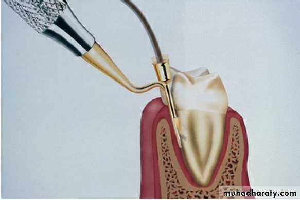
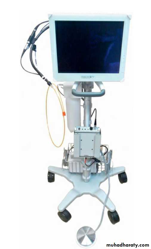
Perioscop (dental endoscope):
Used subg.in diagnosis & treatment of periodontal disease.
Fibroptic endoscope (0.99 mm) with Disposable sterile sheath & fits onto p.d. probe & ultrasonic inst.
Sheath deliver water irrigation.
Attached to video camera & light source to produce image on video monitor.
Magnification range from 24-48 times.
UNIVERSITY OF MOSUL
COLLEGE OF DENTISTRY020-2021
The Perioscopy system can also be used to evaluate subgingival areas for caries, defective restorations, root fractures, and resorption.
It permits operators to detect the presence and location of subgingival deposits and guides them in the thorough removal of these deposits.
UNIVERSITY OF MOSUL
COLLEGE OF DENTISTRYDepartment of:
HERE
UNIVERSITY OF MOSUL
COLLEGE OF DENTISTRY