Definition
• Jaundice came from the French
word “jaune” which means yellow.
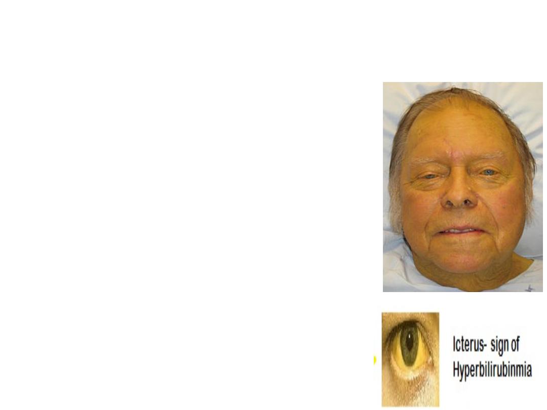
• Yellowish discoloration of sclera,
skin mucous membranes due to
increased serum bilirubin level.
Typically can be detected if serum
bilirubin level above 3 mg/dl (51.3
μmol/L.
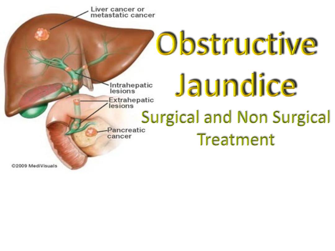
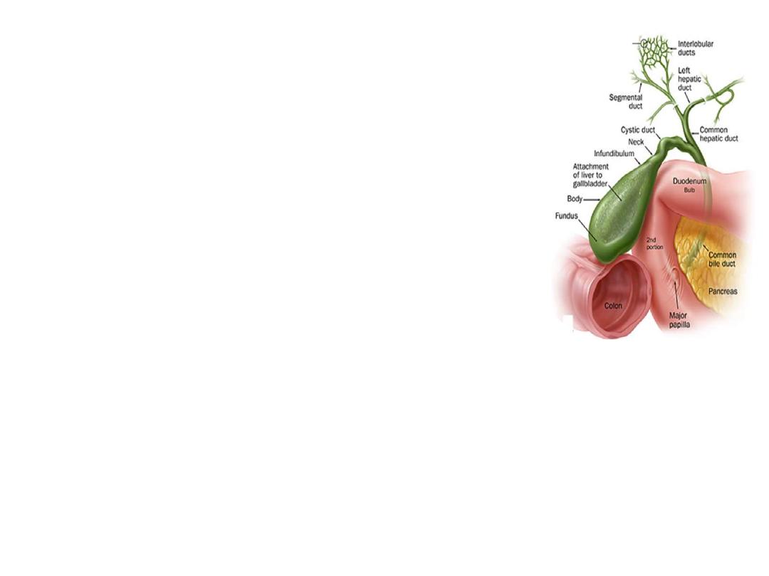
• Obstructive jaundice is interruption
to the drainage of bile in the biliary
system
Classifications:
I.
Prehepatic
II. Hepatic
III. Posthepatic (Obstructive)
• Intraluminal- Transmural- Extramural
• Common- Infrequent- Rare
• Complete (type 1)- Intermittent (Type 2)- Chronic incomplete (Type
3)- Segmental obstruction (Type 4)
• Etiology (congenital, inflammatory, traumatic, neoplastic, parasitic
etc.)
Obstructive Jaundice
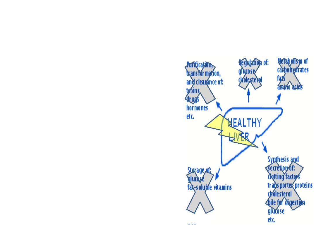
Alteration in:
• Systemic and renal
hemodynamics
• Hepatic function ( protein
synthesis, reticuloendothelial
function,hepatic metabolism)
• Hemostatic mechanism
• Gastointestinal barrier
• Immune function
• Wound healing

Managment
Objectives:
• To identify pts who need relief of obstruction
To establish cause, to plan appropriate
intervention, prevent complications, prevent
recurrence.
S&S for urgent surgical interventions:
• Abdominal pain (70%)
• Jaundice (60%)))
• Tea colored urine/ pale stool
• Altered mental status (10-20)
• Hypotension (30%)
• Fever, persistent (90%)
• RUQ tenderness

Imaging Studies
• Ultrasound
• CT scan, Spiral CT scan
• MRI, MRCP
• Digital substraction angiography
• Cholangiography ERCP, PTC
• IDUS
• PET
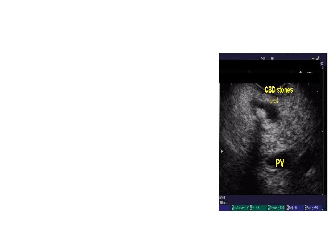
Ultrasonography
• 1
st
choice in O.J.
• Non invasive, cheep, bed side
• Size of bile duct, level of
obstruction, identify the cause in
some cases, liver parenchyma,
• Limitation: obese, Exessive bowel
gases, retroduodenal and
intraduodenal CBD stone
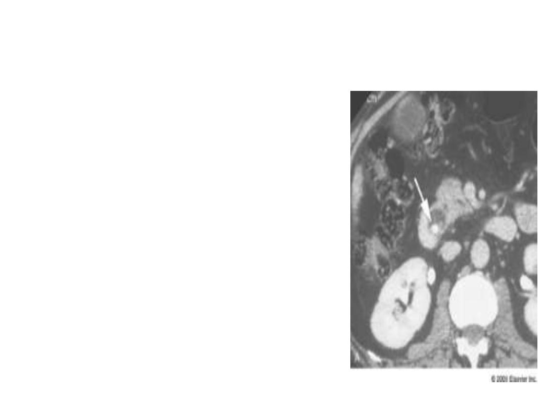
CT scan of Abdomen
• Very useful for assessment of
malignancy
• Intrahepatic biliary dilatations,
• Level of obstruction
• Spiral CT allows : relationship
vascular and bile duct anatomy
at the hilum
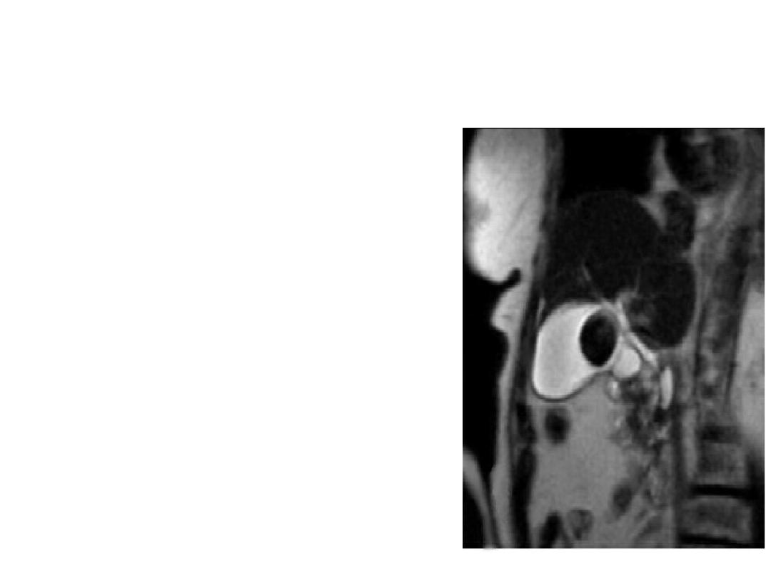
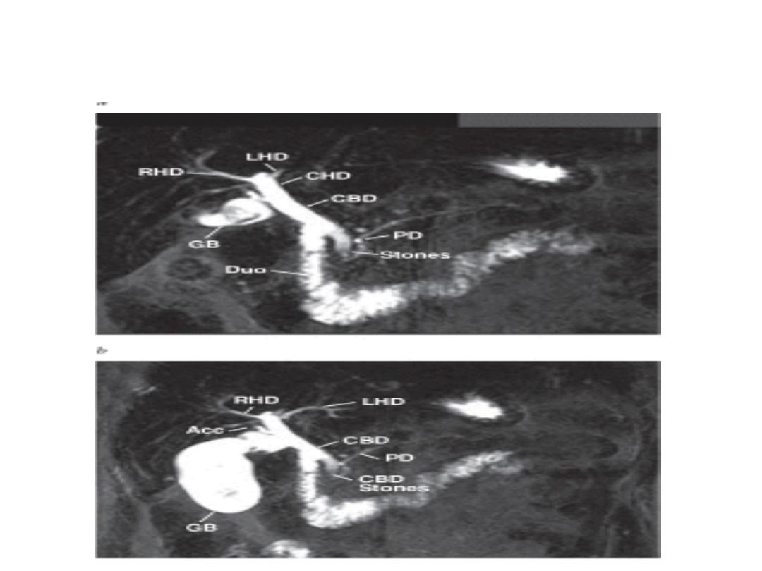
MRCP
• Non invasive
• Useful when ERCP
contraindicated
• No intravenous contrast
• Purely diagnostic
• C/I pt with pacemaker,
cerebral aneurism clips,
other metal implants
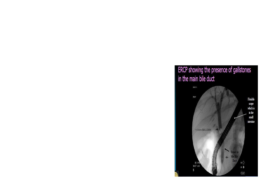
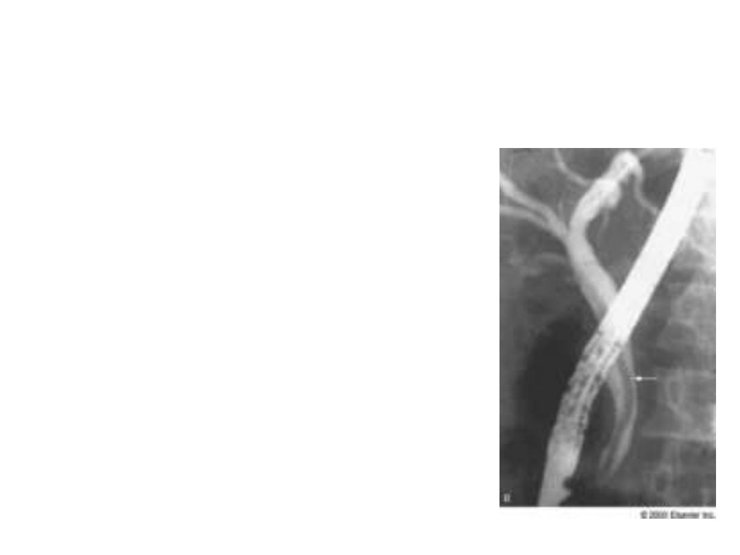
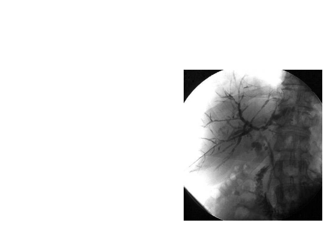
ERCP
• Diagnostic and therapeutic
• Find out obstruction especially in
the lower part of biliary passage
• Invassive
• Cannot reliabily distinguish
between benign and malignant
features
• Opportunity to take tissue sample
• Endoprosthesis
ERCP
• Diagnostic and therapeutic
• Find out obstruction especially in
the lower part of biliary passage
• Invassive
• Cannot reliabily distinguish
betweenbenign and malignant
features
• Opportunity to take tissue sample
• Endoprosthesis
PTC
• Diagnostic and therapeutic
• Best suited for leisions
proximal to the bifurcation
of hepatic duct
• Invasive
• Complications similar to
ERCP

Endoscopic Ultrasound
• Assessment bile duct and
proximal pancreatic
pathology
• Recently IDUS in ERCP
Laparoscopic cholangiography