Gall Bladder
Assist. Prof . Dr Salah aljanaby
General surgeon and laparoscopic surgeon
Babylon medical college
Lecture 2

■
There are clearly important genetic
determinants for cholesterol stone formation.
For example, the prevelance of the disease in
descendents of Chilean, Indians and in
American Indians is extraordinarily high and
not accounted for by environment.
■
There is also an important sex bias in
development of stones - the prevelance in
adult females is two to three times that seen in
males

•
Age >40 years
Bile salt loss (ileal disease or resection)
Female sex (twice risk in men)
Diabetes mellitus
Genetic or ethnic variation
Cystic fibrosis
High fat, low fibre diet
Antihyperlipidaemic drugs(clofibrate)
Obesity
Gallbladder dysmotility
Pregnancy (risk increases with number of pregnancies)
Prolonged fasting
Hyperlipidaemia
Total parenteral nutrition

Pigment Stones
■
Roughly 10% of gallstones are pigment stones
composed of large quantities of bile pigments,
along with lesser amounts of cholesterol and
calcium salts.
■
Black pigment stones
■
consist of 70% calcium bilirubinate and are more
common in patients with haemolytic diseases
(sickle cell anaemia, hereditary spherocytosis,
thalassaemia) and cirrhosis.

■
Brown pigment stones
(accounting for <5% of stones)
■
They form as a result of stasis and infection within the
biliary system, usually in the presence of Escherichia coli
and Klebsiella spp, which produce β glucuronidase that
converts soluble conjugated bilirubin back to the insoluble
unconjugated state leading to the formation of soft,
earthy, brown stones.
■
Ascaris lumbricoides and Opisthorchis senensis have
both been implicated in the formation of these stones,
which are common in South East Asia.

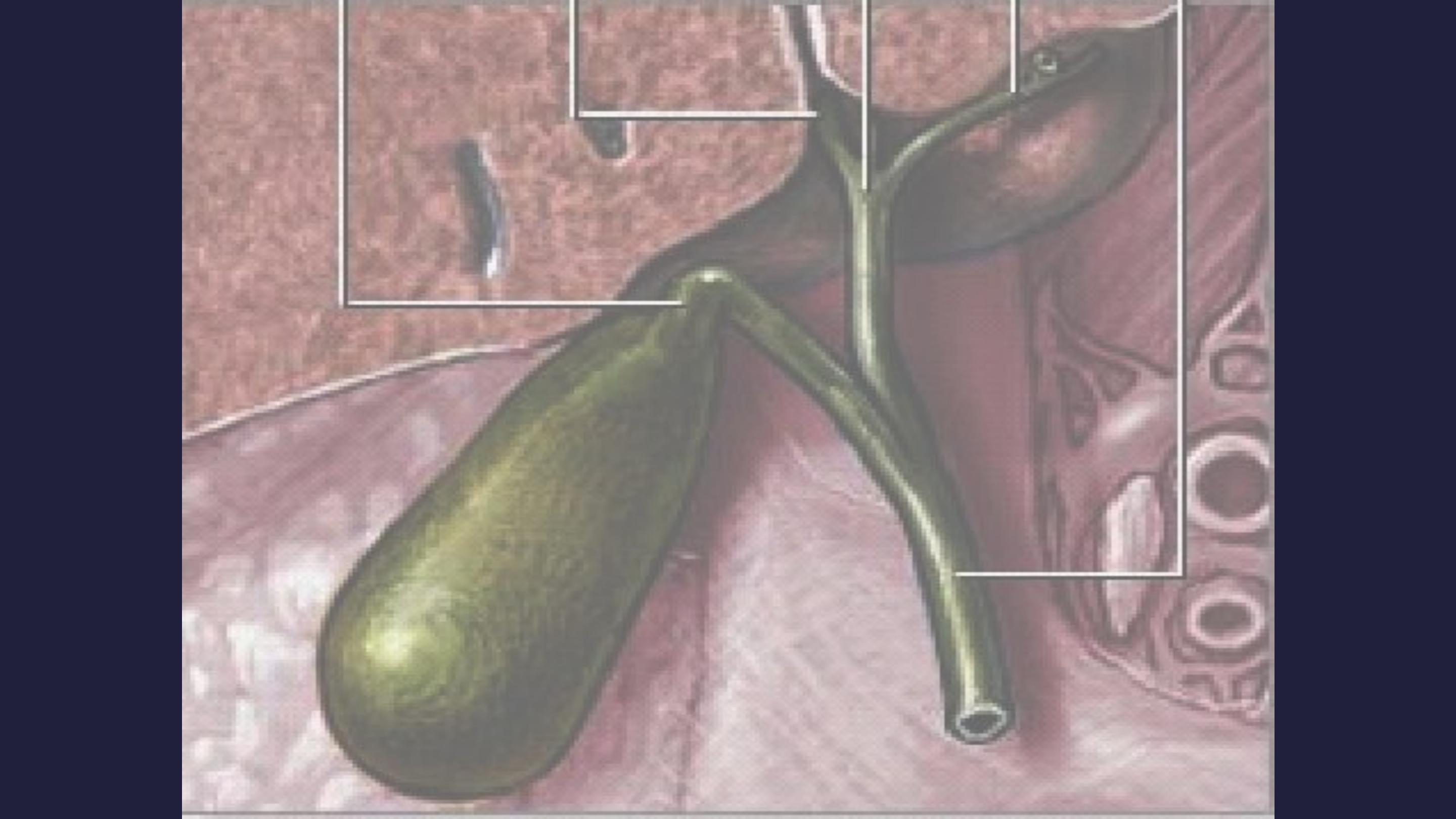
Effects and complications of Gall Stones
In the GB:
■
Silent stones
■
Chronic cholecystitis
■
Acute cholecystitis
■
Gangrene
■
Perforation
■
Empeyma
■
Mucocele
■
carcinoma
In the bile ducts:
■
Obstructive jaundice
■
Cholangitis
■
Acute pancreatitis
In the intestine:
■
Acute intestinal
obstruction (Gall stone
ileus)

Cholecystitis
Definition
■
Cholecystitis refers to a painful inflammation of the
gallbladder's wall. The disorder can occur a single
time (acute), or can recur multiple times (chronic).
■
Cholecystitis is defined as inflammation of the
gallbladder that occurs most commonly because of an
obstruction of the cystic duct from cholelithiasis.
Ninety percent of cases involve stones in the cystic
duct (ie, calculous cholecystitis), with the other 10%
representing acalculous cholecystitis. Although bile
cultures are positive for bacteria in 50-75% of cases,
bacterial proliferation may be a result of cholecystitis
and not the precipitating factor.

Causes
■
Risk factors for calculous cholecystitis mirror
those for cholelithiasis and include the
following:
■
Female sex
■
Certain ethnic groups (Race)
■
Obesity or rapid weight loss
■
Drugs (especially hormonal therapy in women)
■
Pregnancy
■
Increasing age

■
Acalculous cholecystitis is related to
conditions associated with biliary stasis, to
include the following:
■
Critical illness
■
Major surgery or severe trauma/burns
■
Sepsis
■
Long-term TPN
■
Prolonged fasting

■
Other causes of acalculous cholecystitis
include the following:
■
Cardiac events, including myocardial infarction
■
Sickle cell disease
■
Salmonella infections
■
Diabetes mellitus
■
Patients with AIDS with cytomegalovirus,
cryptosporidiosis, or microsporidiosis
■
Idiopathic cases exist.

History
■
Typical gallbladder colic is 1-5 hours of constant pain,
most commonly in the epigastrium or right upper
quadrant. Pain may radiate to the right scapular
region or back. Peritoneal irritation by direct contact
with the gallbladder localizes the pain to the right
upper quadrant. Pain is severe, dull or boring, and
constant (not colicky). Patients tend to move around
to seek relief from the pain. Onset of pain develops
hours after a meal, occurs frequently at night, and
awakens the patient from sleep.
■
Associated symptoms include nausea, vomiting,
pleuritic pain, and fever.

■
Indigestion, belching, bloating, and fatty food
intolerance are thought to be typical
symptoms of gallstones; however, these
symptoms are just as common in people
without gallstones and frequently are not
cured by cholecystectomy.

■
Most gallstones (60-80%) are asymptomatic at
a given time. Smaller stones are more likely to
be symptomatic than larger ones. Almost all
patients develop symptoms prior to
complications.
■
Symptoms of cholecystitis are steady pain in
the right hypochondrium or epigastrium,
nausea, vomiting, and fever. Acute attack
often is precipitated by a large or fatty meal.

Physical
■
Vital signs parallel the degree of illness.
Patients with cholangitis are more likely to
have fever, tachycardia, and/or hypotension.
Patients with gallbladder colic have relatively
normal vital signs.
■
Patients with cholecystitis are usually more ill
appearing than simple biliary colic patients,
and they usually lie still on the examination
table since any movement may aggravate any
peritoneal signs.

■
Abdominal examination;
■
Epigastric
or
RUQ
tenderness and abdominal
guarding.
■
The
Murphy sign
(an inspiratory pause on
palpation of the right upper quadrant) can be
found on abdominal examination.
■
Positive
Murphy sign
was extremely sensitive
(97%) and predictive (PPV, 93%) for
cholecystitis. However, in elderly patients, this
sensitivity may be decreased.

■
peritoneal signs should be taken seriously. Most
uncomplicated cholecystitis does not have peritoneal
signs; thus, search for complications (eg, perforation,
gangrene) or other sources of pain.
■
Gallbladder gangrene can be a complication in up to
20% of cases of cholecystitis and is usually in
diabetics, elderly, or immunocompromised persons.
■
A palpable fullness in the RUQ may be appreciated in
20% of cases.

■
As in all patients with abdominal pain, perform a
complete physical examination, including rectal and
pelvic examinations in women.
■
In elderly patients and those with diabetes, occult
cholecystitis or cholangitis may be the source of fever,
sepsis, or mental status changes.
■
Jaundice is unusual in the early stages of acute
cholecystitis and may be found in fewer than 20% of
patients.
■
A very high bilirubin =think for common bile duct and
pancreatic region disease.

DD
Abdominal Aortic Aneurysm
Acute Mesenteric Ischemia
Amebic Hepatic Abscesses
Appendicitis
Biliary Colic
Biliary Disease
Cholangiocarcinoma
Cholangitis
Choledocholithiasis
Cholelithiasis
Gallbladder Cancer
Gallbladder Mucocele
Gallbladder Tumors
Gastric Ulcers
Gastritis, Acute
Gastroesophageal Reflux Disease
Hepatitis, Viral
Myocardial Infarction
Nephrolithiasis
Pancreatitis, Acute
Peptic Ulcer Disease
Pneumonia, Bacterial
Pregnancy and Urolithiasis
Pyelonephritis, Acute
Renal Disease and Pregnancy
Renal Vein Thrombosis

Lab Studies
■
Labs with cholelithiasis and gallbladder colic should
be completely normal.
■
Because biliary obstruction is limited to the gallbladder
in uncomplicated cholecystitis, elevation in the serum
total bilirubin and alkaline phosphatase concentrations
may not be present.
■
An elevated WBC is expected but not reliable. Only
61% of patients with cholecystitis had a WBC greater
than 11,000. A WBC greater than 15,000 may indicate
perforation or gangrene.

■
Mild elevation of amylase up to 3 times normal may be found in
cholecystitis, especially when gangrene is present.
■
Prothrombin time (PT) and activated partial thromboplastin time
(aPTT) are not expected to be elevated unless sepsis or
underlying cirrhosis is present. Coagulation profiles are helpful if
the patient needs operative intervention.
■
For febrile patients, send 2 sets of blood cultures to attempt to
isolate the organism.
■
Although expected to be normal, urinalysis is essential in the
workup of patients with abdominal pain to exclude pyelonephritis
and renal calculi.
■
Conduct a pregnancy test for women of childbearing age.

Imaging Studies
■
Ultrasound and nuclear medicine studies are
the best imaging studies for the diagnosis of
both cholecystitis and cholelithiasis. Plain
radiography, CT scans, and endoscopic
retrograde cholangiopancreatography (ERCP)
are diagnostic adjuncts.
Abd. radiographs (Plain X-Ray)
■
Adominal radiographs have
low sensitivity and specificity
in evaluating biliary system
pathology, but
■
They can be helpful in
excluding other abdominal
pathology such as renal colic,
bowel obstruction,
perforation. Between 10 and
30% of stones have a ring of
calcium and, therefore, are
radiopaque. A porcelain
gallbladder also may be
observed on plain films.

■
Emphysematous cholecystitis, cholangitis,
cholecystic-enteric fistula, or postendoscopic
manipulation may show air in the biliary tree.
Air in the gallbladder wall indicates
emphysematous cholecystitis due to gas-
forming organisms such as clostridial species
and Escherichia coli.

Computed tomography scan
■
CT scan is recommended only for the evaluation of abdominal
pain if the diagnosis is uncertain. CT scan can demonstrate
gallbladder wall edema, pericholecystic stranding and fluid, and
high-attenuation bile.
■
Advantages: For complications of cholecystitis and cholangitis,
gallbladder perforation, pericholecystic fluid, and intrahepatic
ductal dilation, CT scan may be adequate. CT scan provides
better information of the surrounding structures than sonogram
and HIDA. CT scan is also noninvasive.
■
Disadvantages: CT scan misses 20% of gallstones because the
stones may be of the same radiographic density as bile. CT scan
is also more expensive and takes longer since the patient usually
has to drink oral contrast. Also, given the radiation dose, it may
not be ideal in the pregnant patient.