
Vermiform appendix
Lecture 2
Professor D. Mohanned Alshalah

Recurrent acute appendicitis
Appendicitis is notoriously recurrent.
The attacks vary in intensity and may occur every few months, and the
majority of cases ultimately culminate in severe acute appendicitis.
The appendix in these cases shows fibrosis indicative of previous
inflammation.
Chronic appendicitis, per se, does not exist.

Postoperative complications

Faecal fistula
Adhesive intestinal obstruction
Portal pyaemia (pylephlebitis)
Ileus
Venous thrombosis and embolism
Intra-abdominal abscess
Wound infection

Checklist for unwell patient following appendicectomy
•
■ Examine the wound and abdomen for an abscess
•
■ Consider a pelvic abscess and perform a rectal examination
•
■ Examine the lungs – pneumonitis or collapse
•
■ Examine the legs – consider venous thrombosis
•
■ Examine the conjunctivae for an icteric tinge and the liver for enlargement,
and enquire whether the patient has had rigors (pylephlebitis).
•
■ Examine the urine for organisms (pyelonephritis)
•
■ Suspect subphrenic abscess

When, the greater omentum and loops of small bowel become adherent to
the inflamed appendix, walling off the spread of peritoneal contamination,
and resulting in a phlegmonous mass or paracaecal abscess.
Management of an appendix mass

If patient’s condition is satisfactory, the standard treatment is the
conservative Ochsner–Sherren regimen.
This strategy is based on the premise that the inflammatory process
is already localised and that inadvertent surgery is difficult and may
be dangerous.
It may be impossible to find the appendix and, occasionally, a faecal
fistula may form.

Careful recording of the patient’s condition and the extent of the mass should
be made and the abdomen regularly re examined.
It is helpful to mark the limits of the mass on the abdominal wall using a skin
pencil.
Temperature and pulse rate should be recorded 4-hourly and a fluid
balance record maintained.
A contrast-enhanced CT examination of the abdomen should be performed
Antibiotic therapy instigated.

Clinical improvement is usually evident within 24–48 hours.
Using this regimen, approximately 90 per cent of cases resolve without
incident.
Failure of the mass to resolve should raise suspicion of a carcinoma or
Crohn’s disease.

Criteria for stopping conservative treatment of an appendix mass
■ A rising pulse rate
■ Increasing or spreading abdominal pain
■ Increasing size of the mass

Appendix abscess
Failure of resolution of an appendix mass or continued spiking pyrexia
usually indicates that there is pus within the phlegmonous appendix
mass.
Ultrasound or abdominal CT scan may identify an area suitable for the
insertion of a percutaneous drain.
Rarely, this is unsuccessful and laparotomy through a midline incision is
indicated.

Pelvic abscess
Pelvic abscess formation is an occasional complication of appendicitis.
The most common presentation is a spiking pyrexia several days after
appendicitis; indeed, the patient may already have been discharged from
hospital.
Pelvic pressure or discomfort associated with loose stool or tenesmus is
common.
Rectal examination reveals a boggy mass in the pelvis, anterior to the rectum,
at the level of the peritoneal reflection
Pelvic ultrasound or CT scan will confirm.
Traditionally, treatment has been through transrectal drainage under general
anaesthetic, however increasing availability of radiologically guided
percutaneous drainage has reduced the need considerably.

What is the role of interval appendectomy ?
Question

The need for interval appendicectomy in this
cohort is much debated.
The great majority of patients will not develop
recurrent appendicitis
Studies have identified higher than expected rates of
underlying appendiceal neoplasm in those patients who do
go on to interval appendicectomy, particularly those
patients over the age of 40.

Patients over the age of 40 should have colonoscopy and follow-up
imaging to ensure resolution as a small minority (less than 5 per cent) may
have an underlying appendicular or colonic malignancy.

Carcinoid tumours
Carcinoid tumours (synonym: argentaffinoma) arise in argentaffin tissue
(Kulchitsky cells of the crypts of Lieberkühn) and are most common in the
vermiform appendix.
Carcinoid tumour is found once in every 300–400 appendices subjected to
histological examination and is ten times more common than any other neoplasm
of the appendix.
Neoplasms of the appendix

In many instances, the appendix had been removed because of symptoms of
sub- acute or recurrent appendicitis.
The neoplasm on sectioning the appendix, it can be seen as a yellow tumour
between the intact mucosa and the peritoneum.
Carcinoid tumour of the appendix rarely gives rise to metastases.
Appendicectomy has been shown to be sufficient treatment, unless the caecal
wall is involved, the tumour is 2 cm or more in size or involved lymph nodes
are found, when right hemicolectomy is indicated.

Goblet cell carcinoid tumour exhibits a combination of endocrine and glandular
differentiation.
It has a more aggressive natural history and right hemicolectomy is the main
treatment
Primary adenocarcinoma of the appendix is extremely rare.
It is usually of the colonic type and should be treated by right hemicolectomy.
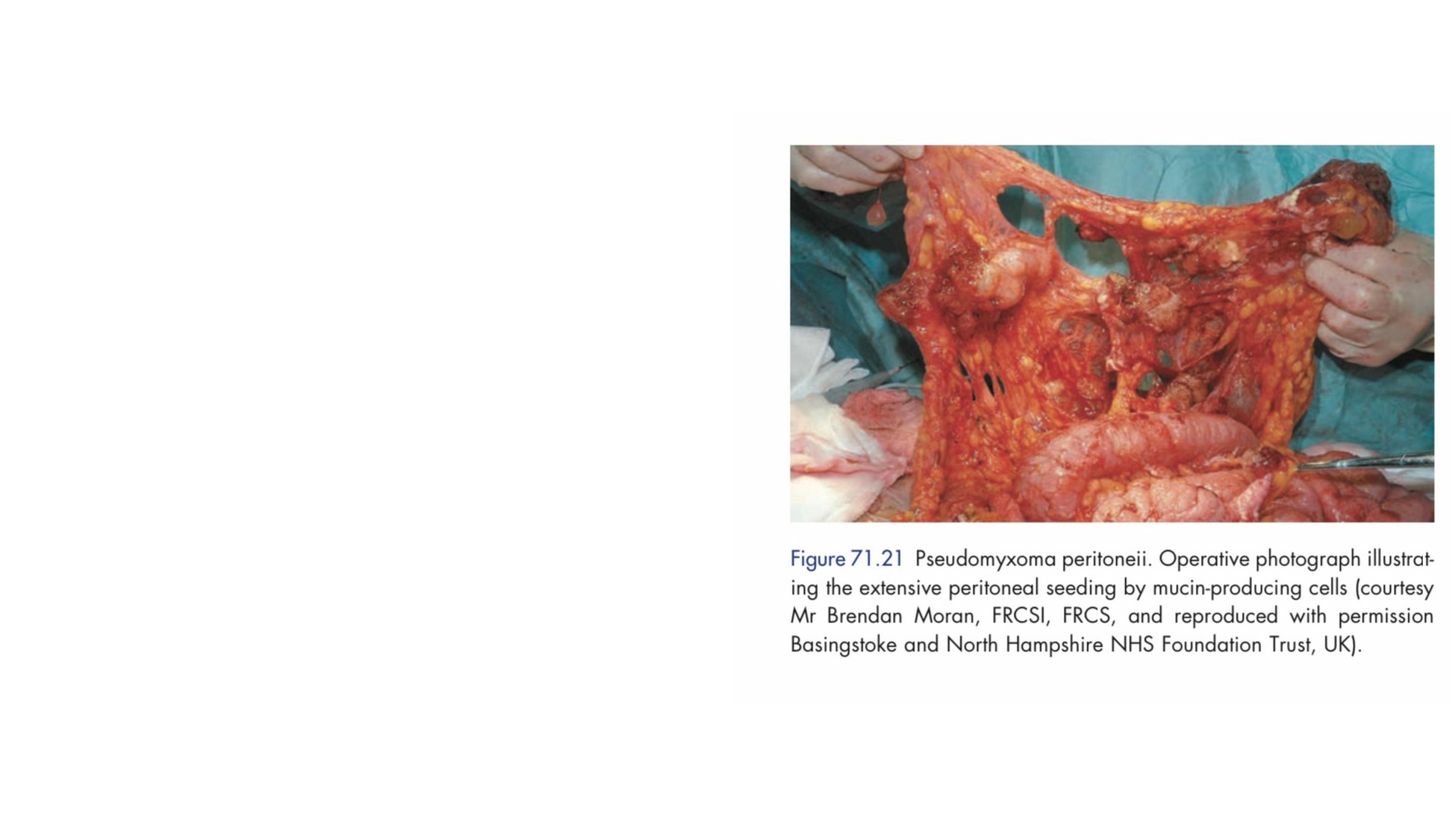
A mucin-secreting adenoma of the
appendix may rupture into the peritoneal
cavity , seeding it with mucus- secreting
cells.
Presentation is often delayed until the
patient has gross abdominal distension
as a result of pseudomyxoma peritoneii,
which may mimic ascites.
Treatment consists of radical resection of
all involved parietal peritoneal surfaces
and aggressive intraperitoneal
chemotherapy.
Mucinous cystadenoma