The development of genital tract
By Dr. Eman fathiThe development of the reproductive system It is a part of the stages of sexual differentiation. Because its location, to a large extent, overlaps the urinary system, the development of them can also be described together as the development of the urinary and reproductive organs.
The reproductive organs are developed
from the intermediate mesodermon each side of the root of the mesentery beneath the epithelium of coelom. The male and female reproductive systems follow a similar pattern of development, with sexual distinction coming about as a result of the influence of hormones.
The permanent organs of the adult are preceded by a set of structures which are purely embryonic, and which with the exception of the ducts disappear almost entirely before the end of fetal life. These embryonic structures are the Wolffian and Müllerian ducts, also known as mesonephric and paramesonephric ducts, respectively. The Wolffian duct remains as the duct in males, and the Müllerian as that of the female.
Contents
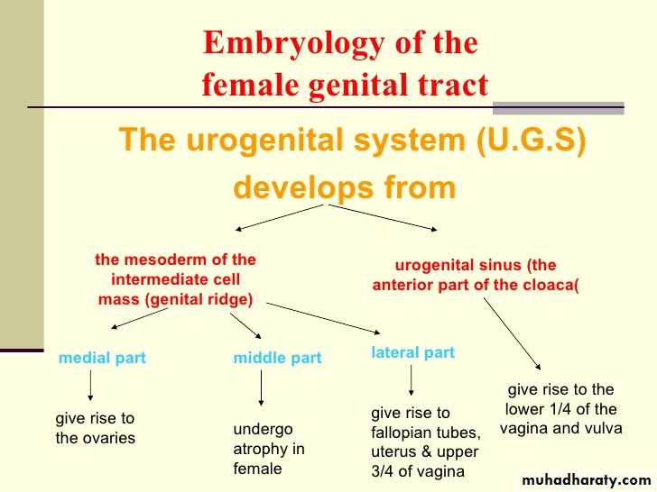
In the outer part of the intermediate mesoderm, between C5-T3 a series of short evaginations from each segment grows dorsally and extends caudally, fusing successively to form the pronephric duct. This continues to grow caudal ward until it opens into the ventral part of the cloaca; The mesonephric duct is what remains of the pronephric duct after the atrophy of the pronephros.Three main stages during development
1.Differentiation of gonad (Sex determination)
2. Differentiation of internal genital organs
3. Differentiation of external genital organs
mesonephric/paramesonephric duct changes are one of the first male/female differences that occur in development, while external genitalia remain indeterminate in appearance for quite a while
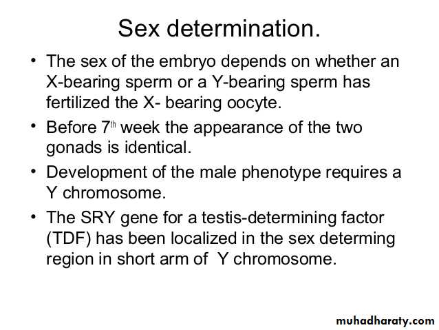
Sex determination takes place at the time of fertilization through the coupling of two gametes, either each with one X chromosome (XX in females) or such with an X and a Y chromosome (XY in males). Primarily, the male (female) phenotype is determined by the presence (or absence) of the Y chromosome with its genes, even though genes on other chromosomes are also involved

.
In addition to the(1) genetic factors, (2)hormonal regulation also plays an important role during the various developmental steps.
During the first 6 weeks the genital system is sex-indifferent and it is only then that the gonads as well as the internal and external genitalia form under hormonal influence
Function of the testis
Normal female development does not depend on gonadal hormones instead it occure due to absence of testisThe testis have 3 endocrine functions :
1.secretion of (MIF)mullerian inhibiting factor
2.secretion of testosterone which directly promotes wolffian development
3. secretion of testosteron which is converted to (DHT) dihydrotetosterone by 5αreductase
in the external genitalia and promotes its development
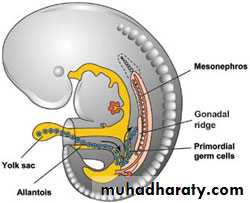
The first sign of development of reproductive organs (male or female) occurs during the fifth week with the formation of the gonadal ridge. The gonadal ridge continues to grow behind the developing peritoneal membrane lining the abdominal cavity. By the sixth week, string like masses called primary sex cords form within the enlarging gonadal ridge. In the female, the primary sex cords will contribute to nurturing tissue of developing ova
At 11th -12th weeks of gestation
9-10 weeks of gestationAt 7th week of gestation
6th week of gestation
5th week of gestation
germ cells enter the leptotene stage of prophase of the 1st meiotic division
Identity of ovary
Identity of the testis
Mass called primary sex cord form with in the genital ridge
Gonadal ridge
It will continue to grow
Development of the ovary
Development of gonads and ova
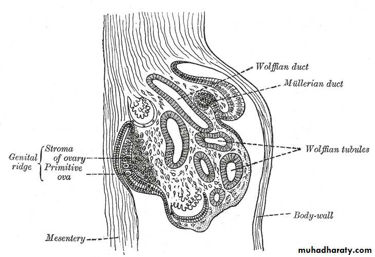
The primitive germ cells are first apparent in the endoderm ) ) the yalk sacFrom which they migrate to the gut and through the mesentery to the genital ridge
The primitive gonads consist of Germ cells thickened coelomic epithelium of genital ridge
By 5-6 weeks the epithelial element and germ cells proliferate vigorously up to 14 weeks
The identity of the gonads is apparent in the testis by 7 weeks, and 9-10 weeks in the ovary
The number of germ cells reach a maximum of (7millions) in each ovary at 15-20 weeks , falling to 2 million at birth , and 400 000 by puberty
At 11-12 weeks the germ cells enter the leptotene stage of prophase of the 1st meiotic division . They remain in the prophase until ovulation occur
Early , ovary is attached to the inguinal fold and uterus by gubernaculum along which it descend to its definitive site
The cranial part of gubernaculum become the ovarian ligament
And the caudal part become the round ligament
Development of uterus and fallopian tubes
At 5-6 weeks the paramesonephric (mullerian) duct develop at the lateral aspect of the mesonephros , it extend caudaly to reach the urogenital sinus at 9 weeks.The wolffian develops before the mullerian and gives out a pouch , the ureteric diverticulum before entering the urogenital sinus and form a separate opening , that’s why the wolffian duct contribute to the ureter and trigon of the bladder.
at 8 weeks both mullerian and wolfian are present , at this point sex differentiation will begin , wolffian system degenerate due to lack of testosterone .
The lower portion of mullerian duct fuse to form uterus and cervix
Separate as the fallopian tubes while the upper portion
Development of uterus and tubes
fifth monthAt 3rd month
• At 9 weeks
at 8 weeks
5 -6 weeks of gestation
a ring-like constriction marks the position of the cervix of the uterus, and after the sixth month the walls of the uterus begin to thicken
Fusion of mullerian duct fuse to form uterus and cervix
Separate as the fallopian tubes while the upper portion
Mullerian extend caudaly to reach the urogenital sinus
Both wolffian and mullerian are present . From this point onwords wolffian disppear due to absence of tetosterone and amullerian will continue
(mullerian) duct develop at the lateral aspect of the mesonephros
Development of the vagina
Paired sinu vaginal bulbs on the posterior aspect of the urogenital sinus fuse with the lower end of the mullerian duct to form the vaginal plate . At 1st the vagina consist of solid mass which grows rapildy and become canalized at 16-18 weeks . In the mature vagina the upper 4/5 (four fifth) derived from the mullerian duct and the lower fifth develop from the urogenital sinusFormation of the vestibule
The urogenital sinus has 3 portions the1.upper portion which will form the bladder
2. pelvic portion form the urethra
3. the lower(phalic portion) which will become increasingly shallow to form the vestibule in female and penile urethra in male.
The genital tubercle form the clitoris and the genital fold form the labia minora , the genital swelling become the labia majora
Male
FemalePrimitive structure
Testis
Ovary
Un differentiated gonads
Appendix testis and prostetic utricleUterus , tubes , and upper 4/5 of the vagina
Mullerian system (paramesonephric)
Epidydimis vas deference , trigone of the bllader , prostetic urethra above the ejaculatory ductOccasional remnant part of the bladder and ureter (
Wolffian system (mesonephric
Penis
ClitorisGenital tubercle
• Penile urethra
Labia minora• Genital fold
Scrotum and bulbo-urethral glands
labia majora and bartholines gland
Genital swelling