
Chronic obstructive pulmonary disease 2
Dr.Ahmed Hussein Jasim F.I.B.M.S (respiratory)
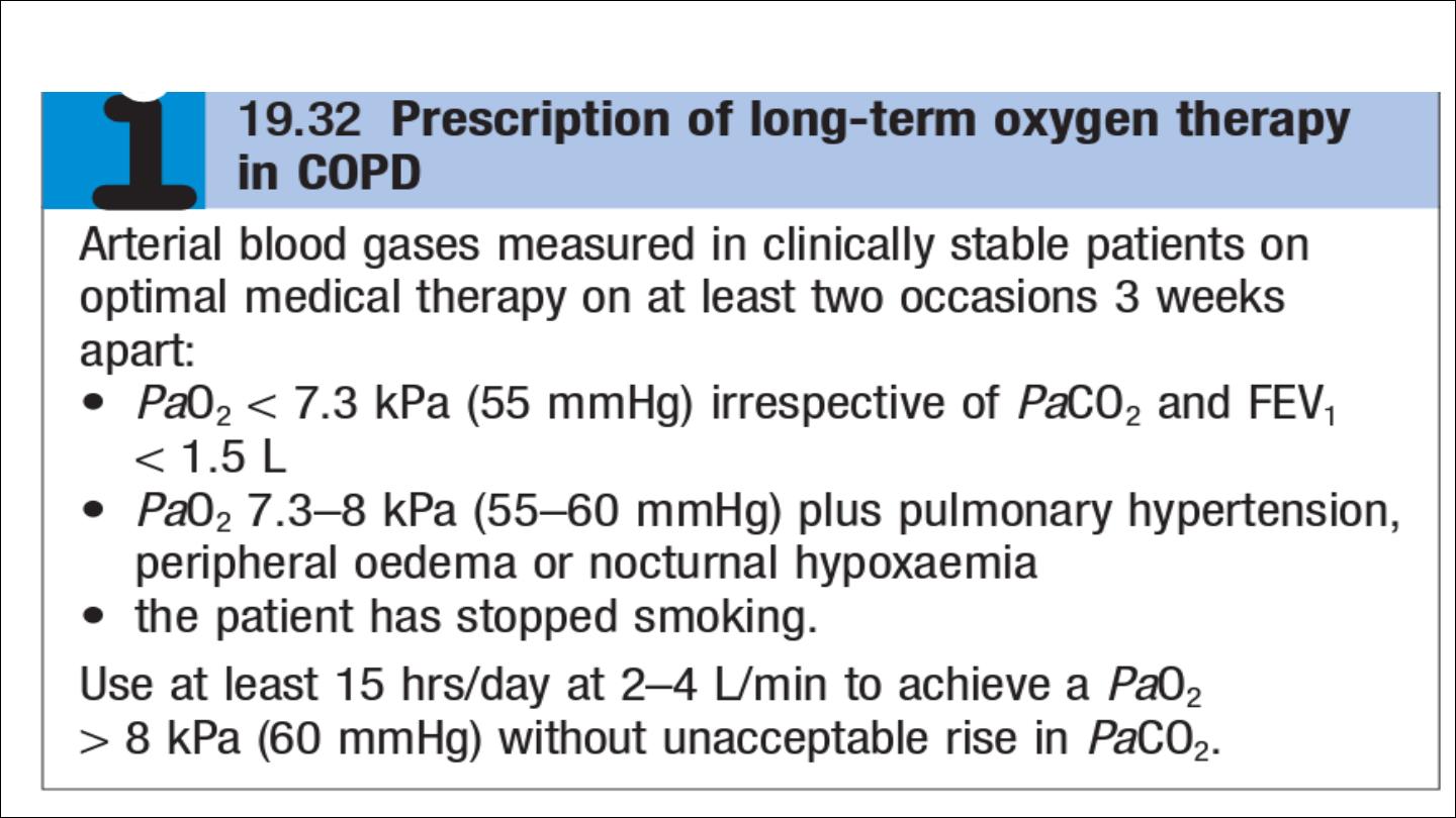
Oxygen therapy

Surgical intervention
Patients in whom large bullae compress surrounding normal lung tissue,
who otherwise have minimal airflow limitation and a lack of generalised
emphysema, may be considered for
bullectomy.
Patients with predominantly upper lobe emphysema, with preserved gas
transfer and no evidence of pulmonary hypertension, may benefit from
lung volume reduction surgery (LVRS).
Lung transplantation
may benefit carefully selected patients with advanced
disease .
Other measures
Patients with COPD should be offered an annual influenza vaccination
and, as appropriate, pneumococcal vaccination.

Prognosis
The prognosis is inversely related to age and directly related to the post-
bronchodilator FEV1
poor
prognostic
indicators
include
weight
loss
and
pulmonary
hypertension
. A composite score comprising the body mass index (B), the
degree of airflow obstruction (O), a measurement of dyspnoea (D) and
exercise capacity (E) may assist in predicting death from respiratory and
other causes
Respiratory failure, cardiac disease and lung cancer represent common
modes of death.
Smoking cessation is the only intervention that is proven to
decrease the smoking-related decline in lung function

Acute exacerbations of COPD
Acute exacerbations of COPD are characterised by an increase in
symptoms and deterioration in lung function and health status.
They become more frequent as the disease progresses and are usually
triggered by bacteria, viruses or a change in air quality.
They may be accompanied by the development of respiratory failure and/
or fluid retention and are an important cause of death.
Many patients can be managed at home with the use of increased
bronchodilator therapy, a short course of oral corticosteroids and, if
appropriate, antibiotics.

The presence of cyanosis, peripheral oedema or an alteration in
consciousness indicates the need for referral to hospital.
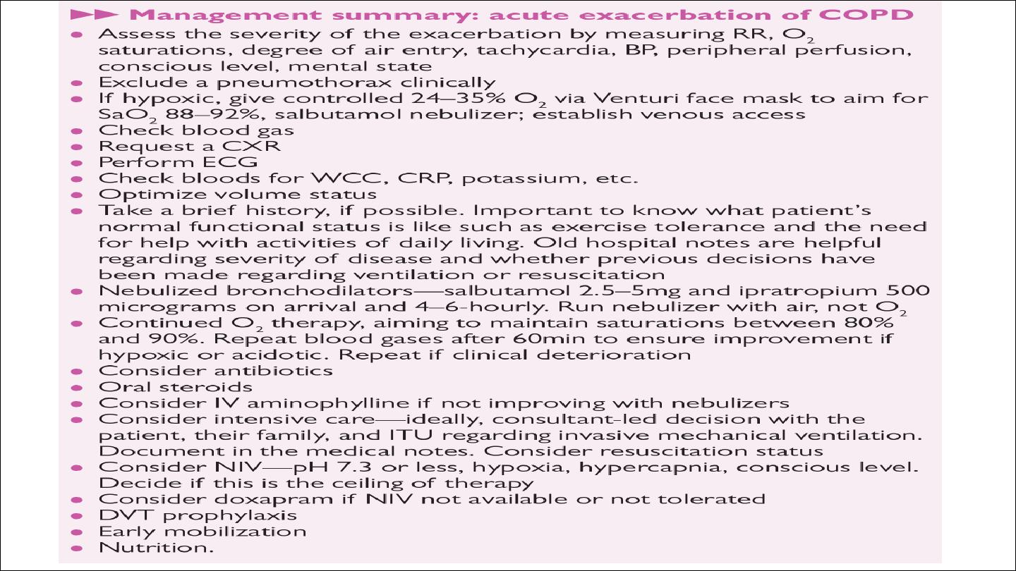
Management
Controlled oxygen at 24% or 28% should be used with the aim of
maintaining a PaO2 above 8 kPa (60 mmHg) (or an SaO2 between 88%
and 92%) without worsening acidosis.
Nebulised short-acting β2-agonists, combined with an anticholinergic
agent (e.g. salbutamol and ipratropium), should be administered.

Oral prednisolone reduces symptoms and improves lung function. Currently,
doses of 30 mg for 10 days are recommended but shorter courses may be
acceptable.
the routine administration of antibiotics They are currently recommended,
however, for patients reporting an increase in sputum purulence, sputum
volume or breathlessness.
In most cases, simple regimens are advised, such
as an aminopenicillin or a macrolide. Co-amoxiclav is only required in
regions where β-lactamase-producing organisms are known to be common.
Exacerbations may be accompanied by the development of peripheral
oedema that usually responds to diuretics.
The use of the respiratory stimulant
doxapram
has been largely
superseded by the development of NIV, but it may be useful for a limited
period in selected patients with a low respiratory rate.

If the patient remains tachypnoeic, hypercapnic and acidotic (PaCO2 > 6 kPa,
H+ ≥ 45 (pH < 7.35)), then NIV should be commenced.Its use is associated with
reduced requirements for mechanical ventilation and reduced mortality.
Mechanical ventilation may be considered in those with a reversible cause for
deterioration (e.g. pneumonia), or if PH <7.25.
Thromboprevention by subcutaneous given heparin or LMWH.

Bronchiectasis
Bronchiectasis means abnormal dilatation of the bronchi.
Chronic suppurative airway infection with sputum production, progressive
scarring and lung damage occur.
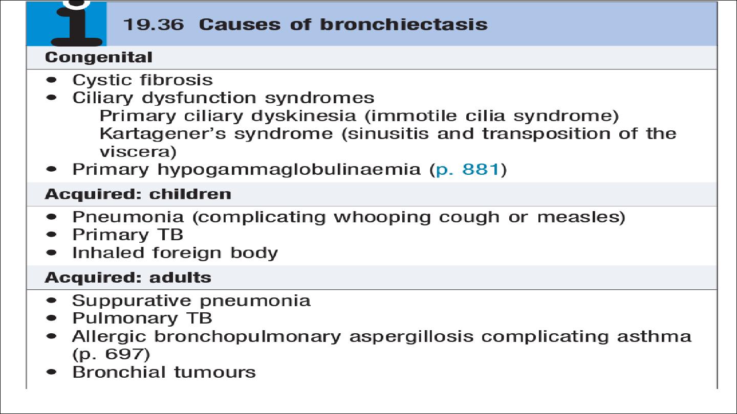
Aetiology and pathology
Tuberculosis is the most common worldwide.
Localized bronchiectasis may occur due to the accumulation of pus beyond an
obstructing bronchial lesion, such as enlarged tuberculous hilar lymph nodes, a
bronchial tumor or an inhaled foreign body (e.g. an aspirated peanut).
The bronchiectatic cavities may be lined by granulation tissue, squamous epithelium
or normal ciliated epithelium. There may also be inflammatory changes in the deeper
layers of the bronchial wall and hypertrophy of the bronchial arteries. Chronic
inflammatory and fibrotic changes are usually found in the surrounding lung tissue,
resulting in progressive destruction of the normal lung architecture in advanced cases.
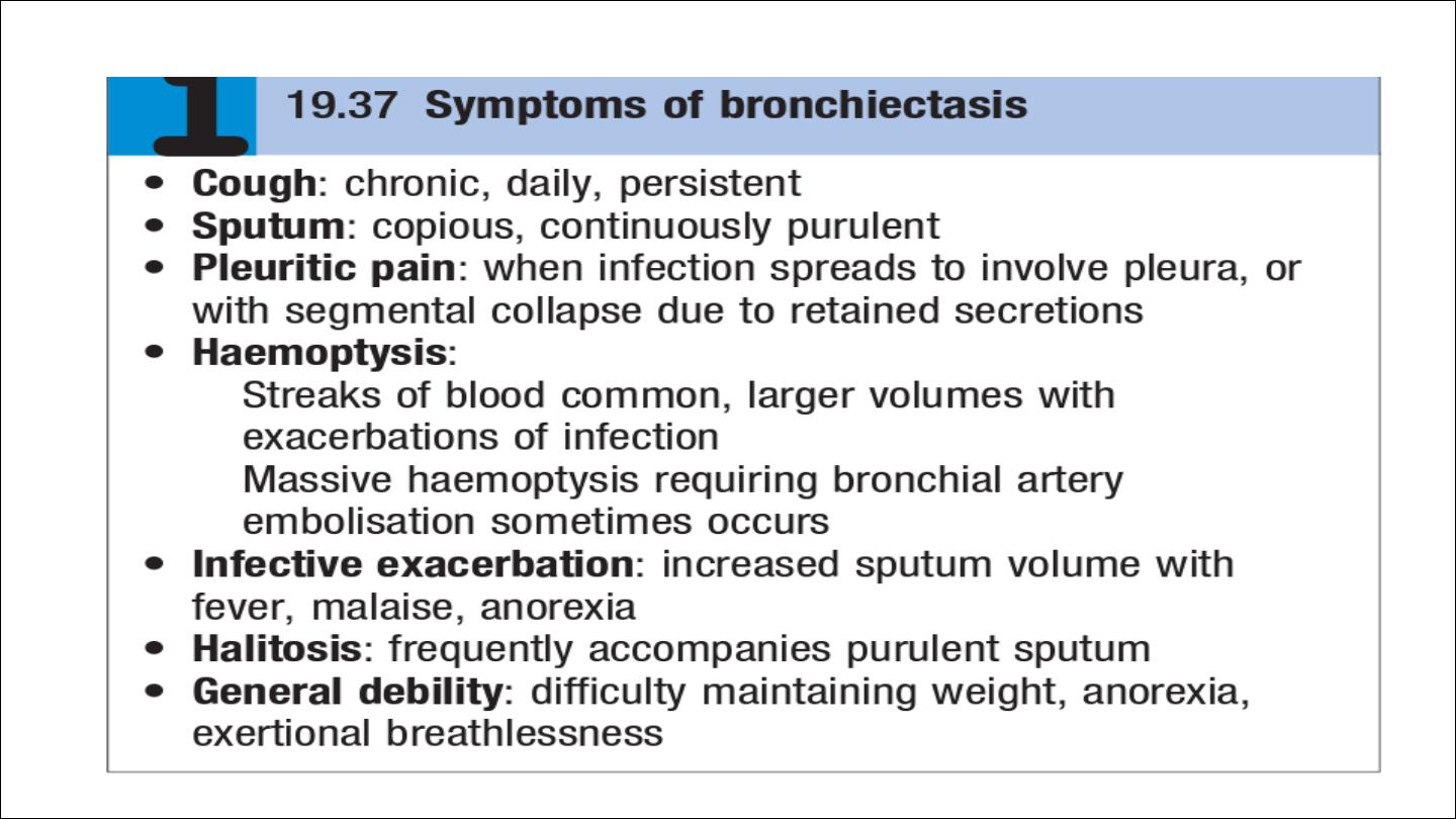
Clinical features

Investigations
common respiratory pathogens, sputum culture may reveal Pseudomonas
aeruginosa, fungi such as Aspergillus and various mycobacteria.
Frequent cultures are necessary to ensure appropriate treatment of
resistant organisms.
chest X-ray thickened airway walls, cystic bronchiectatic spaces, and
associated areas of pneumonic consolidation or collapse may be visible.
CT is much more sensitive, and shows thickened, dilated airways
A screening test can be performed in patients suspected of having a
ciliary dysfunction syndrome by measuring the time taken for a small
pellet of saccharin placed in the anterior chamber of the nose to reach
the pharynx, at which point the patient can taste it. This time should not
exceed 20 minutes but is greatly prolonged in patients with ciliary
dysfunction.

Ciliary beat frequency may also be assessed from biopsies taken from the nose.
Structural abnormalities of cilia can be detected by electron microscopy
Management
Physiotherapy
Deep breathing followed by forced expiratory manœuvres (the ‘active cycle of breathing’
technique) helps to move secretions in the dilated bronchi towards the trachea, from
which they can be cleared by vigorous coughing. Devices that increase airway pressure
either by a constant amount (positive expiratory pressure mask) or in an oscillatory
manner (flutter valve)
Antibiotic therapy
For most patients with bronchiectasis, the appropriate antibiotics are the same as those
used in COPD
Surgical treatment
Excision of bronchiectatic areas is only indicated in a small proportion of cases. These
are usually patients in whom the bronchiectasis is confined to a single lobe or segment
on CT.