
Tuberculosis
(TB)
Tikrit University
College of Medicine
Department of Radiology
Chest Series

Tuberculosis (TB)
• TB caused by Mycobacterium tuberculosis.
• Remains an important disease & significant
problem in developing countries.
• Initial TB - 1
st
exposure:
• 1) Contained disease
• 2) Primary tuberculosis
• Reactivation (post-primary) TB
• Healed TB
• Miliary TB

Initial TB - 1
st
exposure:
• Initial exposure to TB can lead to two
clinical outcomes:
1) Contained disease (90%)
occur in a
patient with normal immunity, results in:
– calcified granulomas and/or
– calcified hilar lymph nodes.
2) Primary tuberculosis
seen more commonly
in
children
and
immunocompromised
patients.
Results when the host cannot contain the
organism.

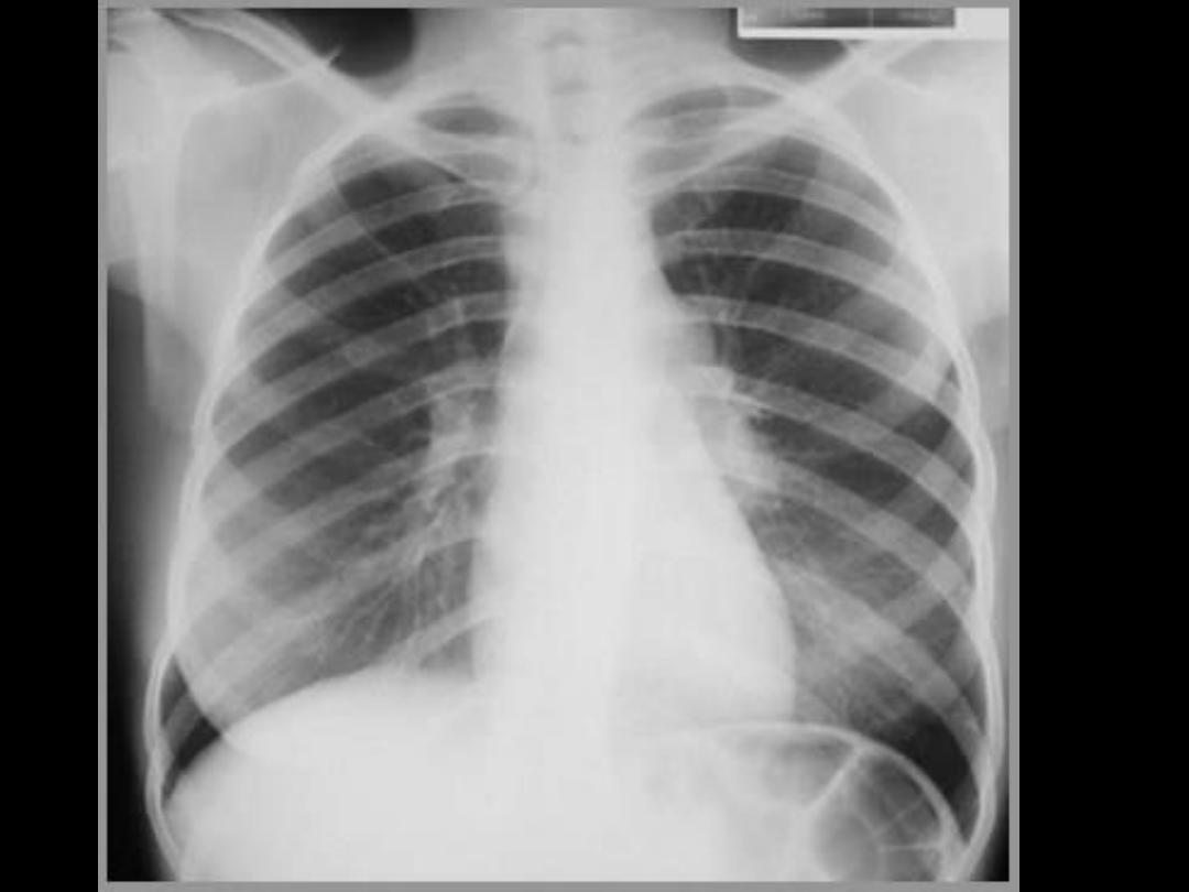
Primary tuberculosis
• Primary tuberculosis represents infection
from the first exposure to TB.
• Primary TB may involve the pulmonary
parenchyma, the airways, and the pleura.
Primary TB often causes adenopathy.
• As many as 15% of patients infected with
primary TB have
no radiographic changes
and the imaging appearance of primary
tuberculosis is
nonspecific
.

• Four imaging manifestations of primary TB (any, none,
or all of them may be seen):
– Ill-defied consolidation
– Lymphadenopathy
: common in primary TB.
– pleural effusion
– Ghon focus
: complex small focal lesion with focal
calcification.
– Miliary disease.
– Cavitation is rare in primary TB
• Primary TB may occur in any lobe, but the most typical
locations are the lower lobes or right middle lobe.
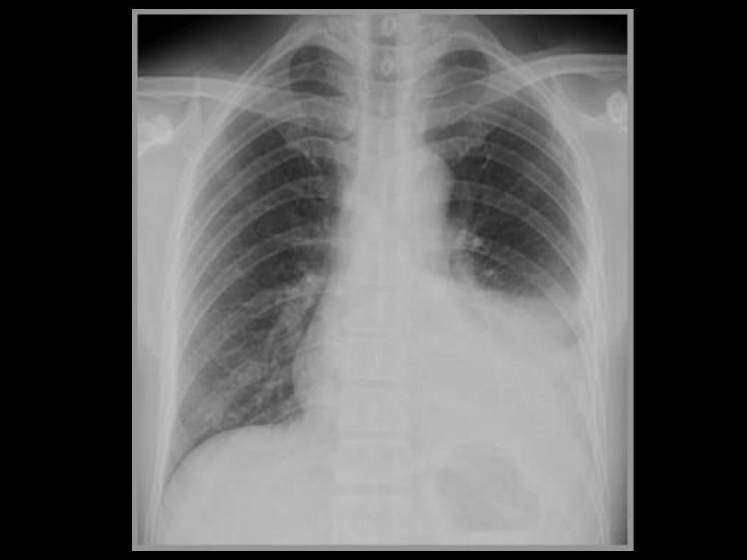

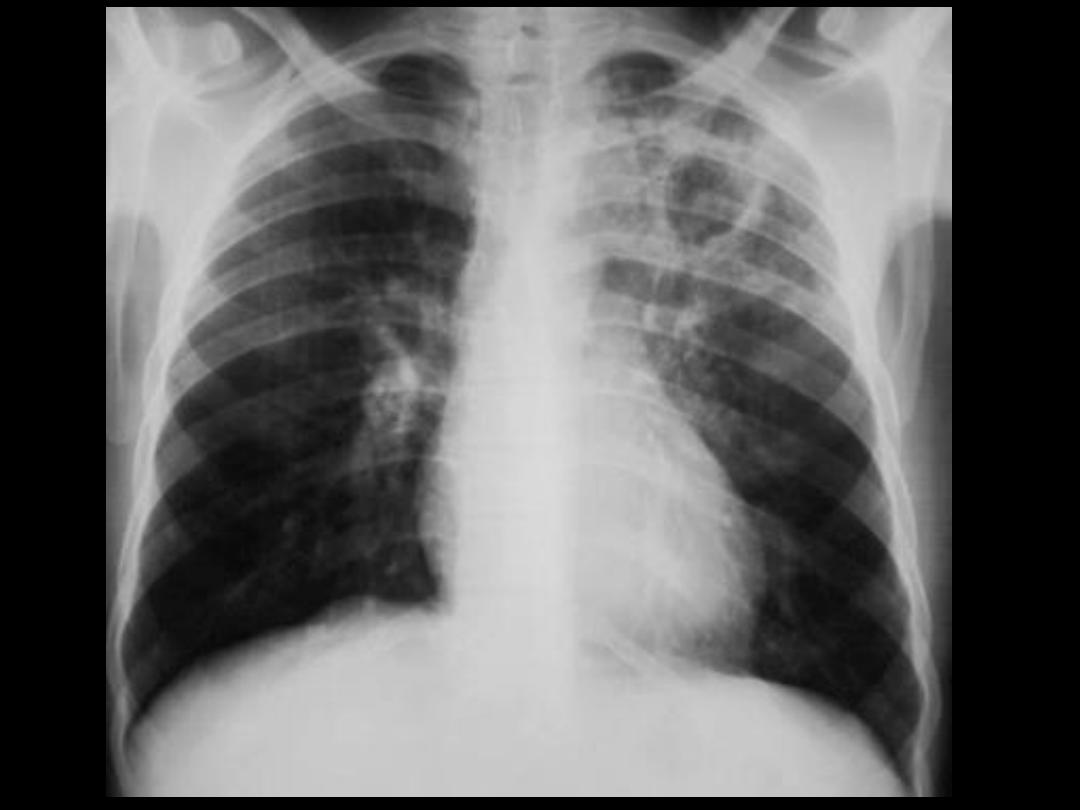
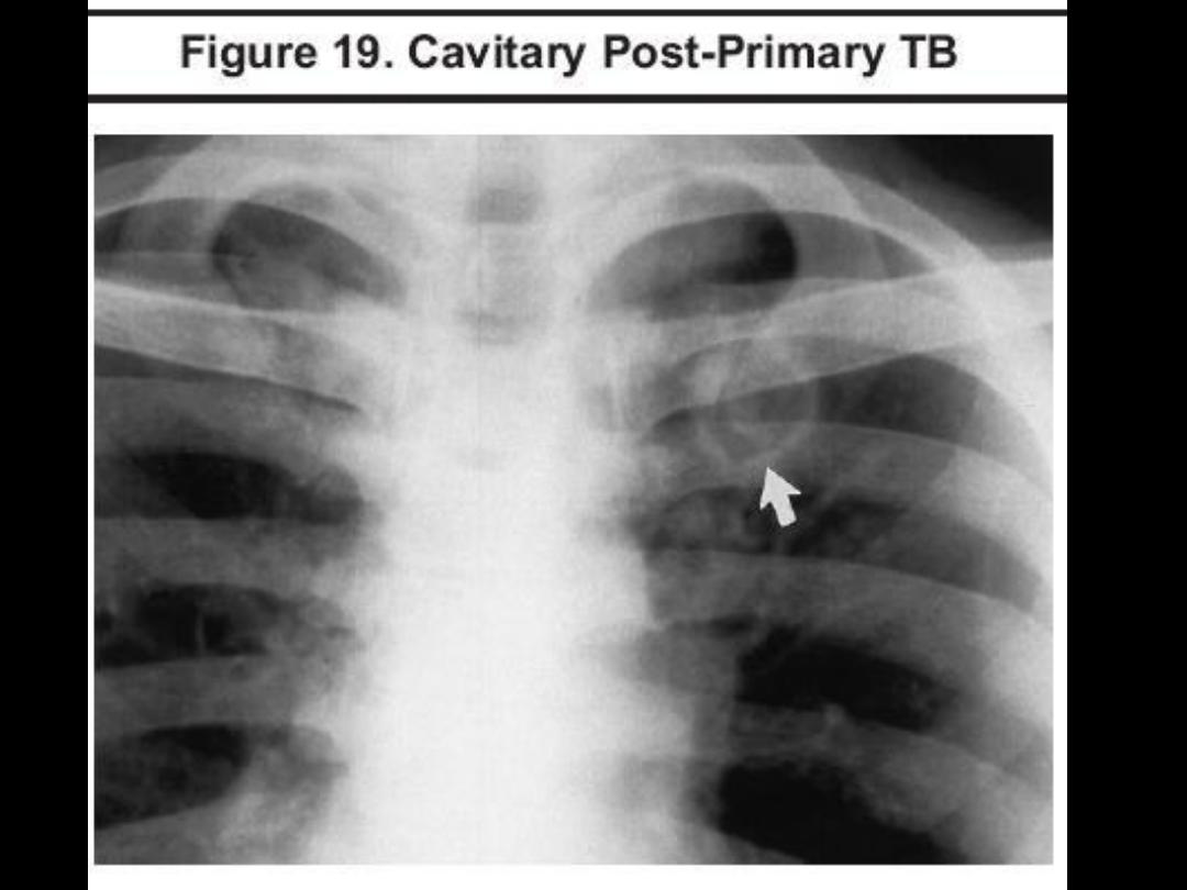
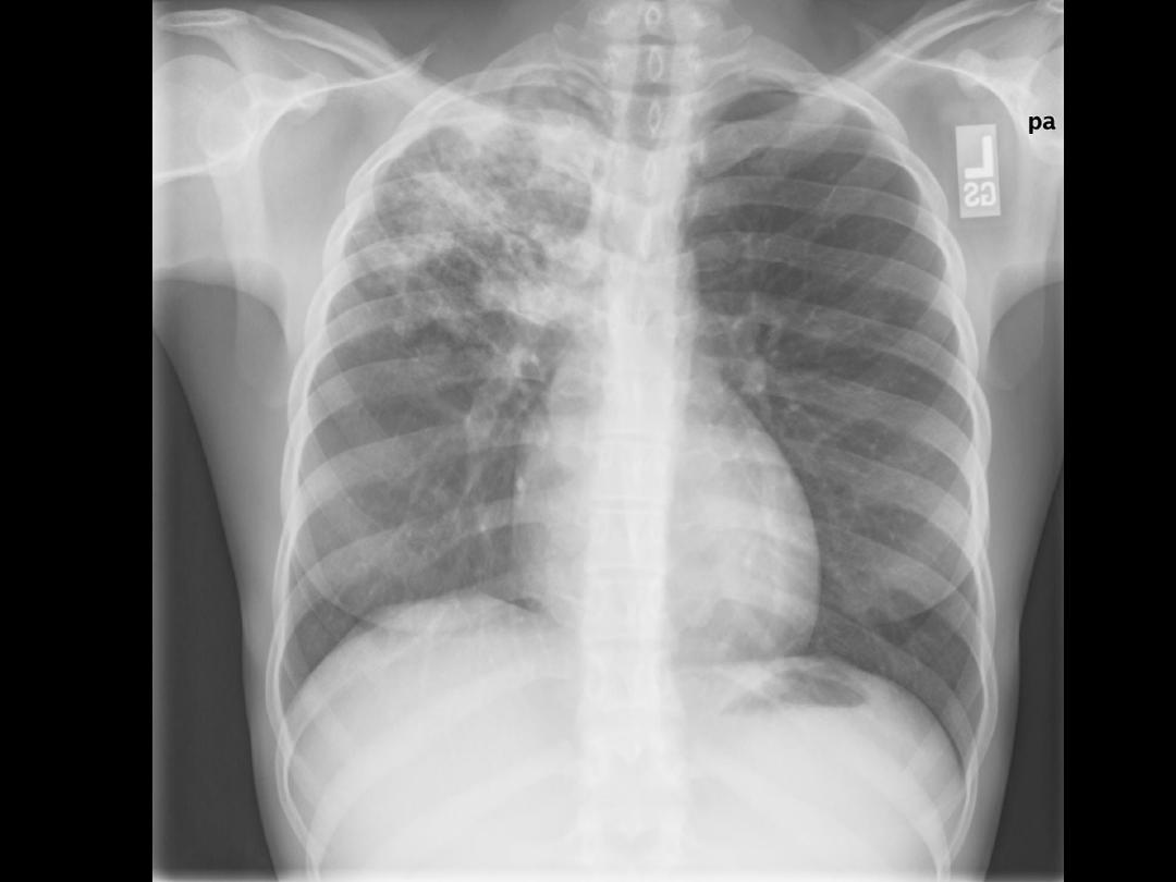
Reactivation (post-primary) TB
• Usually occurs in adolescents and adults and
is caused by reactivation of a dormant
infection acquired earlier in life.
• Clinical manifestations of reactiatin TB
include:
– chronic cough
– low-grade fever
– hemoptysis, and
– night sweats.
• Reactiatin TB most commonly occurs in the
upper lobe
apical
and
posterior segments

Reactivation (post-primary) TB
• In an immunocompetent patient, the
imaging hallmarks of reactivation TB are:
– Focal upper lobe consolidation
– Cavitation
– No lymphadenopathy.
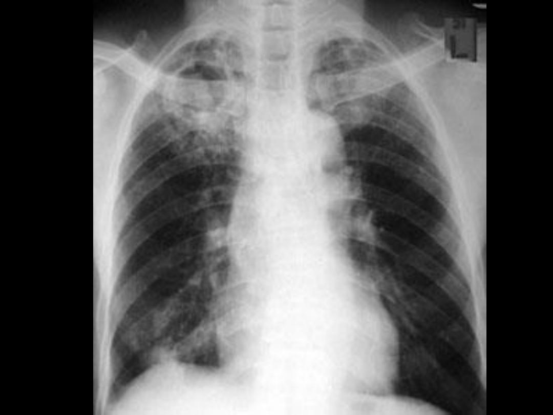

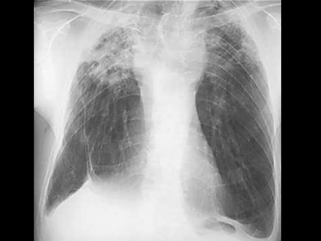
Healed tuberculosis
• Healed TB is evident on radiography as:
• apical scarring, usually with upper
lobe volume loss
• superior hilar retraction.

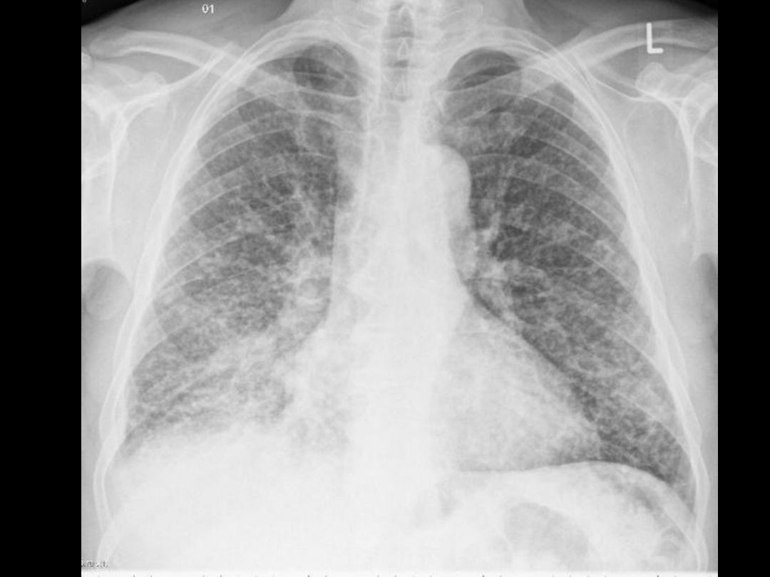
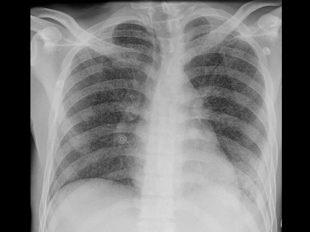
Miliary tuberculosis
• Miliary tuberculosis is a diffuse random
distribution of tiny nodules seen in
hematogenously disseminated TB.
• Miliary TB can occur in
primary
or
reactivation TB
.

DDx of Miliary Shadow
• Miliary TB
• Silicosis
• Coal worker pneumoconiosis
• Sarcoidosis
• Metastasis
