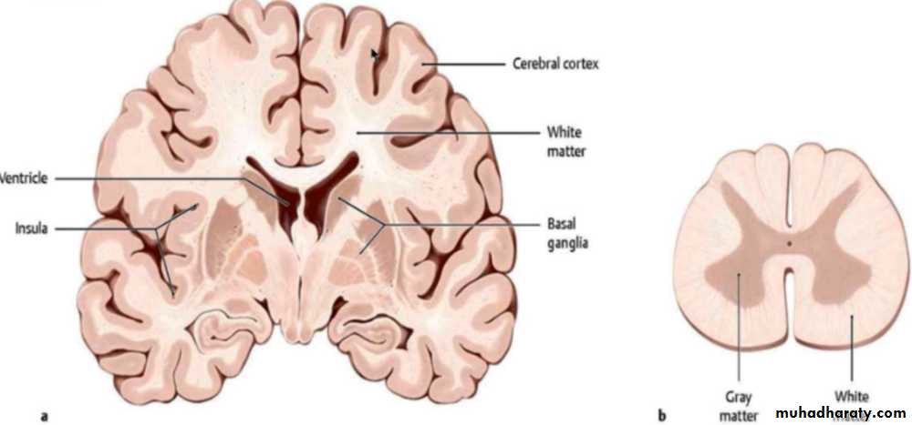
CENTRAL NERVOUS SYSTEM The major regions of the central nervous system (CNS) are the cerebrum, cerebellum, and spinal cord, the CNS is covered by three connective tissue layers, the meninges, but contains very little collagen or fibrous tissue throughout its substance, making it relatively soft and easily damaged by injuries affecting its protective cranium or vertebral bones, The entire CNS displays organized areas of white matter and gray matter, differences caused by the differential distribution of myelin, the main components of white matter are myelinated axons , often grouped together as tracts, and the myelin-producing oligodendrocytes, White matter contains very few neuronal cell bodies, but astrocytes and microglia are present, gray matter contains abundant neuronal cell bodies, dendrites, the initial unmyelinated portions of axons, astrocytes, and microglial cells. Gray matter occupies the thick surface or cortex of both the cerebrum and the cerebellum; most white matter is found in deeper regions. Deep regions of the CNS also have darker aggregates called nuclei consisting of large numbers of neuronal cell bodies and surrounded by white matter.
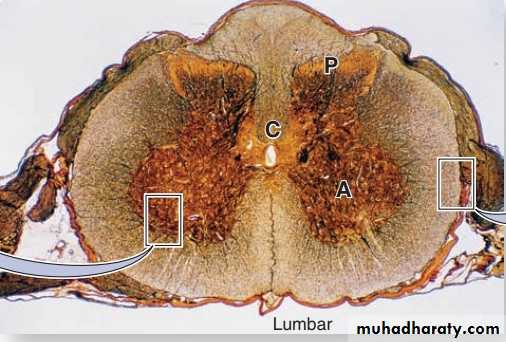
In cross sections of the spinal cord, white matter is peripheral and gray matter is internal and has the general shape of the letter H, in the center is an opening, the central canal which develops from the lumen of the embryonic neural tube. The canal is continuous with the ventricles of the brain, contains CSF, and is lined by ependymal cells. The gray matter forms the anterior horns, which contain motor neurons whose axons make up the ventral roots of spinal nerves, and the posterior horns, which receive sensory fibers from neurons in the spinal (dorsal root) ganglia. Spinal cord neurons are large and multipolar, especially the motor neurons in the anterior horns
Meninges The skull and the vertebral column protect the CNS, but between the bone and nervous tissue are membranes of connective tissue called the meninges. Three meningeal layers are distinguished: the dura, arachnoid, and pia maters.
I- Dura Mater: The thick external dura mater (L., dura mater, tough mother) consists of dense, fibro-elastic connective tissue that is continuous with the periosteum of the skull. The dura mater is always separated from the arachnoid by the thin subdural space. The internal surface of all dura mater, as well as its external surface in the spinal cord, is covered by simple squamous epithelium of mesenchymal origin.
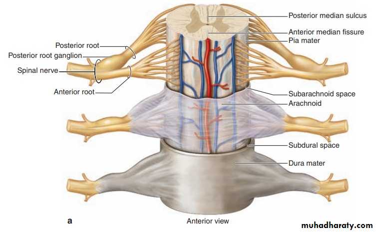
II-Arachnoid The arachnoid (Gr. arachnoeides, spiderweblike) has two components: (1) a sheet of connective tissue in contact with the dura mater and (2) a system of loosely arranged trabeculae composed of collagen and fibroblasts, continuous with the underlying pia mater layer. Surrounding the trabeculae is a large, sponge-like cavity, the subarachnoid space, filled with CSF This fluid-filled space helps cushion and protect the CNS from minor trauma. The subarachnoid space communicates with the ventricles of the brain where the CSF is produced. In some areas, the arachnoid penetrates the dura mater and protrudes into blood-filled venous sinuses located within that layer. These CSF-filled protrusions are called arachnoid villi, which function as a site for absorption of CSF into the blood of the venous sinuses.
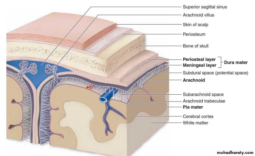
III- Pia Mater The innermost pia mater (L., pia mater, tender mother) consists of flattened, mesenchymally derived cells closely applied to the entire surface of the CNS tissue. The pia does not directly contact nerve cells or fibers, being separated from the neural elements by the very thin superficial layer of astrocytic processes, which adheres firmly to the pia mater. Together, the pia mater and the layer of astrocytic end feet form a physical barrier separating CNS tissue from CSF in the subarachnoid space.
PERIPHERAL NERVOUS SYSTEM The main components of the peripheral nervous system (PNS) are the nerves, ganglia, and nerve endings. Nerves are bundles of nerve fibers (axons) surrounded by Schwann cells and layers of connective tissue.
Nerve Fibers: Nerve fibers are analogous to tracts in the CNS, containing axons enclosed within sheaths of glial cells specialized to facilitate axonal function. In peripheral nerve fibers, axons are sheathed by Schwann cells, the sheath may or may not form myelin around the axons, depending on their diameter.
Ganglia Ganglia are typically ovoid structures containing neuronal cell bodies and their surrounding glial satellite cells supported by delicate connective tissue and surrounded by a denser capsule. Because they serve as relay stations to transmit nerve impulses, at least one nerve enters and another exits from each ganglion. The direction of the nerve impulse determines whether the ganglion will be a sensory or an autonomic ganglion.
Sensory Ganglia : Sensory ganglia receive afferent impulses that go to the CNS. Sensory ganglia are associated with both cranial nerves (cranial ganglia) and the dorsal roots of the spinal nerves (spinal ganglia). The large neuronal cell bodies of ganglia are associated with thin, sheetlike extensions of small glial satellite cells. Sensory ganglia are supported by a distinct connective tissue capsule and an internal framework continuous with the connective tissue layers of the nerves. The neurons of these ganglia are pseudounipolar and relay information from the ganglion’s nerve endings to the gray matter of the spinal cord via synapses with local neurons.
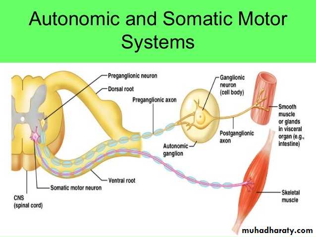
Autonomic Ganglia : Autonomic nerves effect the activity of smooth muscle, the secretion of some glands, heart rate, and many other involuntary activities by which the body maintains a constant internal environment (homeostasis). Autonomic ganglia are small bulbous dilations in autonomic nerves, usually with multipolar neurons. Some are located within certain organs, especially in the walls of the digestive tract, where they constitute the intramural ganglia. The capsules of these ganglia may be poorly defined among the local connective tissue. A layer of satellite cells also envelops the neurons of autonomic ganglia, although these may also be inconspicuous in intramural ganglia. Autonomic nerves use two-neuron circuits. The first neuron of the chain, with the preganglionic fiber, is located in the CNS. Its axon forms a synapse with postganglionic fibers of the second multipolar neuron in the chain located in a peripheral ganglion system
As indicated earlier autonomic nerves make up the autonomic nervous system. This has two parts:
the sympathetic and
the parasympathetic divisions. Neuronal cell bodies of preganglionic sympathetic nerves are located in the thoracic and lumbar segments of the spinal cord and those of the parasympathetic division are in the medulla and midbrain and in the sacral portion of the spinal cord. Sympathetic second neurons are located in small ganglia along the vertebral column, while second neurons of the parasympathetic series are found in very small ganglia always located near or within the effector organs, for example in the walls of the stomach and intestines. Parasympathetic ganglia may lack distinct capsules .