NERVOUS TISSUE
INTRODUCTIONThe nervous tissue is composed of interconnecting network of specialized cells called neurons (nerve cells) supported by neuroglial cells. There are about 10 million neurons in human beings. The function of neurons is to receive stimuli and conduct them to a central site, the central nervous system (CNS), where they are analyzed and integrated to produce a desired response in the effector organs.
CLASSIFICATION OF NEURONS
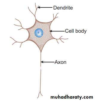
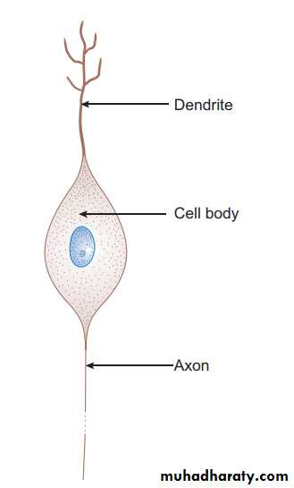
A. Morphological (based on the number of processes) 1. Unipolar neuron—has a single process (rare). 2.Bipolar neuron—has two processes (an axon and a dendrite). 3. Multipolar neuron—has many processes (an axon and many dendrites) 4. Pseudo-unipolar neuron—has a single process that divides into an axon (central process) and a dendrite
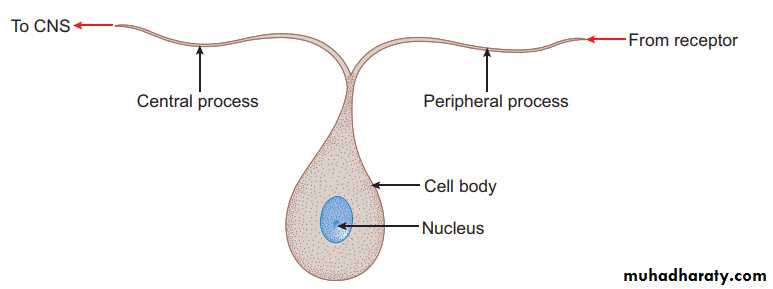
B. Functional (based on the function performed) 1. Sensory neuron—receives stimuli from receptors and conducts impulses to CNS, e.g. sensory ganglia. 2. Motor neuron—conducts impulses from CNS to effector organs (muscles), e.g. ventral horn cells. 3. Interneuron—connects sensory and motor neurons and completes the functional circuit.
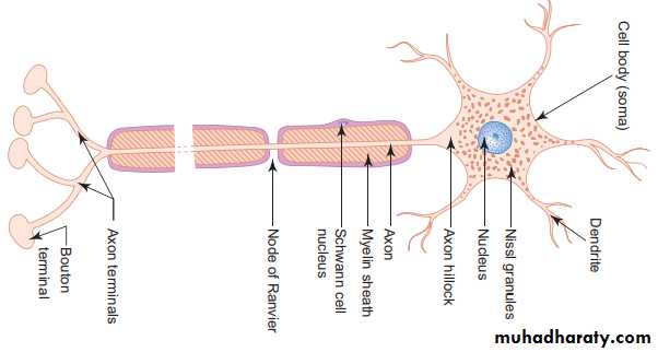
STRUCTURE OF A NEURON (MULTIPOLAR) I- Cell body/Soma/Perikaryon (5–150 nm): The cell bodies of all neurons are situated in the grey matter of the CNS and in the ganglia of PNS. The cell body of a neuron contains the nucleus and the following cytoplasmic organelles and inclusions : 1. Nucleus—is large, euchromatic, spherical and centrally located. 2. Nissl bodies or Nissl substance—are composed of large aggregations of rough endoplasmic reticulum – are observed as basophilic clumps by light microscopy – extend into dendrites but not into axon and axon hillock – disintegrate as a result of injury to axon (chromatolysis). 3. Golgi complex—are found near the nucleus. 4.Mitochondria—are numerous and rod shaped. 5. Neuro-filaments (10 nm dia) and microtubules (25 nm dia)—form neuronal cytoskeleton providing structural support and intracellular transport. 6. Melanin pigments—dark brown granules. 7. Lipofuscin pigments—residual bodies not digested by lysosomes (increase with age).
Structure of multipolar neuron
Ultrastructure of neuron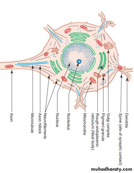
II- Dendrites - Are highly branched, tapering processes of a neuron. So their diameter is not uniform. - Are covered by horny spines (gemmules) which are sites of synaptic contact. -Receive stimuli from sensory cells and other neurons and transmit them towards the soma. So they can be regarded as major sites of information input into neuron.
III- Axon - Single, long, cylindrical process of a neuron. So its diameter is uniform. - Arises from a cone-shaped portion of the cell body called axon hillock, which is devoid of Nissl bodies. - Terminates by dividing into many small branches, axon terminals, ending in small swellings—terminal buttons. --Conducts impulses away from the cell body to the axon terminals from which impulses are transmitted to another neuron or another target cell. -Axons are commonly referred to as nerve fibers. - Are often surrounded by myelin sheath, which is derived either from Schwann cells (PNS) or oligodendrocytes (CNS). -When an axon is cut, peripheral part degenerates. ---Regeneration of the axon is possible only when the cell body of the neuron is intact. -Neurons do not regenerate in the event of cell body death, i.e. they do not multiply.
Myelinated and Unmyelinated Axons
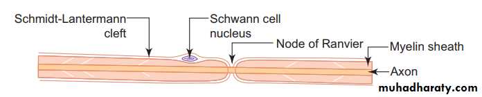
-In the PNS, all axons are enveloped by Schwann cells which provide both structural and metabolic support. -Many axons with small diameter invaginate into one Schwann cell longitudinally and are simply surrounded by the cytoplasm of Schwann cells. ---They are called unmyelinated nerve fibers. -Other axons, especially the ones with larger diameter, invaginate into the Schwann cell and are wrapped by concentric layers of the Schwann cell plasma membrane forming myelin sheath. -These axons are called myelinated nerve fibers. - There are gaps (areas of axon not covered by myelin) along the length of myelin sheath at regular intervals called nodes of Ranvier. - In large myelinated axons, the nerve impulse jumps from node to node resulting in faster conduction (saltatory conduction). -The segment of myelin between two nodes of Ranvier is called internode. -The myelin of one internode is formed by a single Schwann cell.
A myelinated peripheral nerve fiber
Myelination - In the PNS, the myelin sheath of an individual axon is provided by many Schwann cells lying along the length of the axon.GLIAL CELLS & NEURONAL ACTIVITY Neuroglial cells support neuronal survival and activities, and are ten times more abundant in the mammalian brain than the neurons, like neurons, most glial cells develop from progenitor cells of the embryonic neural plate, in the CNS glial cells surround both the neuronal cell bodies, which are often larger than glial cells, and the processes of axons and dendrites occupying the spaces between neurons, except around the larger blood vessels, the CNS has only a very small amount of connective tissue and collagen. Glial cells substitute for cells of connective tissue in some respects, supporting neurons and creating a microenvironment immediately around those cells that is optimal for neuronal activity. The fibrous intercellular network surrounding cells of the CNS may superficially resemble collagen with light microscopy, but it is actually the network of cellular processes emerging from neurons and glial cells. Such processes are collectively called the neuropil
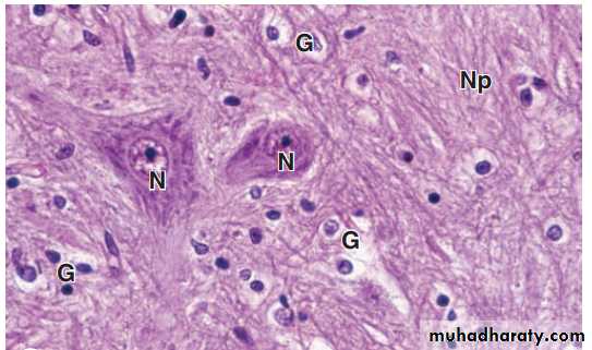
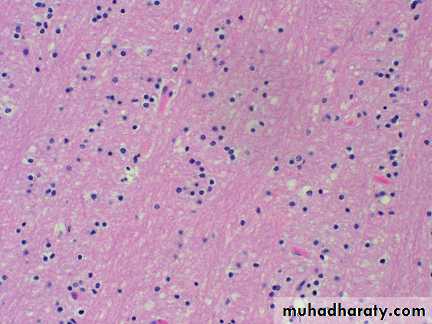
Neuronal tissue stained by routine hematoxylin and eosin stain
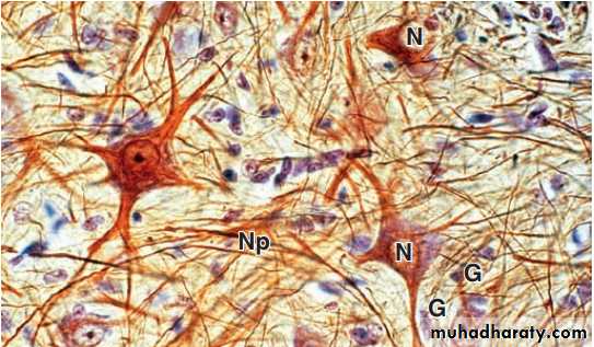
Neuronal tissue stained by gold chloride and hematoxylin
There are six kinds of glial cells:I-Oligodendrocytes:
(oligo it means small, dendro means tree) produce the myelin sheaths around axons that provide the electrical insulation for neurons in the CNS. Oligodendrocytes extend sheetlike processes that wrap around parts of several axons, producing myelin sheaths, these are the predominant glial cells in CNS white matter, which is white because of the lipid concentrated in the wrapped membrane sheaths. The processes and sheaths are not visible by routine light microscope staining, in which oligodendrocytes usually appear as small cells with rounded, condensed nuclei and unstained cytoplasm .
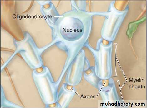
Oligodendrocytes
II-Astrocytes Astrocytes ( astron it means star ) have a large number of radiating processes and are also unique to the CNS, astrocytes are by far the most numerous glial cells of the CNS, as well as the most diverse structurally and functionally, Those with relatively few long processes are called fibrous astrocytes and are typical in white matter; those with many shorter, branched processes are called protoplasmic astrocytes and predominate in the gray matter, terminal branching of astrocytic processes is very extensive, allowing a single astrocyte to associate with over a million synaptic sites. The larger processes of all astrocytes are reinforced with bundles of intermediate filaments .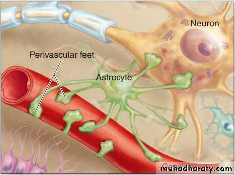
Astrocytes
III: Ependymal Cells:
Ependymal cells are columnar or cuboidal cells that line the ventricles of the brain and central canal of the spinal cord, in some CNS locations, the apical ends of ependymal cells have cilia, which facilitate the movement of cerebrospinal fluid (CSF), and long microvilli, which are likely involved in absorption, ependymal cells are joined apically by junctional complexes similar to those of epithelial cells. However, unlike a true epithelium there is no basal lamina. Instead, the basal ends of ependymal cells are elongated and extend branching processes into the adjacent neuropil.
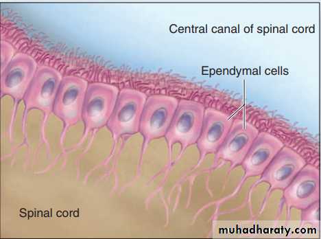
Ependymal cells
IV: Microglia Less numerous than oligodendrocytes or astrocytes but nearly as common as neurons, microglia are small cells with short irregular processes evenly distributed throughout gray and white matter, Unlike other glial cells, microglia migrate through the neuropil, scanning the tissue for damaged cells and invading microorganisms, they secrete a number of immunoregulatory cytokines and constitute the major mechanism of immune defense in the CNS, microglia do not originate from neural progenitor cells like other glia, but from circulating blood monocytes, belonging to the same family as macrophages and other antigen-presenting cells, nuclei of microglial cells can be recognized in routine hematoxylin and eosin (H&E) preparations by their small, dense, elongated structure, which contrasts with the larger, spherical, more lightly stained nuclei of other glial cells.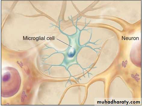
Microglia
V: Schwann Cells : Schwann cells sometimes called neurolemmocytes, are found only in the PNS and differentiate from precursors in the neural crest, schwann cells have trophic interactions with axons and importantly allow for their myelination, like the oligodendrocytes of the CNS, one Schwann cell forms myelin around a segment of one axon, in contrast to the ability of oligodendrocytes to branch and ensheath parts of more than one axon.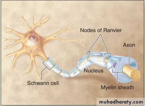
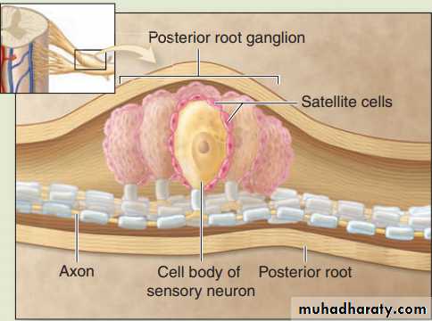
VI: Satellite Cells of ganglia : Also derived from the embryonic neural crest, small satellite cells form an intimate covering layer over the large neuronal cell bodies in the ganglia of the PNS. Satellite cells exert a trophic or supportive effect on these neurons, insulating, nourishing, and regulating their microenvironments.