Done by
Dr.Rafid Remthan Al-Temimi
Clinical Radiology
CAMB,DMRD,M.B.Ch.B.,.
المرحلة
:
الثانية
المادة
:
التشريح
ج
امعة ذي قار
/
كلية الطب
الدكتور
رافد
رمثان التميمي
Veins of the Brain
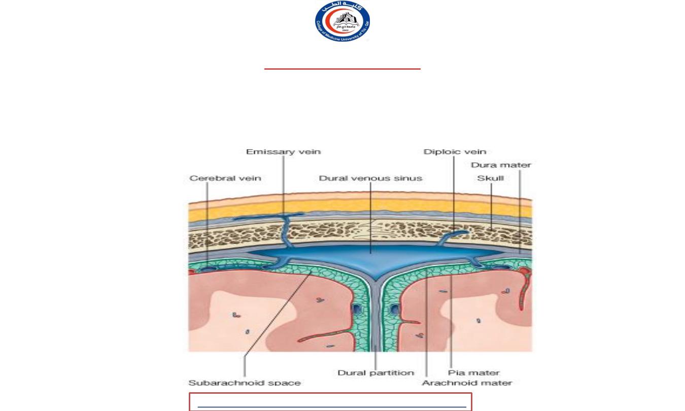
• They have no muscular tissue in their walls, and they have no valves. They lie in subarachnoid
space. They drain into the
cranial (dural) venous sinuses.
• Emissary veins
connect
the
dural venous sinuses
with the
diploic veins
of the skull and with
the
veins of the scalp
University Of Thi-Qar
College Of medicine
2020
,
Radiology,CAMB
Temimi,Clinical
-
AL
emthan
R
Dr.Rafid
Anatomy lecture . 2
nd
stage
Dr.Rafid Al-Temimi
2
University Of Thi-Qar
College Of medicine
2020
,
Radiology,CAMB
Temimi,Clinical
-
AL
emthan
R
Dr.Rafid
Anatomy lecture . 2
nd
stage
Dr.Rafid Al-Temimi
3
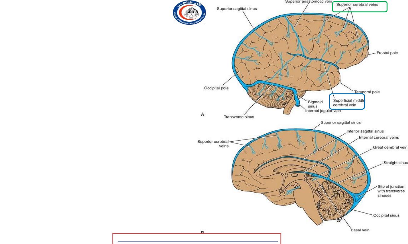
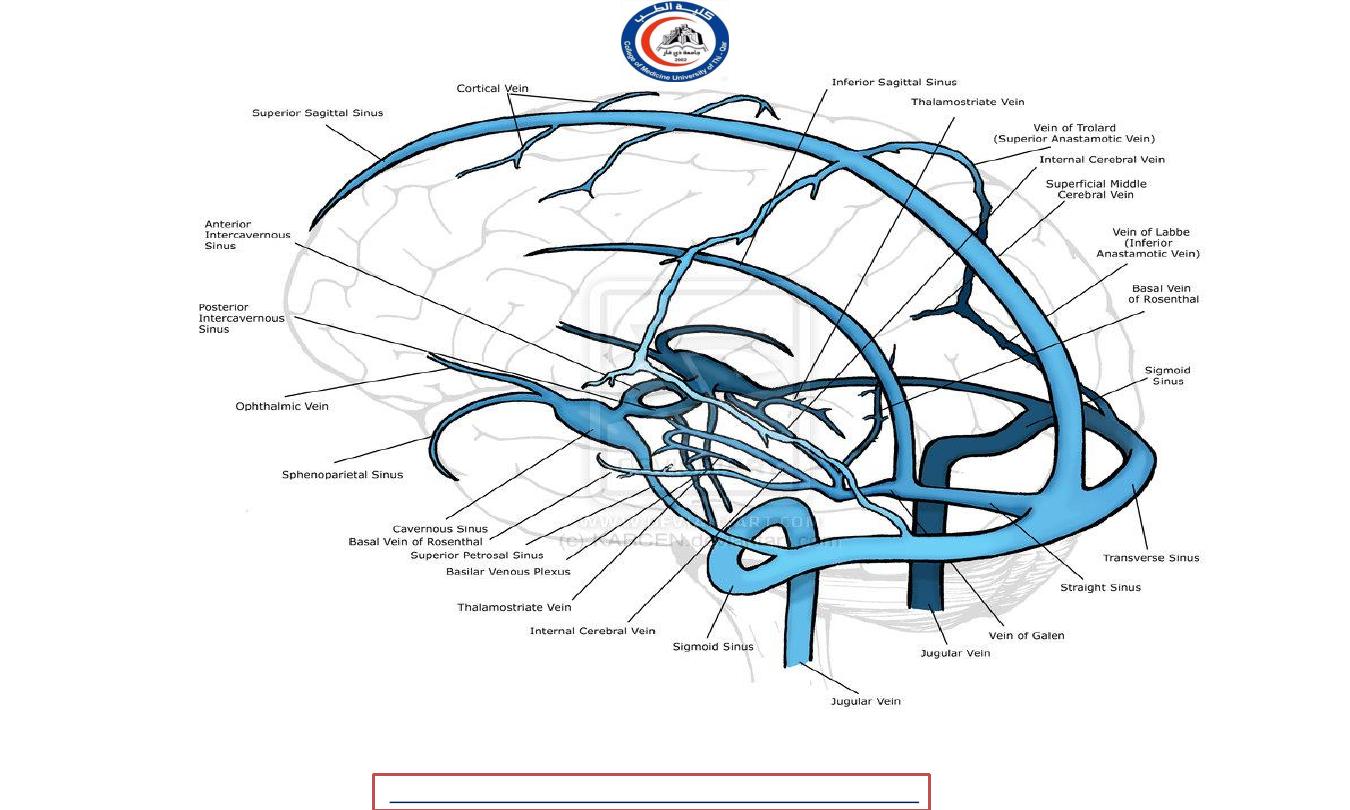
• Cerebral veins divided into:
External Cerebral Veins:
• Superior cerebral veins
drains
superolateral & medial surfaces
and empty into the
superior
sagittal sinus.
• Superficial middle cerebral vein
drains the lateral surface of the
cerebral hemisphere & empties
into the
cavernous sinus
.
2020
,
Radiology,CAMB
Temimi,Clinical
-
AL
emthan
R
Dr.Rafid
Anatomy lecture . 2
nd
stage
Dr.Rafid Al-Temimi
4
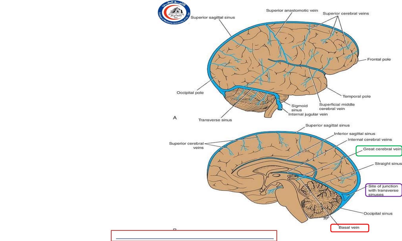
• Deep middle cerebral vein
drains insula and is joined by
anterior cerebral
and
striate
veins
to form the
basal vein
.
• Basal vein
joins the
great
cerebral vein
, which in turn
drains into
straight sinus
.
2020
,
Radiology,CAMB
Temimi,Clinical
-
AL
emthan
R
Dr.Rafid
Anatomy lecture . 2
nd
stage
Dr.Rafid Al-Temimi
5
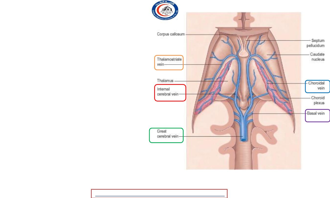
Internal Cerebral Veins
• There are two
internal
cerebral veins.
• They are formed by union of
the
thalamostriate vein
and
choroid vein
• The two unite to form the
great cerebral vein
, which is
joined with
basal veins
&
empties into the
straight
sinus.
University Of Thi-Qar
College Of medicine
2020
,
Radiology,CAMB
Temimi,Clinical
-
AL
emthan
R
Dr.Rafid
Anatomy lecture . 2
nd
stage
Dr.Rafid Al-Temimi
6
Veins of specific brain areas:
• Midbrain
is drained by veins that
open into the
basal
or
great
cerebral
veins.
• Pons
is drained by veins that
open into
basal
vein,
cerebellar
veins.
• Medulla oblongata
is drained by
veins that open into
spinal
veins.
• Cerebellum is drained by veins
that empty into
great cerebral
vein or adjacent
venous sinuses
.
University Of Thi-Qar
College Of medicine
2020
,
Radiology,CAMB
Temimi,Clinical
-
AL
emthan
R
Dr.Rafid
Anatomy lecture . 2
nd
stage
Dr.Rafid Al-Temimi
7
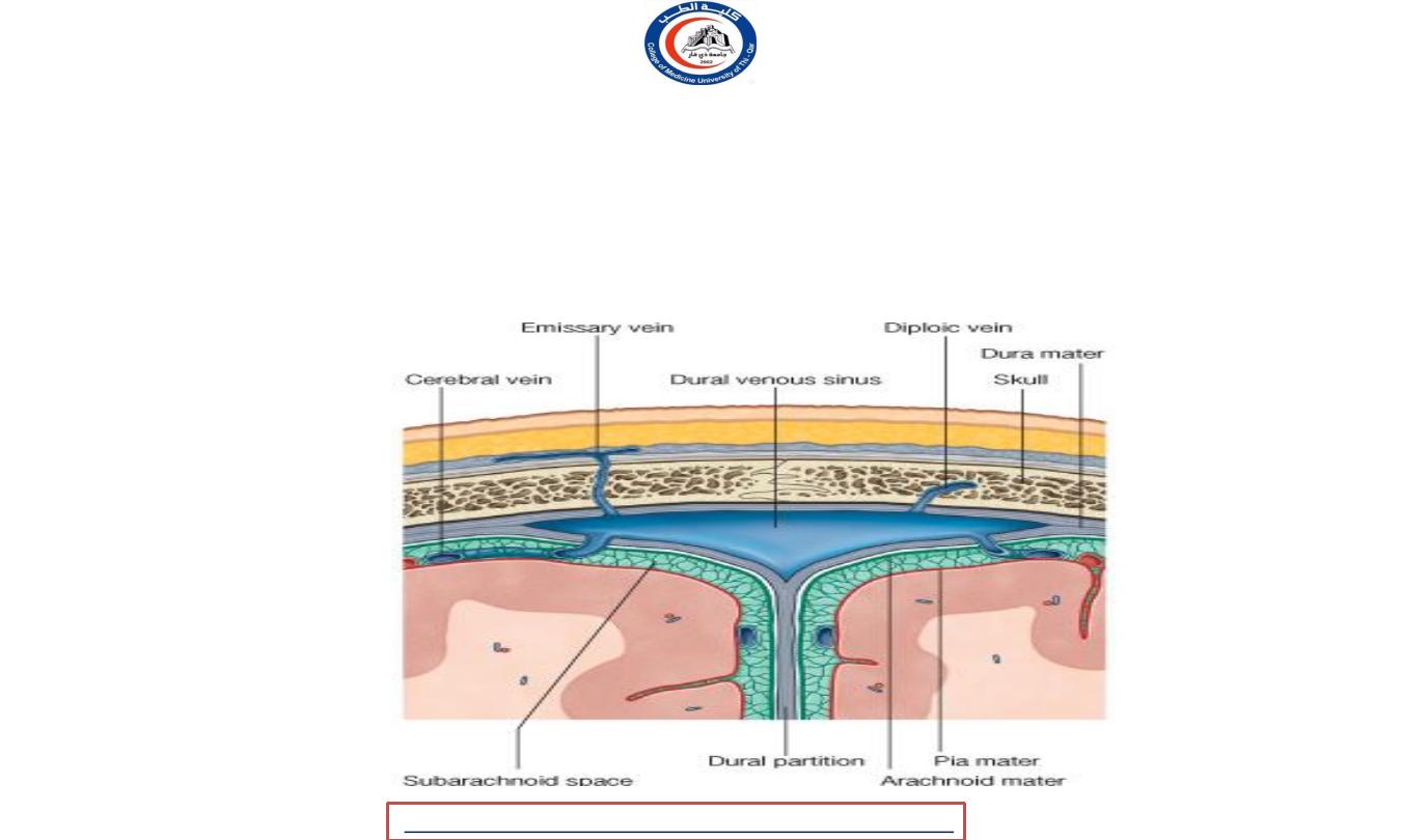
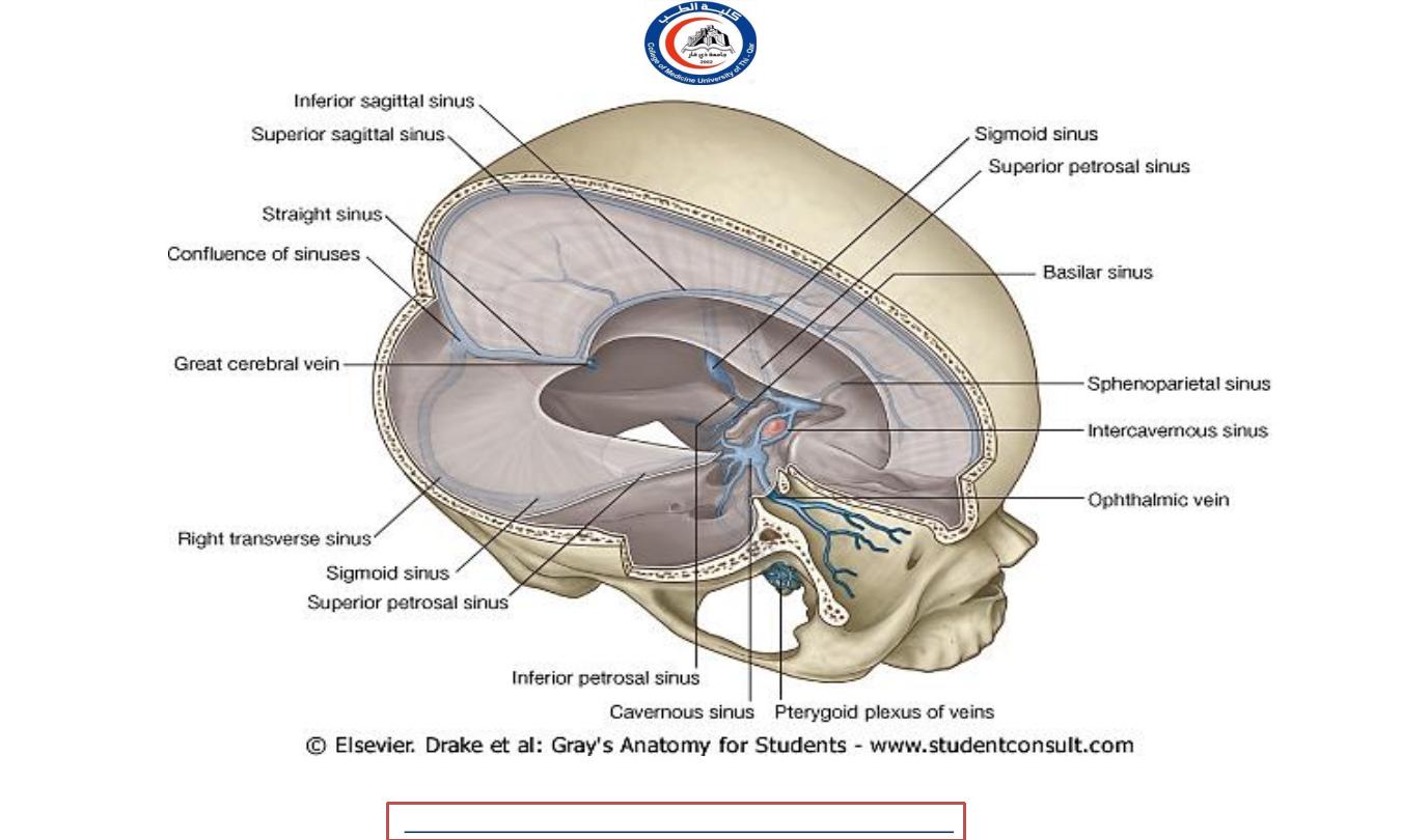
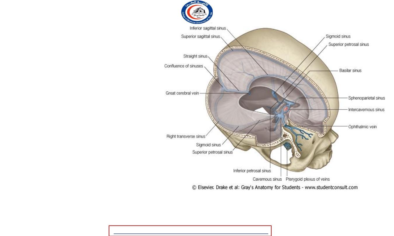
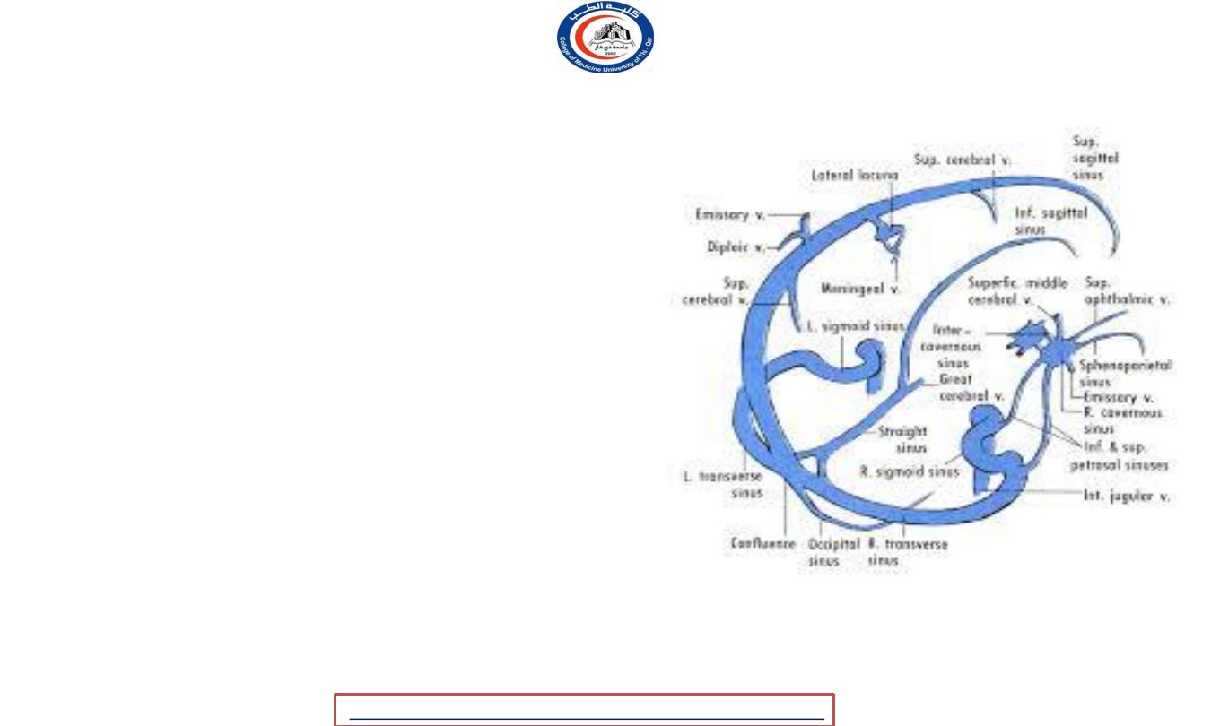
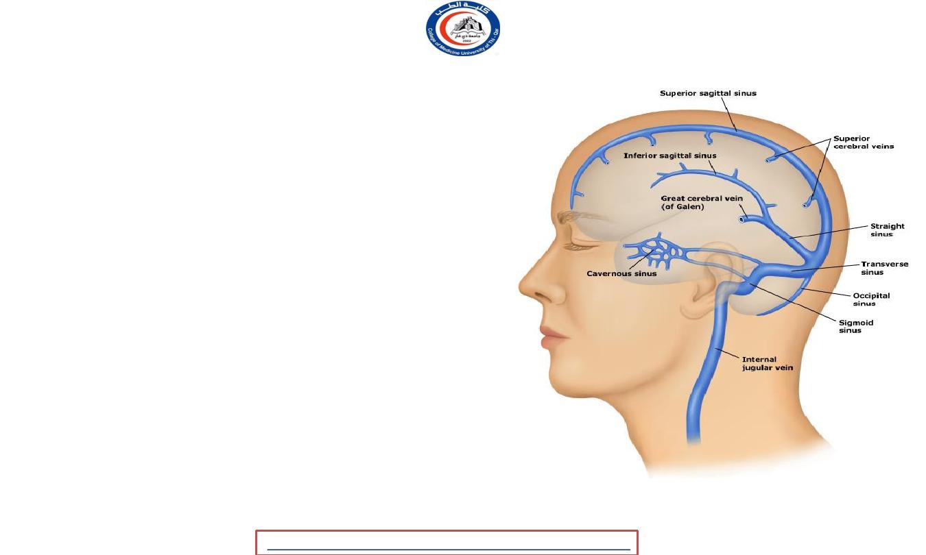
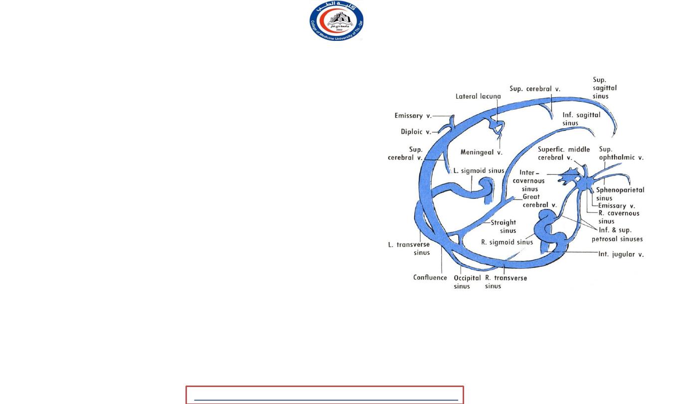
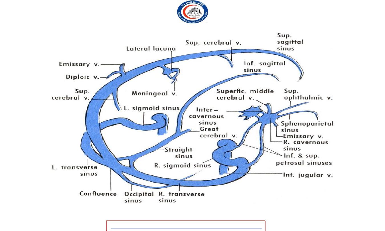
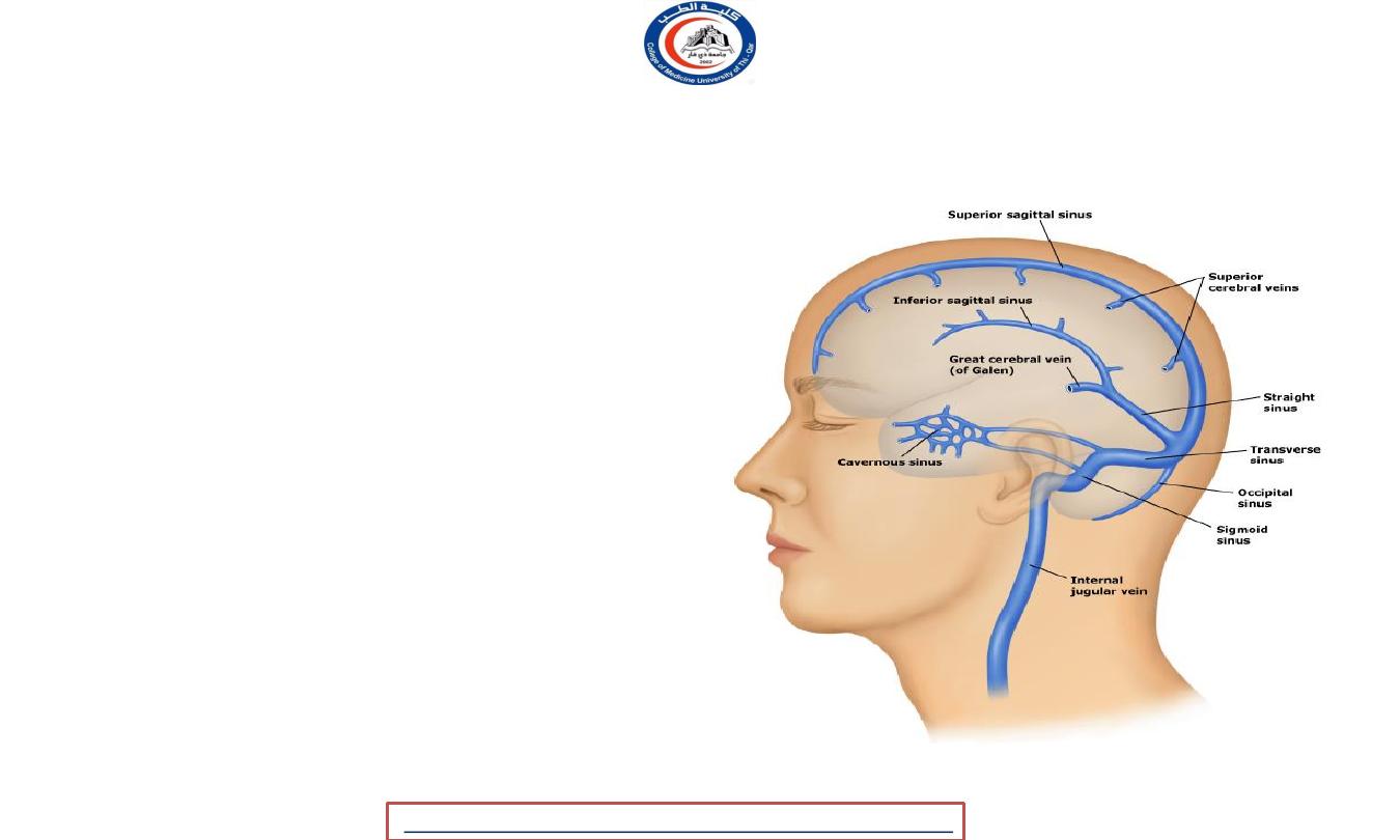
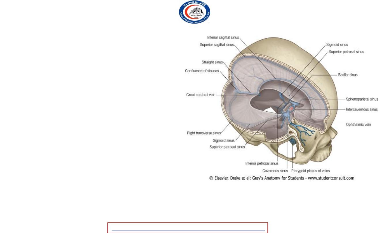
Dural venous sinuses
• The dural venous sinuses are endothelial-lined spaces between the outer periosteal
and the inner meningeal layers of the dura mater
• They finally drain into internal jugular veins
• Emptying into the dural venous sinuses are diploic vein and emissary veins
.
University Of Thi-Qar
College Of medicine
2020
,
Radiology,CAMB
Temimi,Clinical
-
AL
emthan
R
Dr.Rafid
Anatomy lecture . 2
nd
stage
Dr.Rafid Al-Temimi
8
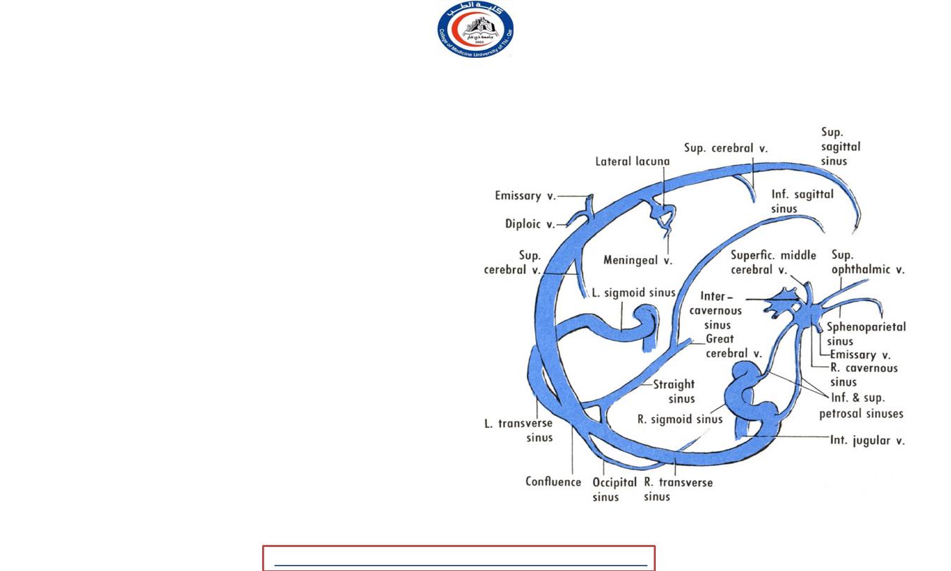
The dural venous sinuses
• Superior sagittal sinus
• Inferior sagittal sinus
• Straight sinus
• Transverse sinuses
• Sigmoid sinuses
• Occipital sinuses
• Confluence of sinuses
• Superior petrosal
• Inferior petrosal
• Cavernous sinuses
University Of Thi-Qar
College Of medicine
2020
,
Radiology,CAMB
Temimi,Clinical
-
AL
emthan
R
Dr.Rafid
Anatomy lecture . 2
nd
stage
Dr.Rafid Al-Temimi
9
University Of Thi-Qar
College Of medicine
2020
,
Radiology,CAMB
Temimi,Clinical
-
AL
emthan
R
Dr.Rafid
Anatomy lecture . 2
nd
stage
Dr.Rafid Al-Temimi
10
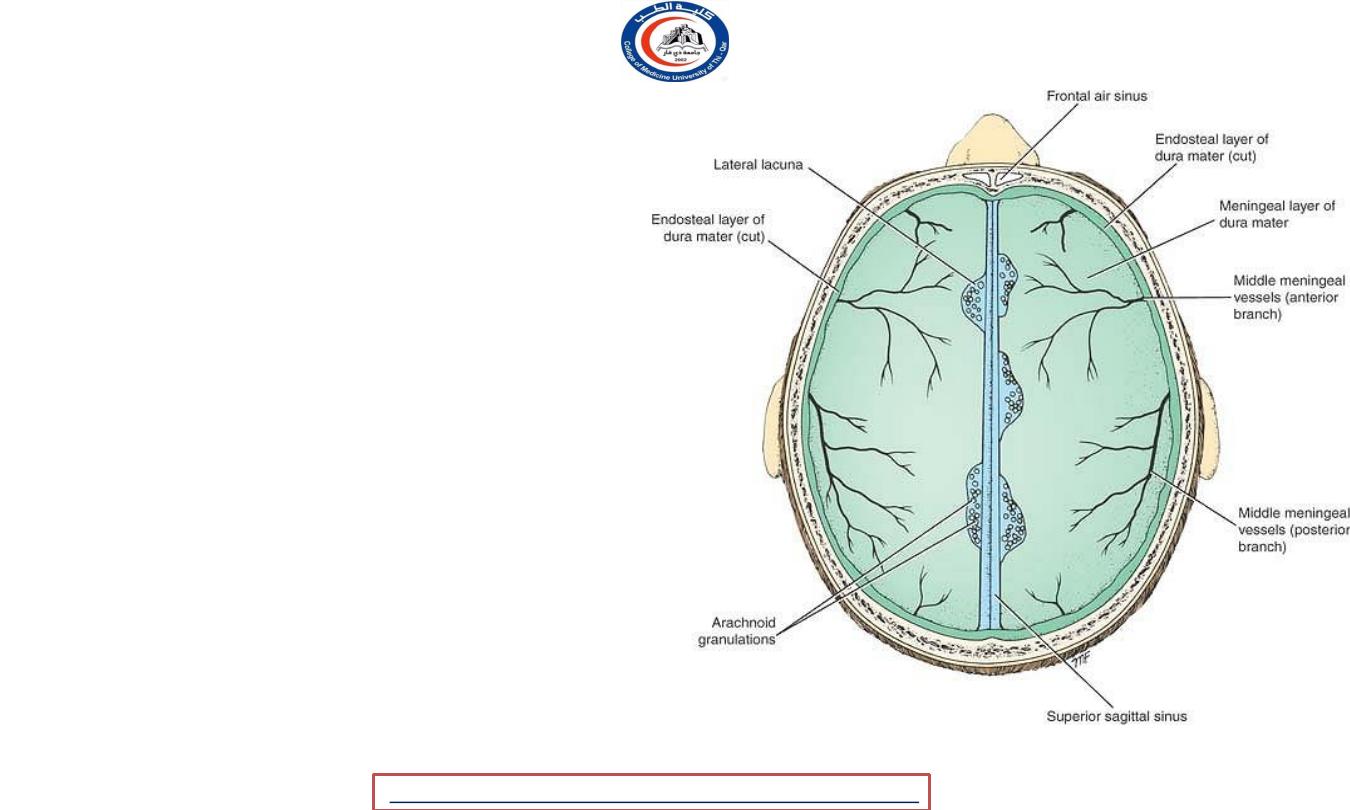
The superior sagittal sinus
• Deviates ,usually the right &
becomes continuous with the
right transverse sinus.
• It communicates with venous
lacunae on each side.
• Numerous arachnoid villi and
granulations project into the
lacunae
• It usually becomes continuous
with the right transverse sinus.
University Of Thi-Qar
College Of medicine
2020
,
Radiology,CAMB
Temimi,Clinical
-
AL
emthan
R
Dr.Rafid
Anatomy lecture . 2
nd
stage
Dr.Rafid Al-Temimi
11
• At the internal occipital
protuberance, it is dilated to
form the confluence of the
sinuses
• It receives :
• The superior cerebral veins
• The occipital sinus
• Diploic & emissary veins
• CSF(through arachnoid
granulations)
University Of Thi-Qar
College Of medicine
2020
,
Radiology,CAMB
Temimi,Clinical
-
AL
emthan
R
Dr.Rafid
Anatomy lecture . 2
nd
stage
Dr.Rafid Al-Temimi
12
The inferior sagittal sinus
• Joins the great cerebral
vein to form the straight
sinus
• It receives
a few
cerebral
veins
from the medial
surface of the cerebral
hemispheres.
University Of Thi-Qar
College Of medicine
2020
,
Radiology,CAMB
Temimi,Clinical
-
AL
emthan
R
Dr.Rafid
Anatomy lecture . 2
nd
stage
Dr.Rafid Al-Temimi
13
The straight sinus
• Is formed by the union of the
inferior
sagittal sinus
with
the great cerebral
vein.
• It
ends
by turning to the
left
(sometimes
to the right) to
form
the
transverse
sinus
.
• It receives:
• Inferior sagittal sinus
• Great cerebral vein, posterior cerebral
veins
• Superior cerebellar veins
• Veins from the falx cerebri
University Of Thi-Qar
College Of medicine
2020
,
Radiology,CAMB
Temimi,Clinical
-
AL
emthan
R
Dr.Rafid
Anatomy lecture . 2
nd
stage
Dr.Rafid Al-Temimi
14
• The transverse sinuses
• Are paired structures that
begins
at the
internal occipital protuberance
• The
right sinus
is usually continuous with
the
superior sagittal sinus
, and the
left
is
continuous with
the straight sinus
.
• The transverse sinuses receive the
:
• Superior petrosal sinuses
• Inferior cerebral
• Cerebellar veins
• Diploic veins.
• They end by turning downward as the
sigmoid sinuses .
University Of Thi-Qar
College Of medicine
2020
,
Radiology,CAMB
Temimi,Clinical
-
AL
emthan
R
Dr.Rafid
Anatomy lecture . 2
nd
stage
Dr.Rafid Al-Temimi
15
• The sigmoid sinuses
• Are a
direc
t continuation of the
transverse sinuses
.
• The sinus then reaches the
posterior
part
of the
jugular foramen
to become
continuous with the superior bulb of
the
internal jugular vein
.
• The occipital sinus
• Is a small sinus occupying the attached
margin of the falx cerebelli.
• It
communicates
with the
internal
vertebral venous plexus
• Drains into the confluence of sinuses.
University Of Thi-Qar
College Of medicine
2020
,
Radiology,CAMB
Temimi,Clinical
-
AL
emthan
R
Dr.Rafid
Anatomy lecture . 2
nd
stage
Dr.Rafid Al-Temimi
16
University Of Thi-Qar
College Of medicine
2020
,
Radiology,CAMB
Temimi,Clinical
-
AL
emthan
R
Dr.Rafid
Anatomy lecture . 2
nd
stage
Dr.Rafid Al-Temimi
17
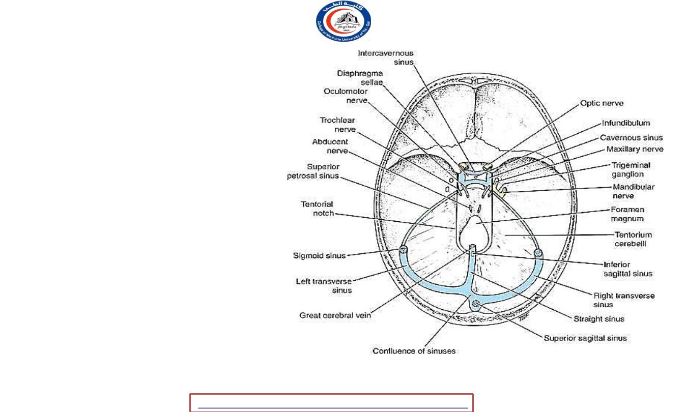
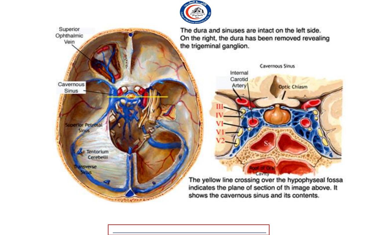
The cavernous sinuses
• Are
situated
in the
middle
cranial fossa
on each side of
the body of the
sphenoid
bone
• The
two sinuses
communicate
with each
other by
anterior and
posterior intercavernous
sinuses cerebri .
University Of Thi-Qar
College Of medicine
2020
,
Radiology,CAMB
Temimi,Clinical
-
AL
emthan
R
Dr.Rafid
Anatomy lecture . 2
nd
stage
Dr.Rafid Al-Temimi
18
University Of Thi-Qar
College Of medicine
2020
,
Radiology,CAMB
Temimi,Clinical
-
AL
emthan
R
Dr.Rafid
Anatomy lecture . 2
nd
stage
Dr.Rafid Al-Temimi
19
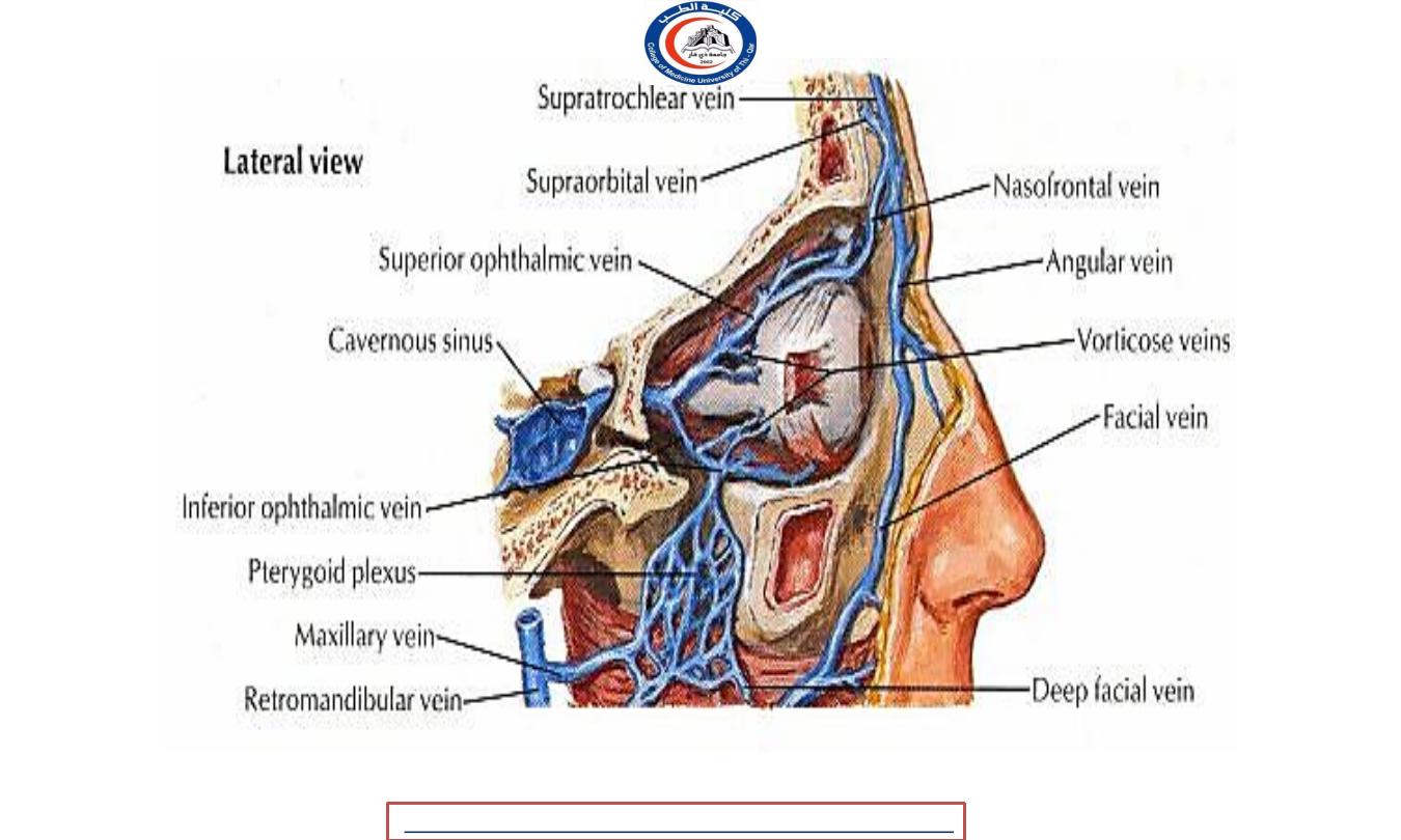
• The cavernous sinus receives:
• Cerebral and ophthalmic veins
• Emissary veins from pterygoid plexus of veins
• Sphenoparietal sinuses
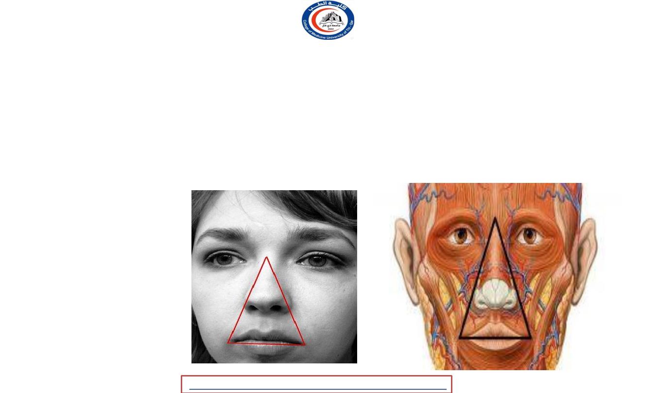
• It has
important communication
with the
facial vein
through the superior
ophthalmic vein. (This is a route by which infection can travel from the facial skin to
the cavernous sinus.)
University Of Thi-Qar
College Of medicine
2020
,
Radiology,CAMB
Temimi,Clinical
-
AL
emthan
R
Dr.Rafid
Anatomy lecture . 2
nd
stage
Dr.Rafid Al-Temimi
20
University Of Thi-Qar
College Of medicine
2020
,
Radiology,CAMB
Temimi,Clinical
-
AL
emthan
R
Dr.Rafid
Anatomy lecture . 2
nd
stage
Dr.Rafid Al-Temimi
21
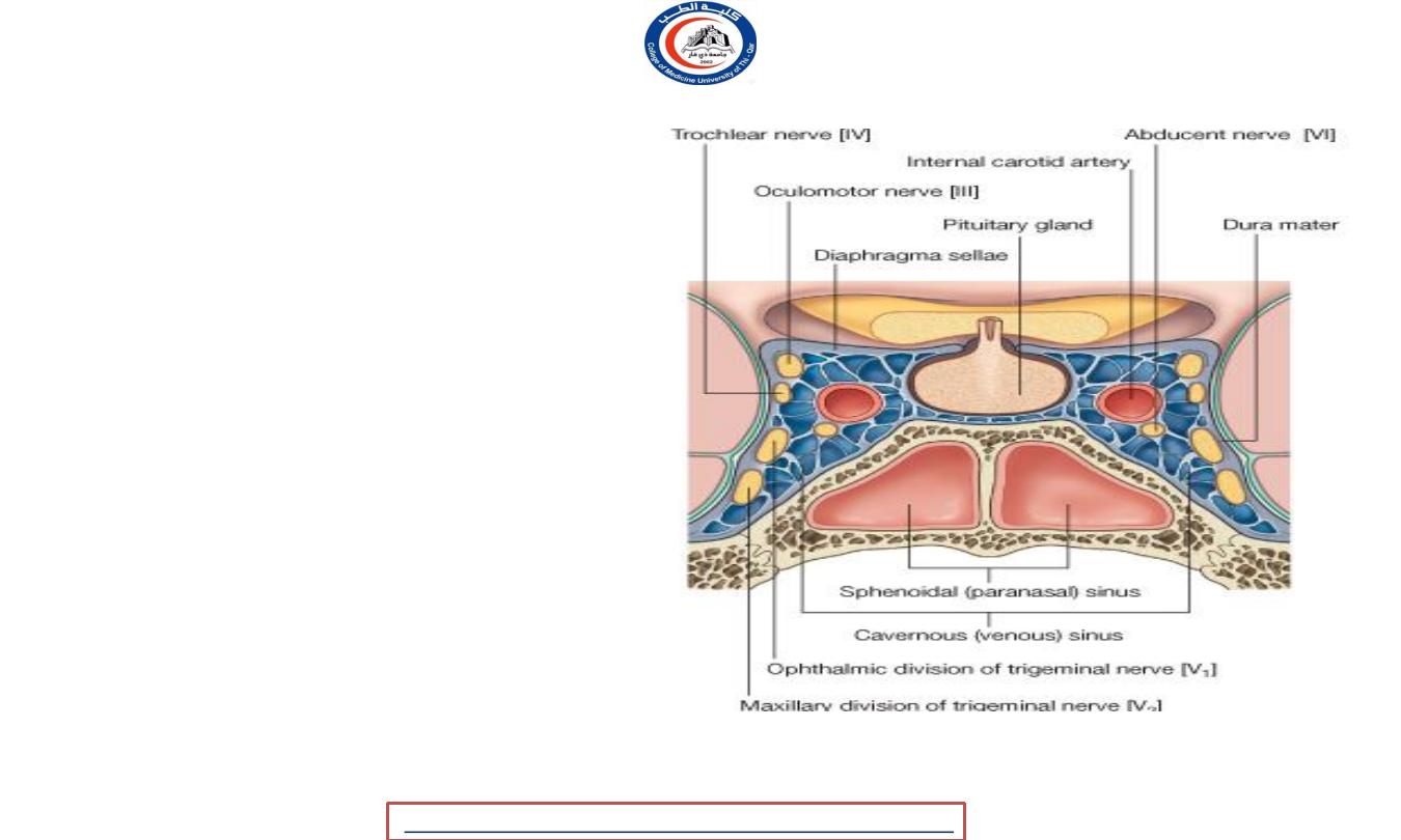
Structures passing through
each cavernous sinus are
:
• The internal carotid artery;
• The abducent nerve [VI].
Structures in the lateral wall of
cavernous sinus are the :
• Oculomotor nerve [III];
• Trochlear nerve [IV];
• Ophthalmic nerve [V
1
];
• Maxillary nerve [V
2
].
University Of Thi-Qar
College Of medicine
2020
,
Radiology,CAMB
Temimi,Clinical
-
AL
emthan
R
Dr.Rafid
Anatomy lecture . 2
nd
stage
Dr.Rafid Al-Temimi
22
• The superior and inferior
petrosal sinuses
• Are
small sinuses
situated on
the superior and inferior
borders of the petrous part of
the temporal bone .
• Superior sinuses
drain the
cavernous sinus into the
transverse sinus.
• Inferior sinuses
drains the
cavernous sinus into the internal
jugular vein.
University Of Thi-Qar
College Of medicine
2020
,
Radiology,CAMB
Temimi,Clinical
-
AL
emthan
R
Dr.Rafid
Anatomy lecture . 2
nd
stage
Dr.Rafid Al-Temimi
23
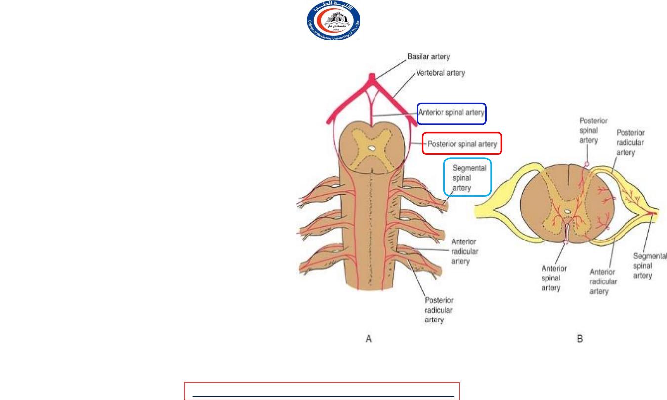
Blood Supply of the Spinal Cord
• It is supplied by
three arteries
:
•
Two posterior spinal
arteries
from vertebral arteries.
•
Anterior spinal
artery from
union of two arteries from
vertebral artery .
• Spinal arteries are reinforced
by
segmental arteries
that arise
from arteries outside the
vertebral column.
University Of Thi-Qar
College Of medicine
2020
,
Radiology,CAMB
Temimi,Clinical
-
AL
emthan
R
Dr.Rafid
Anatomy lecture . 2
nd
stage
Dr.Rafid Al-Temimi
24
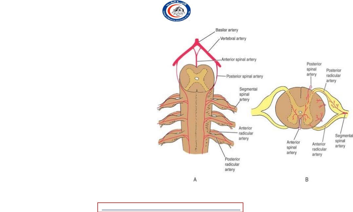
• Each segmental spinal artery
gives rise to
anterior and
posterior radicular arteries
• The
great anterior
medullary artery
of
Adamkiewicz
,
that
arise
from the
aorta
, enters the
vertebral canal and
anastomoses
with the
anterior and posterior spinal
arteries.
University Of Thi-Qar
College Of medicine
2020
,
Radiology,CAMB
Temimi,Clinical
-
AL
emthan
R
Dr.Rafid
Anatomy lecture . 2
nd
stage
Dr.Rafid Al-Temimi
25

Veins of the Spinal Cord
• The veins of the spinal cord drain into six tortuous
longitudinal channels that
communicate
within the skull
with
the veins of the brain
and
the venous sinuses.
• They drain
mainly
into the internal vertebral venous
plexus
.
University Of Thi-Qar
College Of medicine
2020
,
Radiology,CAMB
Temimi,Clinical
-
AL
emthan
R
Dr.Rafid
Anatomy lecture . 2
nd
stage
Dr.Rafid Al-Temimi
26
THANK YOU
University Of Thi-Qar
College Of medicine
Anatomy lecture . 2
nd
stage
Dr.Rafid Al-Temimi
27