
Tumours of the oropharynx :
Tonsillar tumours
Benign tumours are very rare. But tonsillar
stones (tonsiliths) with surrounding
ulceration, mucus retention cysts, herpes
simplex or giant aphthous ulcers, may
mimic the more common malignant
tumours of the tonsil. These tumours are
becoming increasingly common, and
unusually, are now being found in younger
patients and in non-smokers.

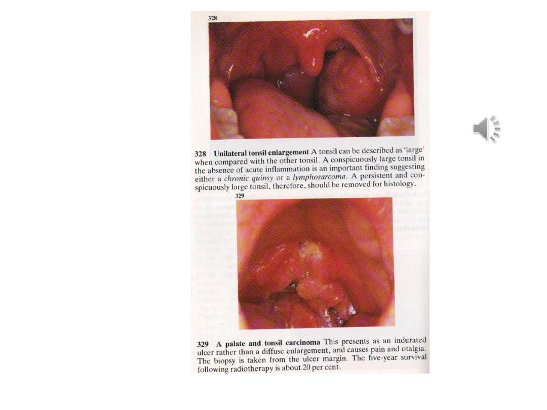
Squamous cell carcinoma (SCC)
This is the commonest tumour of the tonsil. It tends to occur in
middle-aged and elderly people, but in recent years tonsillar
SCC has become more frequent in patients under the age of 40.
Many of these patients are also unusual candidates for SCC
because they are non-smokers and non-drinkers.
Signs and symptoms
•Pain in the throat
•Referred otalgia
•Ulcer on the tonsil
•Lump in the neck.
As the tumour grows it may affect the patient's ability to
swallow and it may lead to an alteration in the voice—this is
known as ‘hot potato speech’.
Diagnosis is usually confirmed with a biopsy taken at the time
of the staging pan endoscopy. Fine needle aspiration of any
neck mass is also necessary. Imaging usually entails CT and/or a
MRI scan. It is important to exclude any bronchogenic
synchronous tumour with a chest X-ray and/or a chest CT scan
and bronchoscopy where necessary.

Treatment
Decisions about treatment should be made in a
multidisciplinary clinic, taking into account the size and
stage of the primary tumour, the presence of nodal
metastasize, the patient's general medical status and the
patient's wishes.
Treatment options include:
• Radiotherapy alone
• Chemoradiotherapy
• Trans oral laser surgery
• En-bloc surgical excision—this removes the primary
and the affected nodes from the neck. It will often be
necessary to reconstruct the surgical defect to allow
for adequate speech and swallowing afterwards. This
will often take the form of free tissue transfer such as
a radial forearm free flap.

Lymphoma
This is the second most common tonsil tumour.
Signs and symptoms
•Enlargement of one of the tonsils
•Lymphadenopathy in the neck—may be large
•Mucosal ulceration—less common than in SCC.
Investigations
-Fine needle aspiration cytology may suggest lymphoma, but it
rarely confirms the diagnosis. It is often necessary to perform an
excision biopsy of one of the nodes.
-Staging is necessary with CT scanning of the neck chest,
abdomen, and pelvis. Further surgical intervention is not
required other than to secure a threatened airway.
Treatment
Usually consists of chemotherapy and/or radiotherapy.

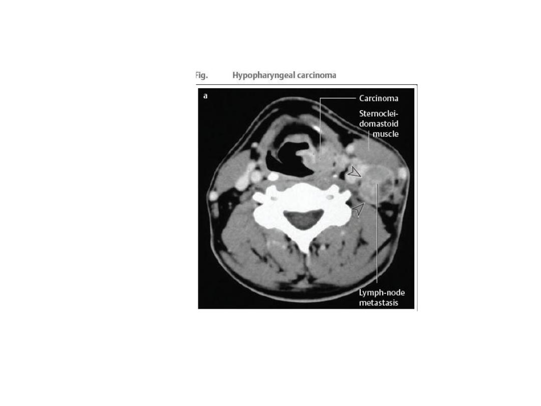
-Tumors of Hypopharynx:
Benign Tumors
Benign tumors of the hypopharynx are considered
ararity. They may present clinically with dysphagia,
regurgitation,or retrosternal pain. The diagnosis is
established with an incisional biopsy taken
endoscopically under general endotracheal anesthesia.
Treatment consists of surgical removal, depending on
the tumor size.
Plummer-Vinson syndrome :It is a pre malignant .Most
common in nonsmoking Scandinavian women
characterized by iron deficiency anemia , dysphagia
associated with postcricoid web and angular cheilitis .
There is increased incidence of oral leukoplakia and
s.c.c( 10% ). Treatment with iron will improve the
anemia but will not reverse the mucosal changes .

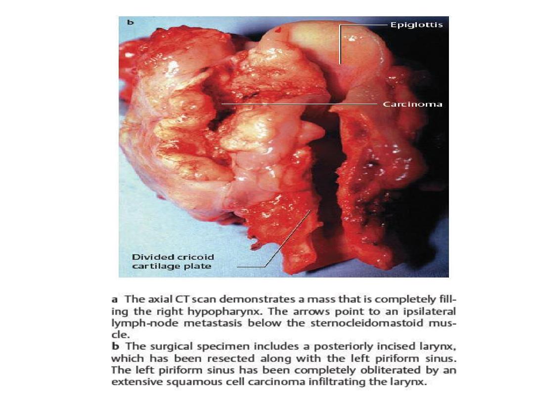
Malignant Tumors
The hypopharynx is divided into 3 parts :
1.
Postcricoid
2.
Pyriform fossae
3.
Posterior pharyngeal wall
Histologically, almost all of these tumors are squamous cell
carcinomas. As with oral and oropharyngeal carcinomas, there is an
etiologic link to chronic alcohol and nicotine abuse.
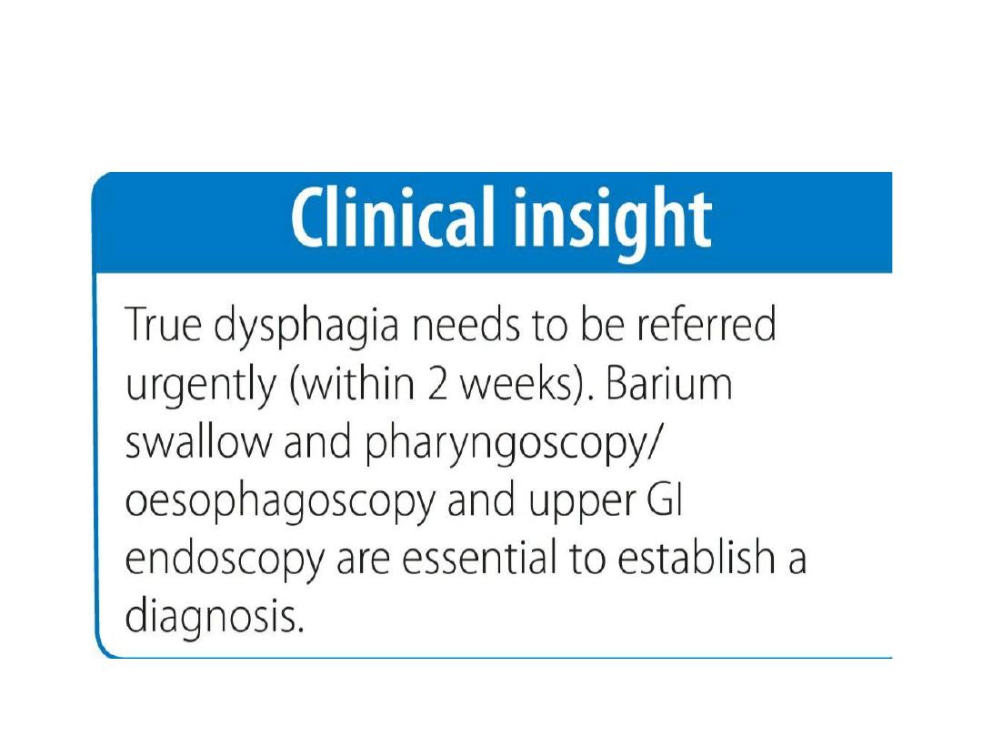
Symptoms: Most malignant tumors of the hypopharynx are
diagnosed at an advanced stage because earlier lesions do not
produce symptoms. Initial complaints tend to be nonspecific,
depending on tumor size and location, and consist of dysphagia and a
fetid breath odor. Later there may be pain radiating to the ear.
Hoarseness and possible
dyspnea signify tumor extension to the larynx.
In many cases, cervical lymph-node metastasis is noted as the earliest
sign of disease.

Diagnosis: Besides the mirror examination or indirect laryngoscopy,
the diagnostic workup
should include endoscopic examination under general endotracheal
anesthesia, as this is the best way to evaluate tumor extent. A biopsy
can also be taken in the same sitting for histologic confirmation.
Additionally, sectional imaging modalities can help to define the
tumor size and check for involvement of adjacent structures while
also evaluating the cervical lymphnode status
Treatment: Treatment depends on tumor size but usually consists of
local surgical excision with a concomitant neck dissection Many
malignant tumors of the hypopharynx have already spread to the
larynx, making it necessary to perform a laryngectomy in the same
sitting. The tissue defect is closed primarily whenever possible. This
cannot be done with extensive hypopharyngeal resections due to the
high risk of stricture formation, and larger defects should be
reconstructed by means of a free jejunum transfer with microvascular
anastomosis. Surgery should be followed by radiation to the tumor
site
And lymphatics. Alternative treatments for advanced hypopharyngeal
cancers are primary radiotherapy and combined radiation and
chemotherapy.
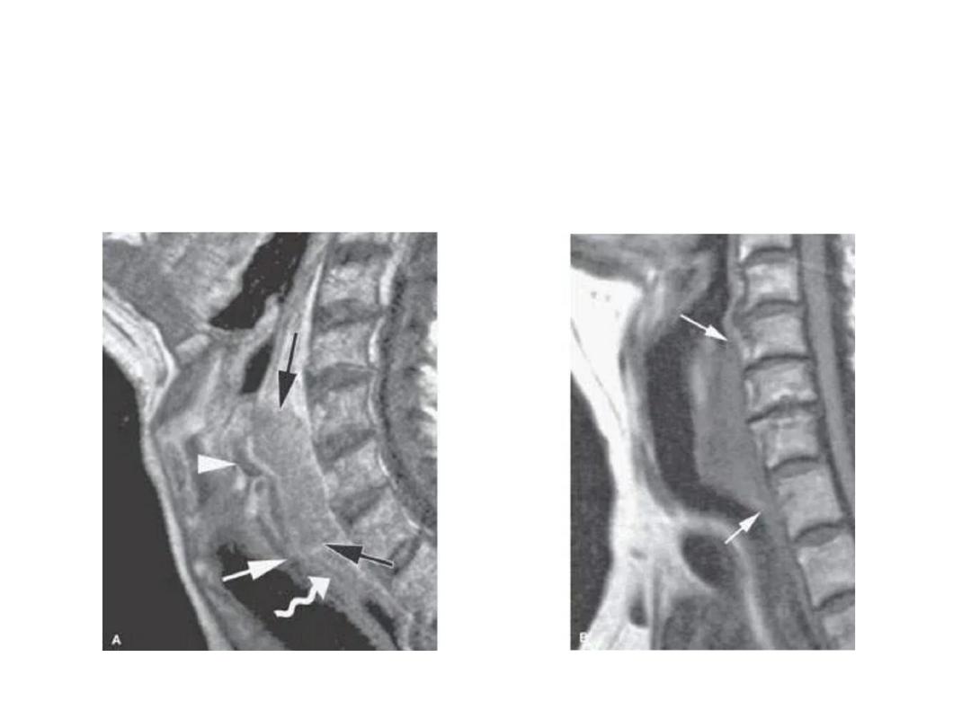
TUMOUR OF HYPOPHARYNX