
CHEST ANATOMY THI-QAR UNIVERSITY
COLLEGE OF MEDICINE
LECTURE 6 2019/2020
Dr. Rafid AL-Temimi; Clinical radiology (CABM)
Page
1
Dr. Ahmed Abdulameer Daffar ; Thoracic & Vascular Surgeon ( FIBMS )
THE CHEST
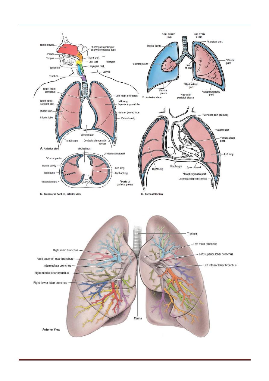
PLEURAE
The pleurae and lungs lie on either side of the mediastinum within the chest cavity.
Each pleura has two parts:
1. Parietal layer, which lines the thoracic wall.
2. Visceral layer, which completely covers the outer surfaces of the lungs.
The two layers become continuous with one another by means of a cuff of pleura that
surrounds the structures entering and leaving the lung at the hilum of each lung (lung
root).
To allow for movement of the pulmonary vessels and large bronchi during respiration,
the pleural cuff hangs down as a loose fold called the pulmonary ligament.
The parietal and visceral layers of pleura are separated from one another by a space, the
pleural cavity or pleural space.
The pleural cavity normally contains a small amount of tissue fluid, the pleural fluid,
which covers the surfaces of the pleura as a thin film and permits the two layers to move
on each other with the minimum of friction.
For purposes of description, it is customary to divide the parietal pleura according to the
region in which it lies or the surface that it covers.
The cervical pleura extend up into the neck, lining the undersurface of the
suprapleural membrane.
The costal pleura lines the inner surfaces of the ribs, the costal cartilages, the
intercostal spaces, the sides of the vertebral bodies, and the back of the sternum.
The diaphragmatic pleura cover the thoracic surface of the diaphragm.
The mediastinal pleura cover and form the lateral boundary of the mediastinum.
CHEST ANATOMY THI-QAR UNIVERSITY
COLLEGE OF MEDICINE
LECTURE 6 2019/2020
Dr. Rafid AL-Temimi; Clinical radiology (CABM)
Page
2
Dr. Ahmed Abdulameer Daffar ; Thoracic & Vascular Surgeon ( FIBMS )

CHEST ANATOMY THI-QAR UNIVERSITY
COLLEGE OF MEDICINE
LECTURE 6 2019/2020
Dr. Rafid AL-Temimi; Clinical radiology (CABM)
Page
3
Dr. Ahmed Abdulameer Daffar ; Thoracic & Vascular Surgeon ( FIBMS )
Bronchi
The trachea bifurcates behind the arch of the aorta into the right and left principal
(primary or main) bronchi.
The bronchi divide dichotomously, giving rise to several million terminal bronchioles
that terminate in one or more respiratory bronchioles.
Each respiratory bronchiole divides into 2 to 11 alveolar ducts that enter the alveolar
sacs. The alveoli arise from the walls of the sacs as diverticula.
Principal Bronchi:
1.
The right principal (main) bronchus
: is wider, shorter, and more vertical than
the left and is about 1 in. (2.5 cm) long.
Before entering the hilum of the right lung, the principal bronchus gives off the
superior lobar bronchus. On entering the hilum, it divides into a middle and an
inferior lobar bronchus.
2.
The left principal (main) bronchus
: is narrower, longer, and more horizontal than
the right and is about 2 in. (5 cm) long.
It passes to the left below the arch of the aorta. On entering the hilum of the left lung,
the principal bronchus divides into a superior and an inferior lobar bronchus.
CHEST ANATOMY THI-QAR UNIVERSITY
COLLEGE OF MEDICINE
LECTURE 6 2019/2020
Dr. Rafid AL-Temimi; Clinical radiology (CABM)
Page
4
Dr. Ahmed Abdulameer Daffar ; Thoracic & Vascular Surgeon ( FIBMS )