
CHEST ANATOMY THI-QAR UNIVERSITY
COLLEGE OF MEDICINE
LECTURE 2 2019/2020
Dr. Rafid AL-Temimi ; Clinical radiology ( CABM)
Page
1
Dr. Ahmed Abdulameer Daffar ; Thoracic & Vascular Surgeon ( FIBMS )
THE CHEST
JOINTS OF THE CHEST WALL:
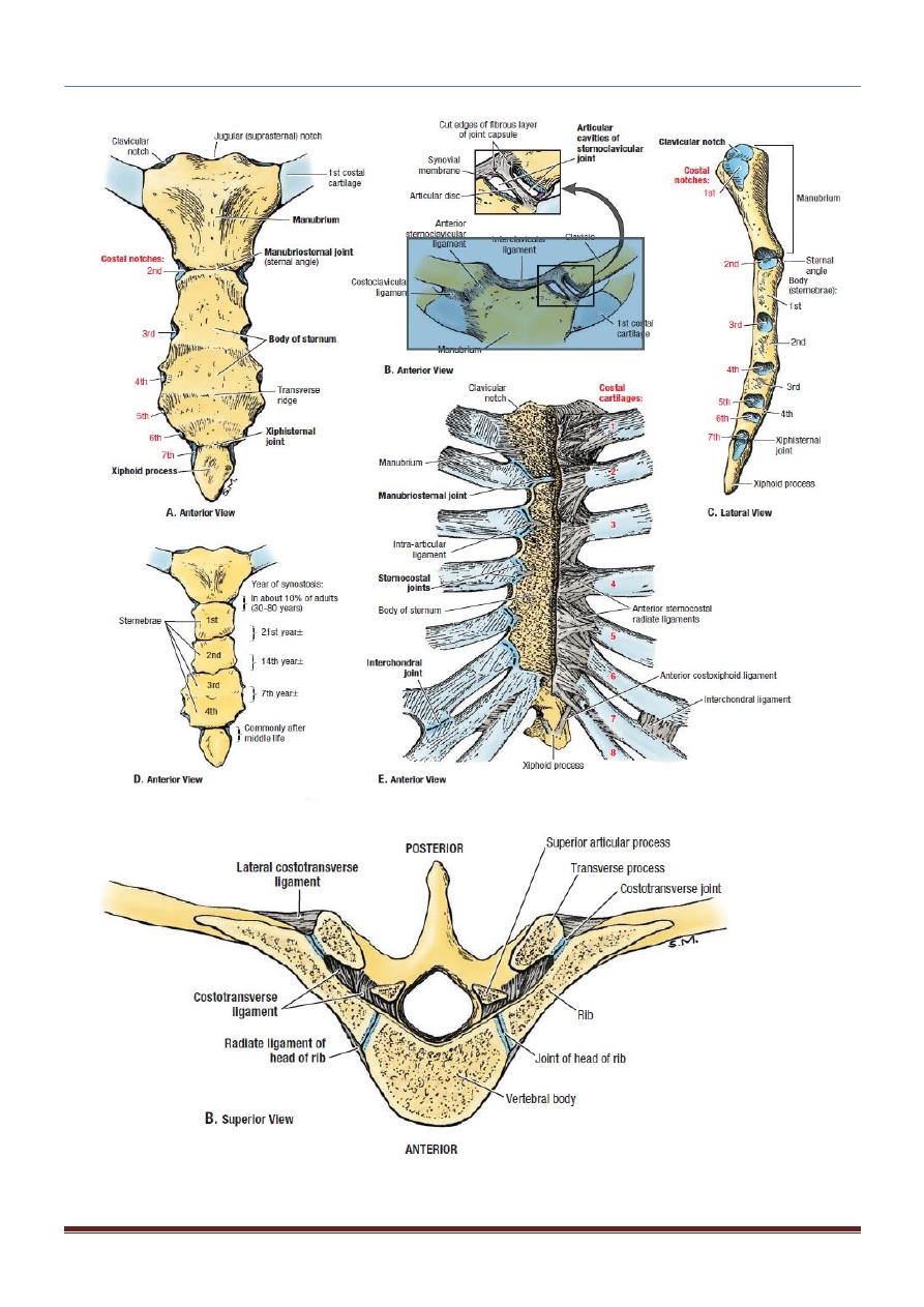
JOINTS OF THE STERNUM:
The manubriosternal joint is a cartilaginous joint between the manubrium and the
body of the sternum. A small amount of angular movement is possible during
respiration.
The xiphisternal joint is a cartilaginous joint between the xiphoid process
(cartilage) and the body of the sternum.
The xiphoid process usually fuses with the body of the sternum during middle age.
JOINTS OF THE RIBS:
-Joints of the Heads of the Ribs:
The articulation is between the head of rib with vertebral body
-Joints of the Tubercles of the Ribs:
The articulation is between the tubercle of rib with the transverse process of the
corresponding vertebra.
This joint is absent on the 11th and 12th ribs.
-Joints of the Ribs and Costal Cartilages:
These joints are cartilaginous joints.
No movement is possible.
-Joints of the Costal Cartilages with the Sternum:
The 1st costal cartilages articulate with the manubrium, by cartilaginous joints that
permit no movement.
The 2nd to 7th costal cartilages articulate with the lateral border of the sternum by
synovial joints. In addition, the 6th, 7th, 8th, 9th, and 10th costal cartilages articulate
with one another along their borders by small synovial joints.
The cartilages of the 11th and 12th ribs are embedded in the abdominal musculature.
CHEST ANATOMY THI-QAR UNIVERSITY
COLLEGE OF MEDICINE
LECTURE 2 2019/2020
Dr. Rafid AL-Temimi ; Clinical radiology ( CABM)
Page
2
Dr. Ahmed Abdulameer Daffar ; Thoracic & Vascular Surgeon ( FIBMS )

CHEST ANATOMY THI-QAR UNIVERSITY
COLLEGE OF MEDICINE
LECTURE 2 2019/2020
Dr. Rafid AL-Temimi ; Clinical radiology ( CABM)
Page
3
Dr. Ahmed Abdulameer Daffar ; Thoracic & Vascular Surgeon ( FIBMS )
OPENINGS OF THE THORAX:
Superiorly:
The chest cavity communicates with the root of the neck through an opening called
the thoracic outlet.
It is called an outlet because important vessels and nerves emerge from the thorax
here to enter the neck and upper limbs.
Inferiorly:
The thoracic cavity communicates with the abdomen through a large opening.
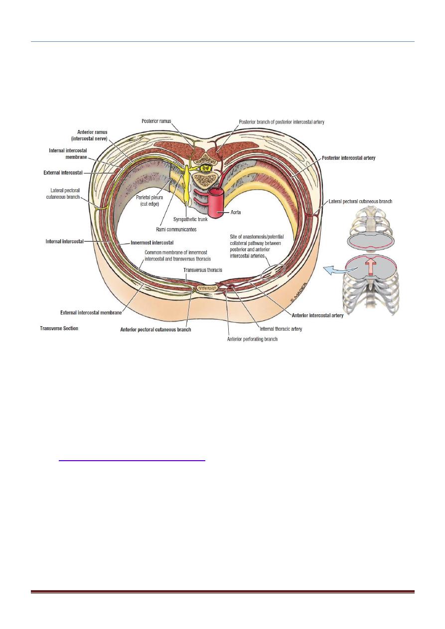
INTERCOSTAL SPACES:
The spaces between the ribs contain three muscles of respiration:
The external intercostal, the internal intercostal, and the innermost intercostal
muscle.
The innermost intercostal muscle is lined internally by the endothoracic fascia,
which is lined internally by the parietal pleura.
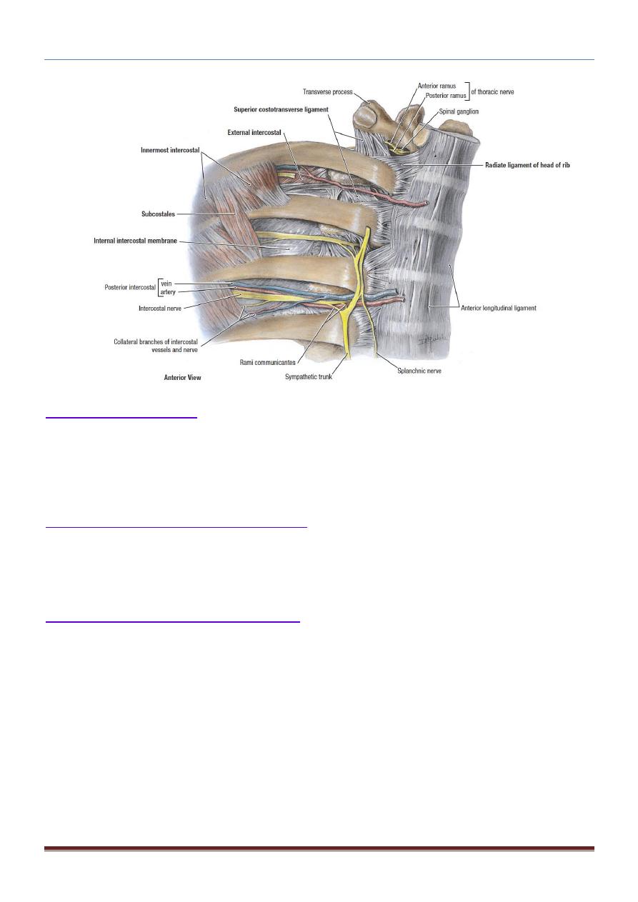
The intercostal nerves and blood vessels run between the intermediate and deepest
layers of muscles.
They are arranged in the following order from above downward: intercostal vein,
intercostal artery, and intercostal nerve (i.e., VAN).
Intercostal Muscles:
The external intercostal muscle: forms the most superficial layer.
Its fibers are directed downward and forward from the inferior border of the
rib above to the superior border of the rib below.
The muscle extends forward to the costal cartilage where it is replaced by an
aponeurosis, the anterior (external) intercostal membrane.
The internal intercostal muscle forms the intermediate layer.
Its fibers are directed downward and backward from the subcostal groove of
the rib above to the upper border of the rib below.
The muscle extends backward from the sternum in front to the angles of the
ribs behind, where the muscle is replaced by an aponeurosis, the posterior
(internal) intercostal membrane.
The innermost intercostal muscle forms the deepest layer and corresponds to the
transversus abdominis muscle in the anterior abdominal wall.
CHEST ANATOMY THI-QAR UNIVERSITY
COLLEGE OF MEDICINE
LECTURE 2 2019/2020
Dr. Rafid AL-Temimi ; Clinical radiology ( CABM)
Page
4
Dr. Ahmed Abdulameer Daffar ; Thoracic & Vascular Surgeon ( FIBMS )
It is an incomplete muscle layer and crosses more than one intercostal space within the
ribs.
It is related internally to fascia (endothoracic fascia) and parietal pleura and externally
to the intercostal nerves and vessels.
Nerve &vascular Supply:
The intercostal muscles are supplied by the corresponding intercostal nerves.
The intercostal nerves and blood vessels (the neurovascular bundle), as in the abdominal
wall, run between the middle and innermost layers of muscles .
They are arranged in the following order from above downward: intercostal vein,
intercostal artery, and intercostal nerve (i.e., VAN).
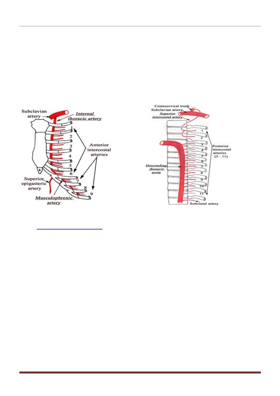
Intercostal Arteries and Veins:
Each intercostal space contains a large single posterior intercostal artery and two small
anterior intercostal arteries.
The posterior intercostal arteries of the first two spaces are branches from the superior
intercostal artery,(a branch of the costocervical trunk of the subclavian artery).
The posterior intercostal arteries of the lower nine spaces are branches of the descending
thoracic aorta.
CHEST ANATOMY THI-QAR UNIVERSITY
COLLEGE OF MEDICINE
LECTURE 2 2019/2020
Dr. Rafid AL-Temimi ; Clinical radiology ( CABM)
Page
5
Dr. Ahmed Abdulameer Daffar ; Thoracic & Vascular Surgeon ( FIBMS )
The anterior intercostal arteries of the first six spaces are branches of the internal
thoracic artery, which arises from the first part of the subclavian artery.
The anterior intercostal arteries of the lower spaces are branches of the musculophrenic
artery, one of the terminal branches of the internal thoracic artery.
The corresponding posterior intercostal veins drain backward into the azygos veins.
The anterior intercostal veins drain forward into the internal thoracic and the
musculophrenic veins.
Intercostal Nerves:
The intercostal nerves are the anterior rami of the first 11 thoracic spinal nerves.
The anterior ramus of the 12th thoracic nerve lies in the abdomen and runs forward
in the abdominal wall as the subcostal nerve.
It then runs forward inferiorly to the intercostal vessels in the subcostal groove of the
corresponding rib, between the innermost intercostal and internal intercostal
muscle.
The first six nerves are distributed within their intercostal spaces.
The 7th to 9
th
intercostal nerves leave the anterior ends of their intercostal spaces by
passing deep to the costal cartilages, to enter the anterior abdominal wall.
The 10th and 11th nerves, since the corresponding ribs are floating, pass directly into
theabdominal wall.
CHEST ANATOMY THI-QAR UNIVERSITY
COLLEGE OF MEDICINE
LECTURE 2 2019/2020
Dr. Rafid AL-Temimi ; Clinical radiology ( CABM)
Page
6
Dr. Ahmed Abdulameer Daffar ; Thoracic & Vascular Surgeon ( FIBMS )
Levatores Costarum:
There are 12 pairs of muscles.
Each levator costa is triangular in shape and arises by its apex from the tip of the
transverse process and is inserted into the rib below.
Nerve supply: Posterior rami of thoracic spinal nerves.
Serratus Posterior Superior Muscle:
It is a thin, flat muscle that arises from the lower cervical and upper thoracic spines.
Its fibers pass downward and laterally and are inserted into the upper ribs.
Nerve supply: Intercostal nerves.
Serratus Posterior Inferior Muscle:
It's a thin, flat muscle that arises from the upper lumbar and lower thoracic spines.
Its fibers pass upward and laterally and are inserted into the lower ribs.
Nerve supply: Intercostal nerves.