CHEST ANATOMY THI-QAR UNIVERSITY
COLLEGE OF MEDICINE
LECTURE 1 2019/2020
Dr. Rafid AL-Temimi ; Clinical radiology ( CABM)
Page
1
Dr. Ahmed Abdulameer Daffar ; Thoracic & Vascular Surgeon ( FIBMS )
THE CHEST
B
ASIC
A
NATOMY:
The thorax (or chest) is the region of the body between the neck and the abdomen.
It is flattened in front and behind but rounded at the sides.
The framework of the walls of the thorax, which is referred to as the
thoracic
cage
, is formed by the vertebral column behind, the ribs and intercostal spaces on
either side, and the sternum and costal cartilages in front.
Superiorly, the thorax communicates with the neck, and inferiorly it is separated
from the abdomen by the diaphragm.
The cavity of the thorax can be divided into a median partition, called the
mediastinum
, and the laterally placed
pleurae
and
lungs
.
The lungs are covered by a thin membrane called the
visceral pleura
while
parietal pleura
lines the inner surface of the chest wall.
CHEST ANATOMY THI-QAR UNIVERSITY
COLLEGE OF MEDICINE
LECTURE 1 2019/2020
Dr. Rafid AL-Temimi ; Clinical radiology ( CABM)
Page
2
Dr. Ahmed Abdulameer Daffar ; Thoracic & Vascular Surgeon ( FIBMS )
Structure of the Thoracic Wall
o
The thoracic wall is covered on the outside by skin and by muscles attaching
the shoulder girdle to the trunk. It is lined with parietal pleura.
o
The thoracic wall is formed :
Posteriorly: the thoracic part of the vertebral column.
Anteriorly: the sternum and costal cartilages.
Laterally : the ribs and intercostal spaces
Superiorly : the suprapleural membrane
Inferiorly : the diaphragm, which separates the thoracic cavity from
the abdominal cavity.
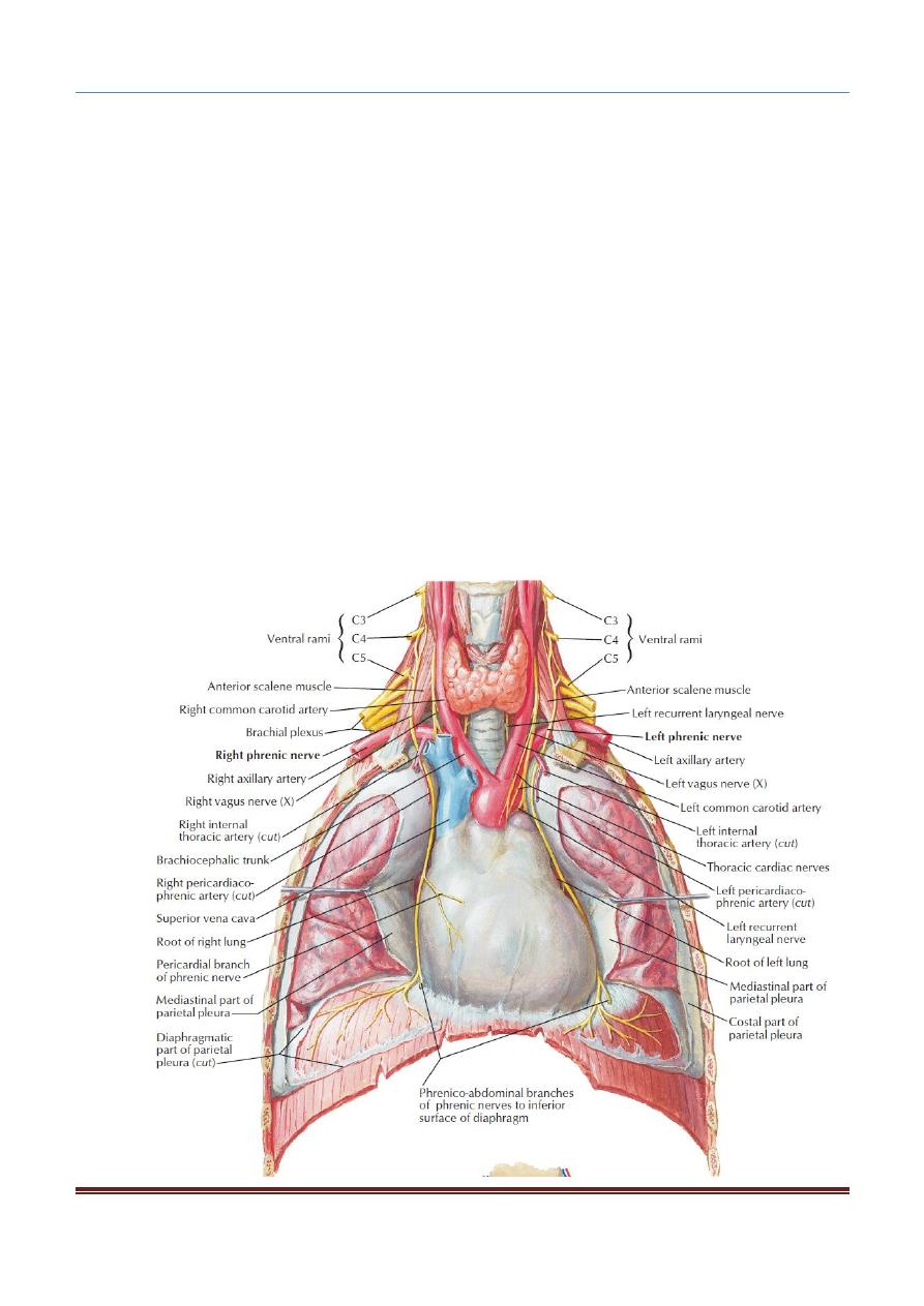
Supra-pleural Membrane:
Superiorly, the thorax opens into the root of the neck by a narrow aperture, the
thoracic outlet
which transmits structures (esophagus, trachea, blood vessels,
etc.) and for the most part lie close to the midline.
On either side of these structures, the outlet is closed by a dense fascial layer
called the
supra-pleural membrane.
This tent-shaped fibrous sheet is attached laterally to the medial border of the
1st rib and costal cartilage.

CHEST ANATOMY THI-QAR UNIVERSITY
COLLEGE OF MEDICINE
LECTURE 1 2019/2020
Dr. Rafid AL-Temimi ; Clinical radiology ( CABM)
Page
3
Dr. Ahmed Abdulameer Daffar ; Thoracic & Vascular Surgeon ( FIBMS )
It is attached at its apex to the tip of the transverse process of the seventh
cervical vertebra and medially to the fascia investing the structures passing
from the thorax into the neck.
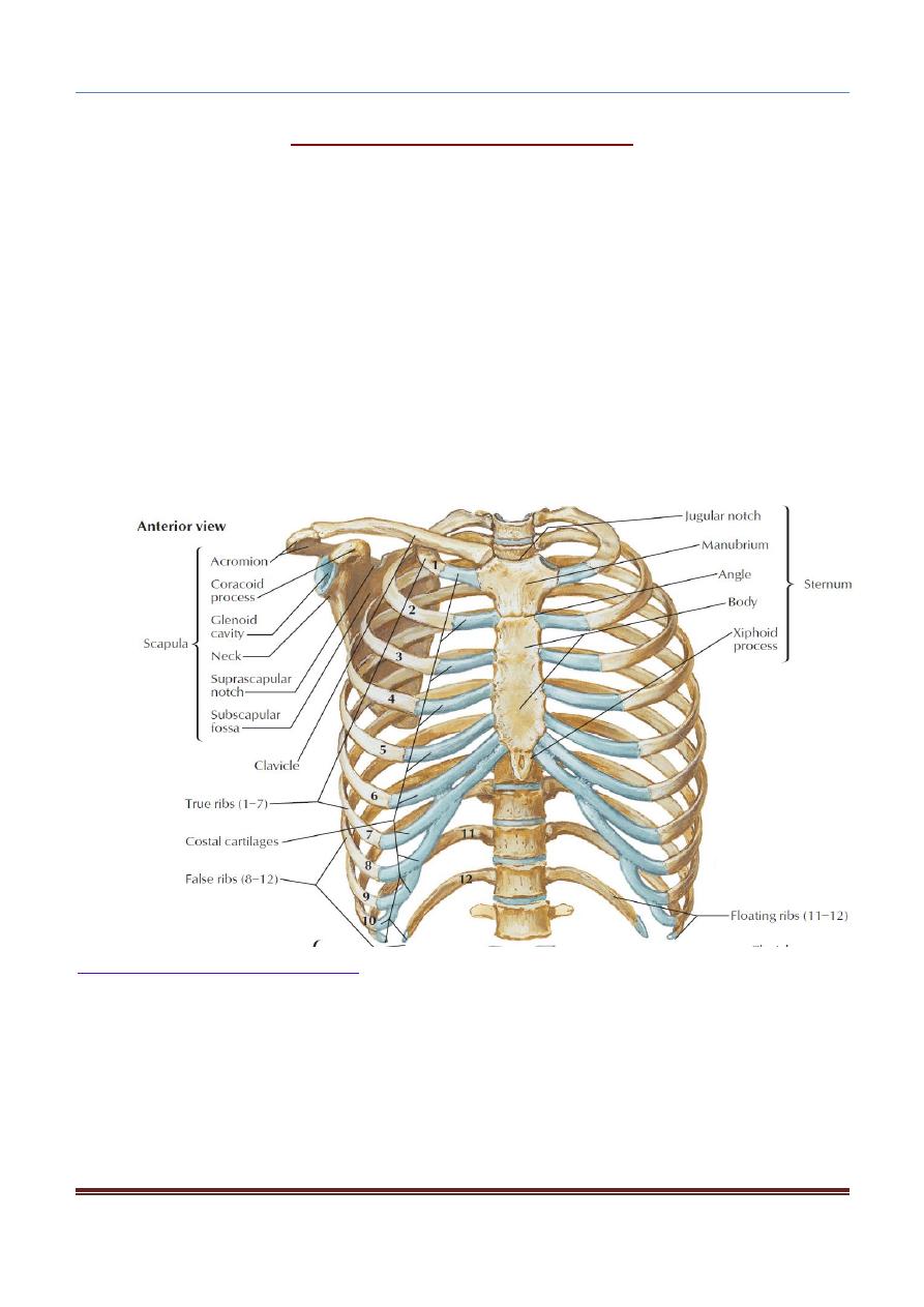
STERNUM:
The sternum lies in the midline of the anterior chest wall.
It is a flat bone that can be divided into three parts: manubrium sterni, body
of the sternum, and xiphoid process.
The sternal angle (angle of Louis):
Formed by the articulation of the manubrium with the body of the sternum, can be
recognized by the presence of a transverse ridge on the anterior aspect of the
sternum.
The transverse ridge lies at the level of the 2nd costal cartilage, the point from
which all costal cartilages and ribs are counted.
The sternal angle lies opposite the intervertebral disc between the 4th and 5th
thoracic vertebrae.
RIBS:
There are 12 pairs of ribs, all of which are attached posteriorly to the thoracic vertebrae.
The ribs are divided into three categories:
1. True ribs: The upper seven pairs are attached anteriorly to the sternum by their
costal cartilages.
2. False ribs: The 8th, 9th, and 10th pairs of ribs are attached anteriorly to each
other and to the 7th rib by means of their costal cartilages and small synovial
joints.
3. Floating ribs: The 11th and 12th pairs have no anterior attachment.
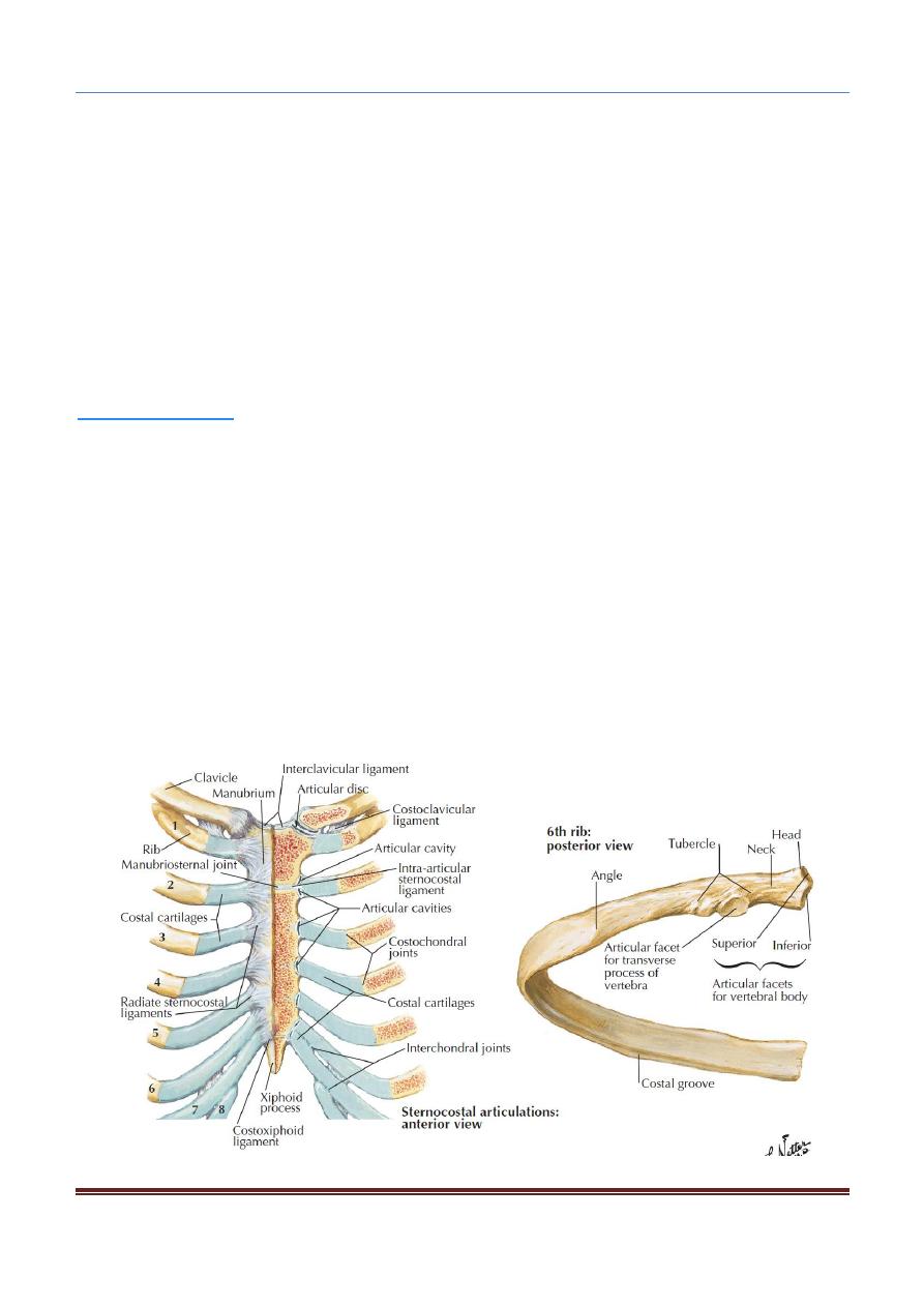
TYPICAL RIB:
Example of Typical rib : 3-9
A typical rib is a long, twisted, flat bone having a rounded, smooth superior border
and a sharp, thin inferior border.
The inferior border overhangs and forms the costal groove, which accommodates
the intercostal vessels and nerve.
The anterior end of each rib is attached to the corresponding costal cartilage.
CHEST ANATOMY THI-QAR UNIVERSITY
COLLEGE OF MEDICINE
LECTURE 1 2019/2020
Dr. Rafid AL-Temimi ; Clinical radiology ( CABM)
Page
4
Dr. Ahmed Abdulameer Daffar ; Thoracic & Vascular Surgeon ( FIBMS )
A rib has a head, neck; tubercle, shaft, and angle.
a) Head: has two facets for articulation with the numerically corresponding vertebral
body and that of the vertebra immediately above.
b) Neck: is a constricted portion situated between the head and the tubercle.
c) Tubercle: is a prominence on the outer surface of the rib at the junction of the
neck with the shaft.It has a facet for articulation with the transverse process of the
numerically corresponding vertebra.
d) Shaft: is thin and flattened and twisted on its long axis. Its inferior border has the
costal groove.
e) Angle: is where the shaft of the rib bends sharply forward.
ATYPICAL RIB:
Example of Atypical rib: 1; 2; 10; 11; 12
The 1st rib is important clinically because of its close relationship to the lower
nerves of the brachial plexus and the main vessels to the arm, namely, the
subclavian artery and vein. This rib is small and flattened from above
The scalenus anterior muscle is attached to its upper surface and inner border.
Anterior to the scalenus anterior, the subclavian vein crosses the rib; posterior to
the muscle attachment, the subclavian artery and the lower trunk of the
brachial plexus cross the rib and lie in contact with the bone.