
Date 28/5/2020
Subject :histology
Lecture 1 (one)
Epithelial Tissue
First year
Time(2-3)
Dr. Sabreen Saleem AL –Sayigh(Ph.D.)

Epithelial Tissue
General objective:
The aims of this lecture are :
The main function of the epithelial tissue
list the type of the epithelial tissue
Explain the structure and fined of the organs

Epithelial Tissue
The human body is composed of only four basic types of
tissue: epithelial, connective, muscular, and nervous.
These tissues, which are formed by cells and molecules of
the extracellular matrix, exist not as isolated units but
rather in association with one another and in variable
proportions, forming different organs and systems of the
body.

Epithelial tissues : are composed of closely aggregated
polyhedral cells with very little extracellular substance.
These cells have strong adhesion and form cellular sheets
that cover the surface of the body and line its cavities. The
principal functions of epithelial tissues are:
1-Covering, lining, and protecting surfaces (eg, skin).
2- Absorption (eg, the intestines).
3- Secretion (eg, the epithelial cells of glands).
4-Contractility (eg, myoepithelial cells).

Characteristic Features of Epithelial Cells :
The forms and dimensions of epithelial cells range from
high columnar to cuboidal to low squamous cells.
Their common polyhedral form results from their close
juxtaposition in cellular layers or masses and is similar
to what would be observed if a large number of inflated
balloons were compressed into a limited space.
Epithelial cell nuclei have a distinctive shape, varying
from spherical to elongated or elliptic. The nuclear
form often corresponds roughly to the cell shape; thus,
cuboidal cells have spherical nuclei, and squamous cells
have flattened nuclei. Most epithelia rest on connective
tissue
The macromolecular
components of basal
laminae form precise
three-dimensional
arrays, of these
include:
1-Laminin: These are
large glycoprotein
molecules
2- Type IV collagen:
Monomers of type IV
collagen contain three
polypeptide chains.
3-Entactin (nidogen):
a glycoprotein,

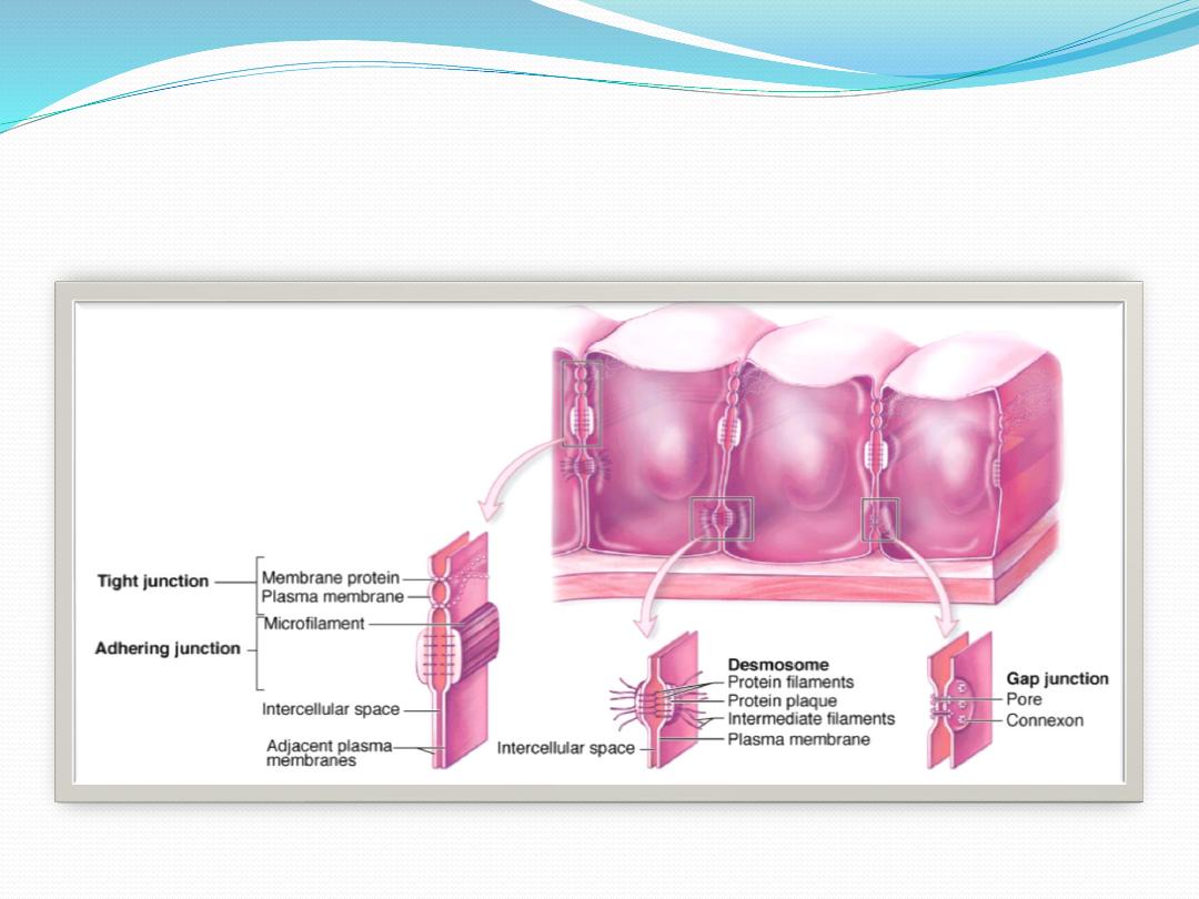
2-Intercellular Adhesion & Other Junctions
Several membrane-associated structures contribute to
adhesion and communication between cells. They are
present in most tissues but are particularly numerous
and prominent in epithelia and will be described here:
1-Tight junctions, or zonulae occludens: are the most
apical of the junctions. The Latin terminology gives
important information about the geometry of the
junction. "Zonula" indicates that the junctions form
bands completely encircling each cell, and "occludens"
refers to the membrane fusions that close off the space
between the cells

2-adherent junction or zonula adherens: The next type of
junction This junction also encircles the cell, usually
immediately below the zonula occludens, and provides for
the firm adhesion of one cell to its neighbors. Adhesion is
mediated by ,transmembrane glycoproteins of each cell,
3-desmosome or macula adherens. As the names imply,
this junctional type resembles a single "spot-weld" and
does not form a belt around the cell. The desmosome is a
disk-shaped structure at the surface of one cell that is
matched with an identical structure at the surface of an
adjacent cell desmosomes provide firm adhesion among
the cells.

4-Gap or communicating junctions can occur almost
anywhere along the lateral membranes of epithelial cells,
but are also found between cells in nearly all mammalian
tissues.
5- hemidesmosomes In the contact area between epithelial
cells and the subjacent basal lamina, can often be observed
ultratructurally. These adhesive structures resemble a half-
desmosome and bind the cell to the basal lamina the
plaques contain abundant integrins, transmembrane
proteins that are receptor sites for the extracellular
macromolecules laminin and collagen type IV

3-Specializations of the Apical cell Surface
The free or apical surface of many types of epithelial
cells has specialized structures to increase the cell
surface area or to move substances or particles bound
to the epithelium.
1- Microvilli: When viewed in the electron microscope,
many cells are seen to have cytoplasmic projections.
These projections may be short or long fingerlike
extensions or folds that pursue a sinuous course, and
they range in number from a few to many. Most are
temporary, reflecting cytoplasmic movements and the
activity of actin filaments.

2-Stereocilia : are long apical processes of cells in other
absorptive epithelia such as that lining the epididymis and
ductus deferens. These structures are much longer and less
motile than microvilli, are branched, and should not be
confused with true cilia. Like microvilli, stereocilia also
increase the cells' surface area, facilitating the movement of
molecules into and out of the cell
3-Cilia : Cilia are elongated, highly motile structures on the
surface of some epithelial cells, 5–10 m long and 0.2 m in
diameter, which is much longer and two times wider than a
typical microvillus

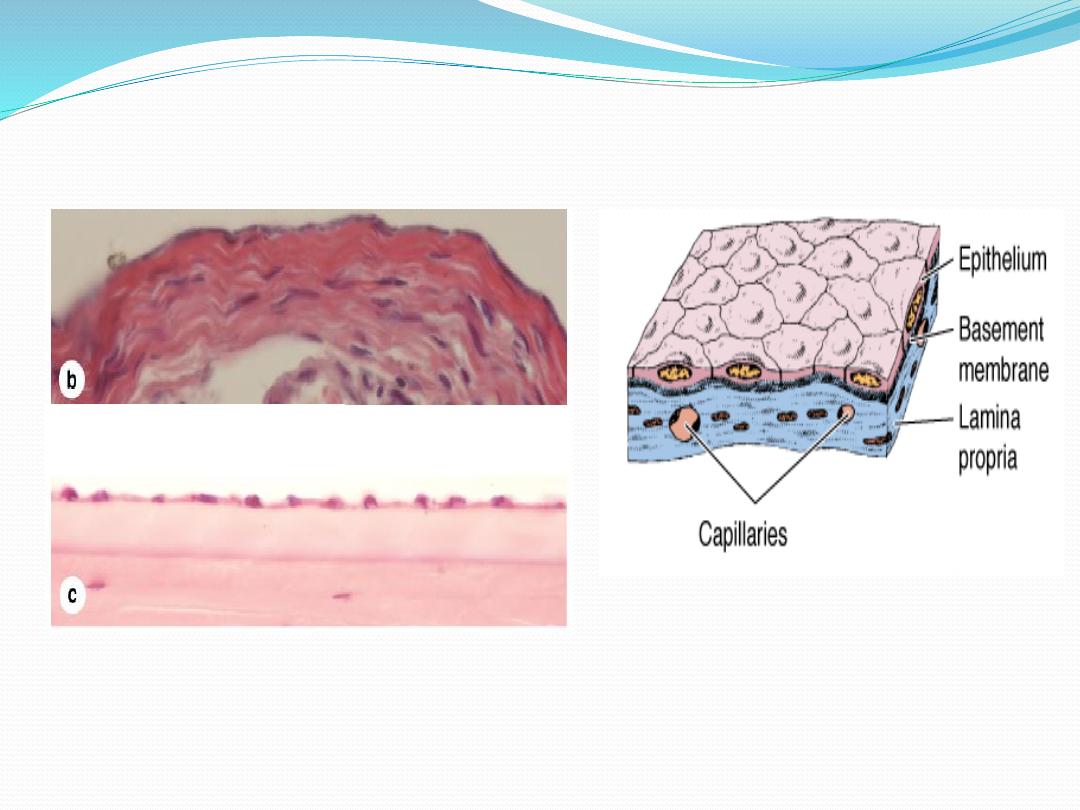
Epithelial Tissue
In the case of epithelia lining the
cavity of internal organs (especially in
the digestive, respiratory, and urinary
systems) this layer of connective
tissue is often called the lamina
propria. The lamina propria not only
serves to support the epithelium but
also provides nutrition and binds it to
underlying structures

1- Covering or Lining Epithelia
Covering epithelia are tissues in which the cells are
organized in layers that cover the external surface or
line the cavities of the body. They are classified
according to the number of cell layers and the
morphologic features of the cells in the surface layer.

A - Simple epithelia contain only one layer of cells and
stratified epithelia contain more than one layer.
1-Simple squamous epithelia
In simple squamous epithelium, cells of the single layer
are flat and usually very thin, with only the thicker cell
nucleus appearing as a bulge to denote the cell. Simple
epithelia are typically specialized as lining of vessels and
cavities and regulate substances which can enter
underlying tissue from the vessel or cavity. The thin cells
often exhibit trans cytosis. Examples shown here are those
lining the renal loops of Henle
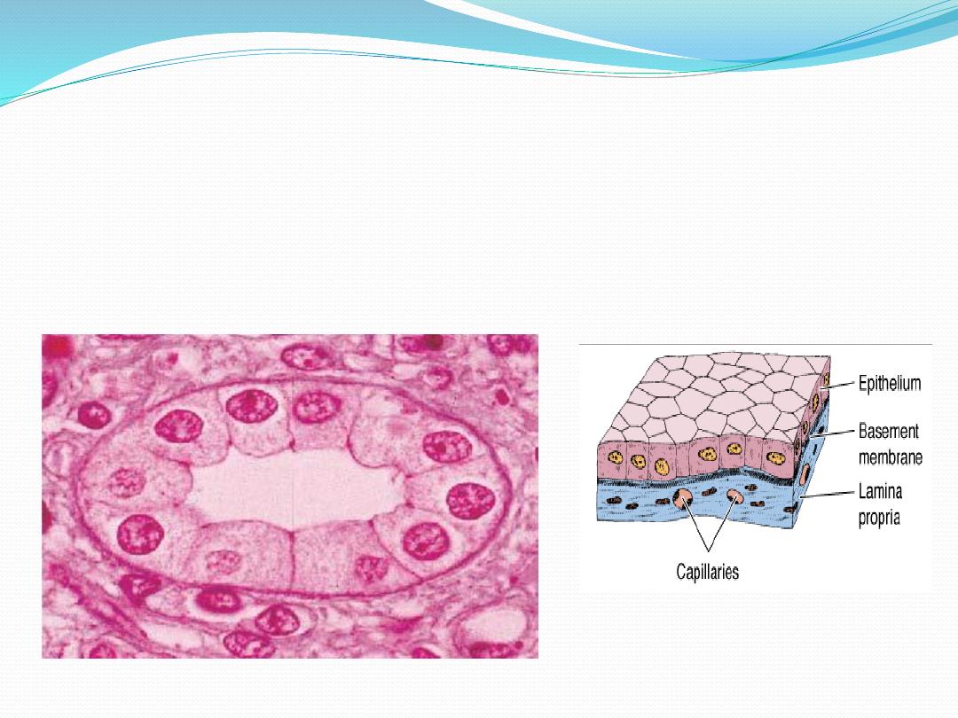
2-Simple cuboidal epithelium.
Cells of simple cuboidal epithelia vary in their height but are roug hly as tall as
they are wide. Their g reater thickness often includes cytoplasm rich in
mitochondria providing energ y for a high level of active transport of substances
across the epithelium. E xamples of simple cuboidal epithelia sh own here are
from a renal collecting tubule (a), a pancreatic duct (b), and the mesothelium
covering an ovary (c).
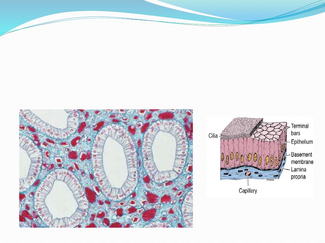
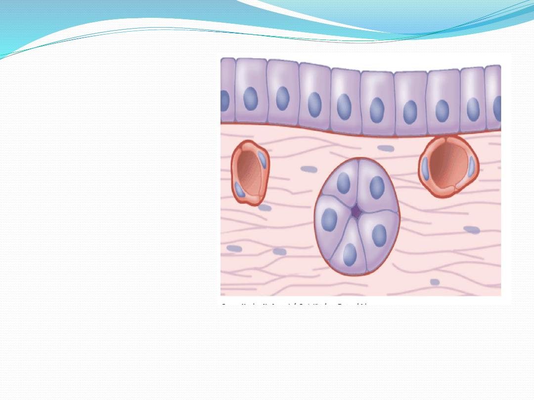
3-Simple columnar epithelium. Cells of simple columnar epithelia are taller than they are
wide. Such cells are usually highly specialized for absorption, with microvilli, and often
have interspersed secretory cells or ciliated cells. The additional cytoplasm in columnar
cells allows additional mitochondria and other organelles needed for absorption and
processing. The examples shown here are from a renal collecting duct (a), the oviduct
lining, with both secretory and ciliated cells (b), and the lining of the gall bladder (c).

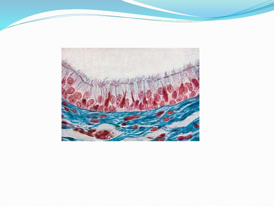
4- Single-Layered Pseudostratified
ColumnarEpithelium
In single-layered pseudostratified epithelium, the
longitudinal axis of the cells is always oriented vertical
to the tissue surface. The cells appear polygonalin
cross-sections. A row of oval nuclei mostly occupy the
basal part of the cell, while most of the cell ganelles are
located in the supra nuclear cell region

B- Stratified epithelia are classified according to the
cell shape of the superficial layers: squamous,
cuboidal, columnar, and transitional. The very thin
surface cells of stratified squamous epithelia can be
"keratinized" (rich in keratin intermediate filaments) or
"nonkeratinized" (with relatively sparse amounts of
keratin).

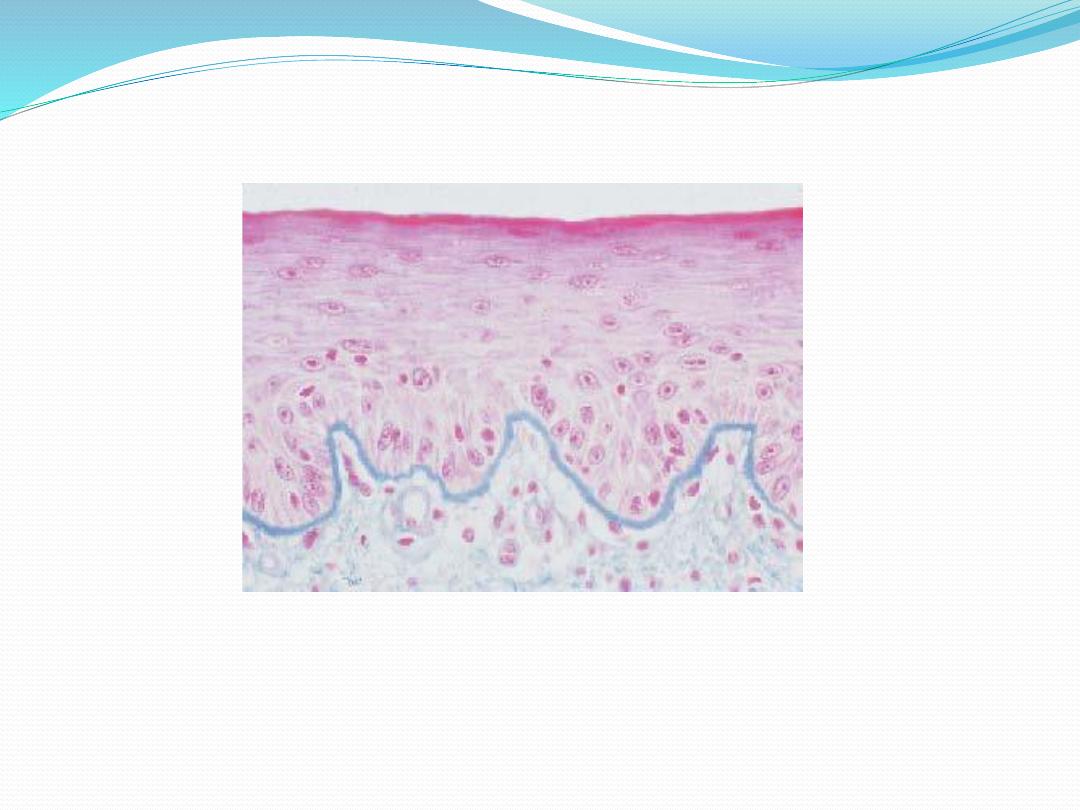
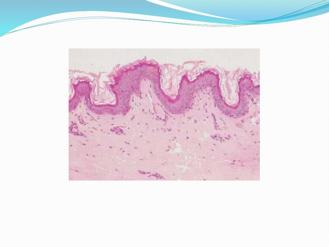
1-Stratified squamous keratinized epithelium is
found mainly in the epidermis of skin. Its cells form
many layers, and the cells closer to the underlying
connective tissue are usually cuboidal or low columnar.
The cells become irregular in shape and flatten as they
accumulate keratin in the process of keratinization and
are moved progressively closer to the surface, where they
become thin, metabolically inactive packets (squames)
of keratin lacking nuclei. This surface layer of cells helps
protect against water loss across this epithelium.
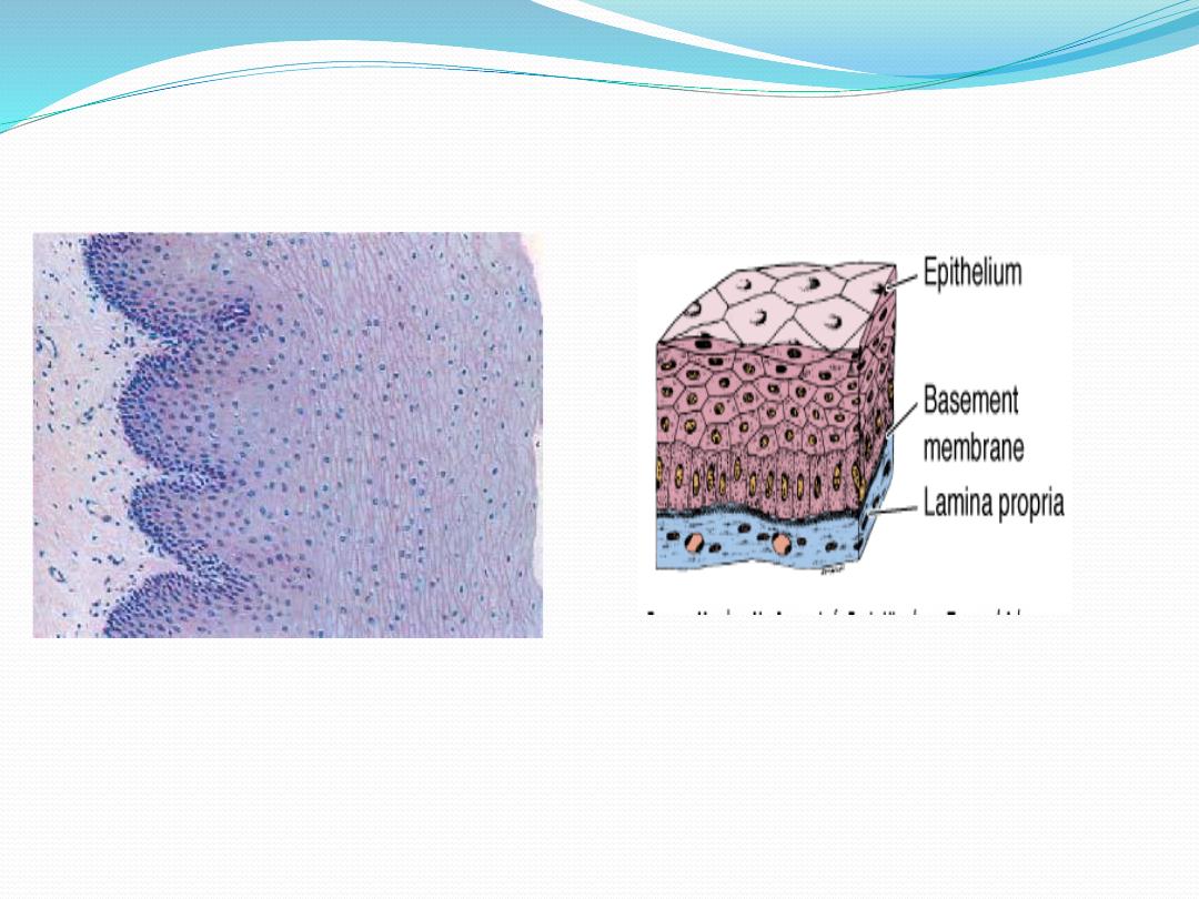
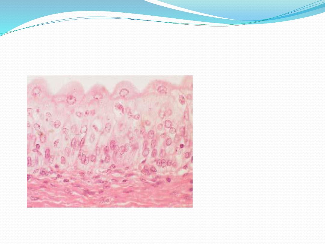
Stratified squamous nonkeratinized epithelium
(Figure 4–14) lines wet cavities (eg, mouth, esophagus,
and vagina). In such areas where water loss is not a
problem, the flattened cells of the epithelial surface layer
are living cells containing much less keratin and
retaining their nuclei.

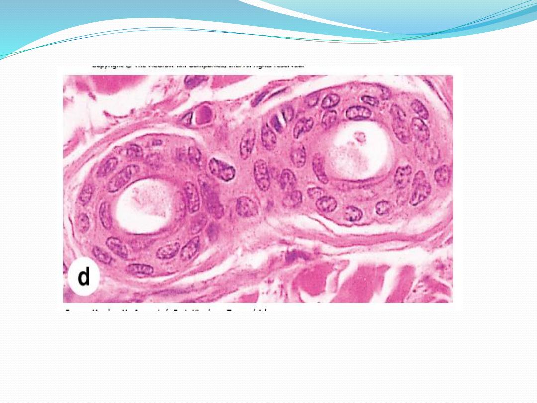
2-Stratified
cuboidal
and
stratified
columnar
epithelia are rare. Stratified columnar epithelium can
be found in the conjunctiva lining the eyelids, where it
is both protective and mucus secreting. Stratified
cuboidal epithelium is restricted to large excretory
ducts
of
sweat
and
salivary
glands,
where
it
apparently provides a lining more robust than that of a
simple epithelium
.
3-Transitional epithelium or urothelium, which lines only the urinary bladder, the ureter,
and the upper part of the urethra, is characterized by a superficial layer of domelike cells
that are neither squamous nor columnar