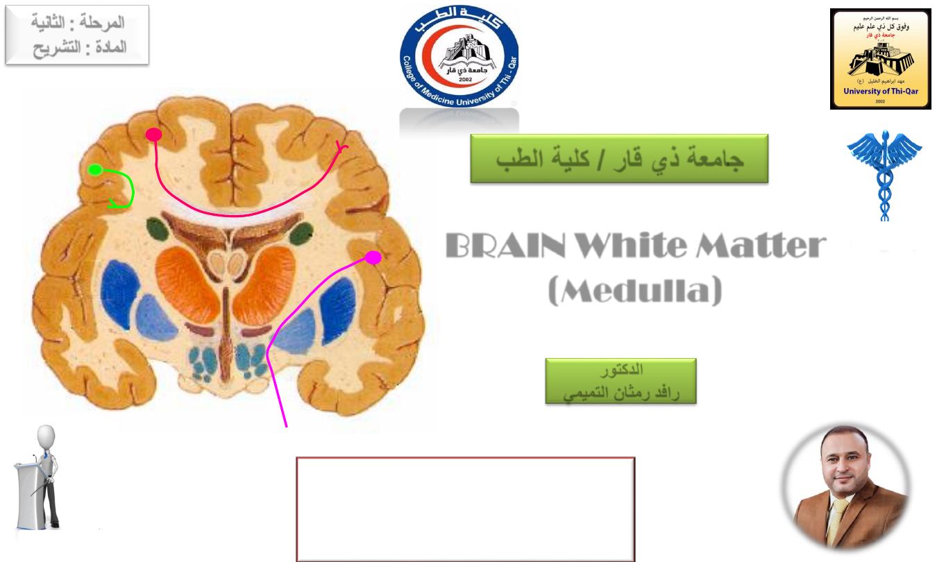
BRAIN White Matter
(
Medulla
)
Done by
Dr.Rafid Remthan Al-Temimi
Clinical Radiology
CAMB,DMRD,M.B.Ch.B.,.
المرحلة
:
الثانية
المادة
:
التشريح
ج
امعة ذي قار
/
كلية الطب
الدكتور
رافد
رمثان التميمي
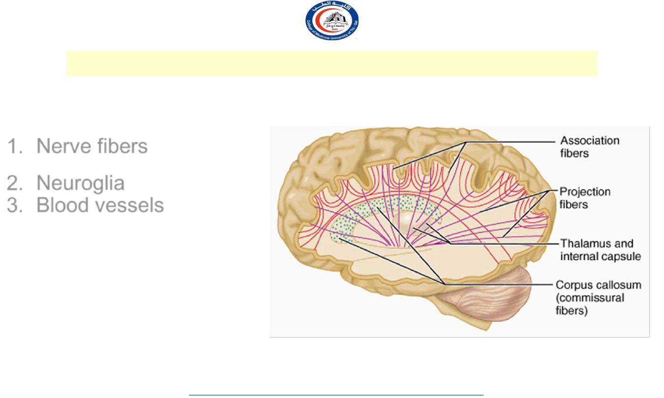
Underlies the cortex
Contains:
1. Nerve fibers (
predominantly
myelinated)
2. Neuroglia
3. Blood vessels
The nerve fibers originate,
terminate or sometimes
both, within the cortex
Cerebral hemisphere
– white mater
2
University Of Thi-Qar
College Of medicine
Anatomy lecture . 2
nd
stage
Dr.Rafid Al-Temimi
Dr.Rafid Remthan AL-Temimi,Clinical Radiology,CAMB, 2020
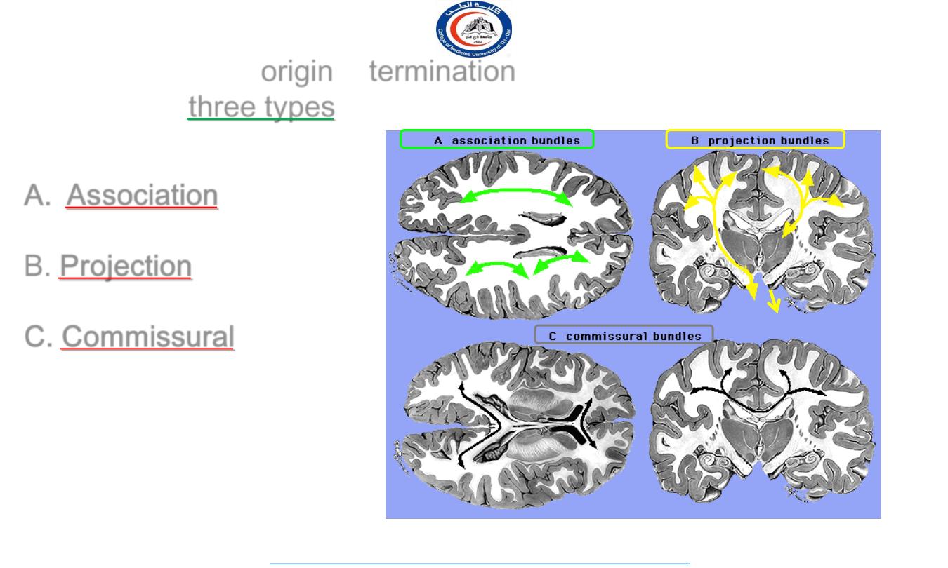
Depending on their
origin
&
termination
, these nerve fibers are
classified into
three types
:
A. Association
B. Projection
C. Commissural
3
University Of Thi-Qar
College Of medicine
Anatomy lecture . 2
nd
stage
Dr.Rafid Al-Temimi
Dr.Rafid Remthan AL-Temimi,Clinical Radiology,CAMB, 2020
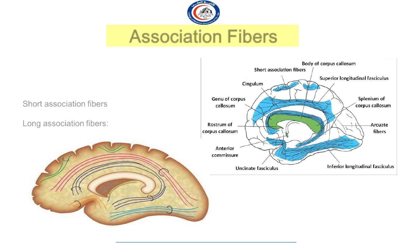
Association Fibers
Unite different parts of the
same
hemisphere
Are of
two kinds:
Short association fibers
:
those
connecting
adjacent gyri,
Long association fibers:
those connecting
more distant gyri
4
University Of Thi-Qar
College Of medicine
Anatomy lecture . 2
nd
stage
Dr.Rafid Al-Temimi
Dr.Rafid Remthan AL-Temimi,Clinical Radiology,CAMB, 2020
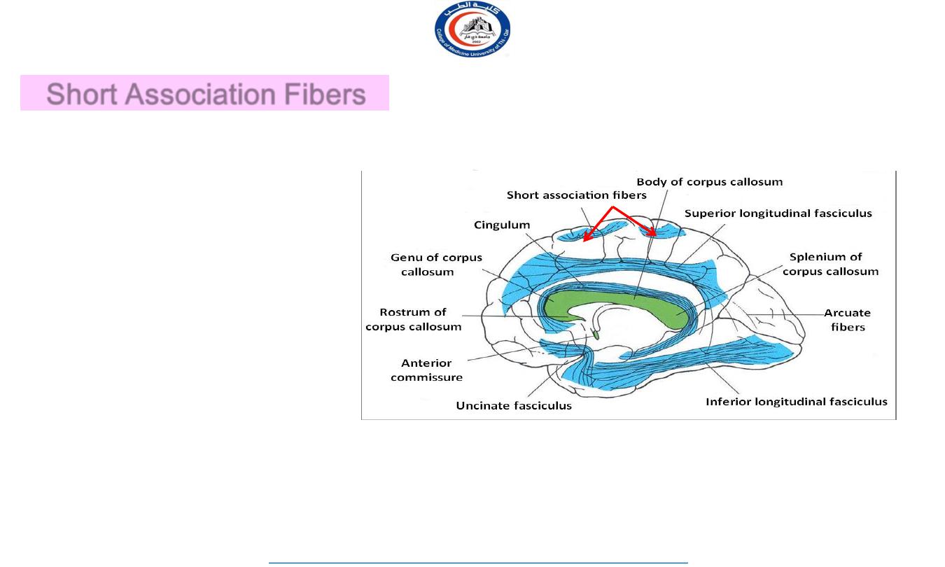
Short Association Fibers
Lie immediately beneath the
gray substance of the cortex
Connect together the
adjacent
gyri.
5
University Of Thi-Qar
College Of medicine
Anatomy lecture . 2
nd
stage
Dr.Rafid Al-Temimi
Dr.Rafid Remthan AL-Temimi,Clinical Radiology,CAMB, 2020
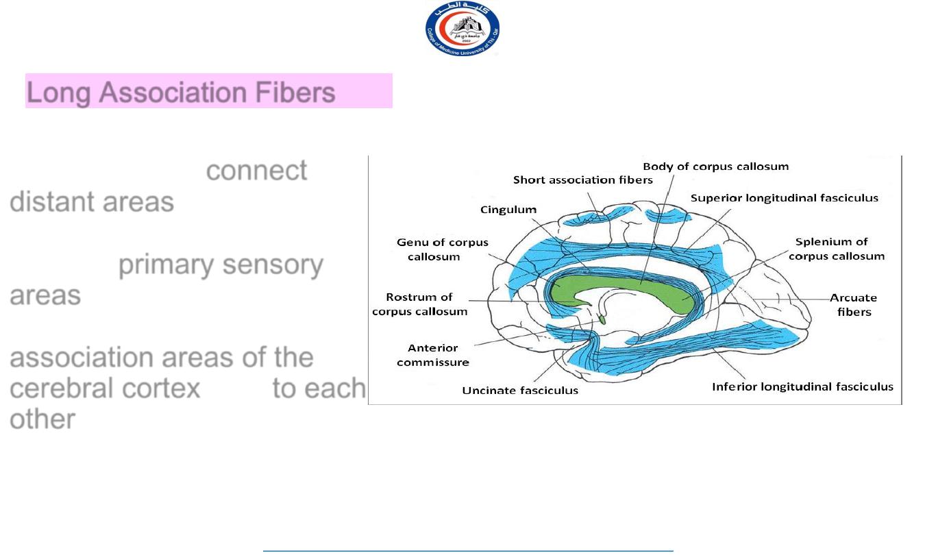
Long Association Fibers
Long fibers travel through
white matter to
connect
distant areas
of cerebral
cortex
Link the
primary sensory
areas
in parietal, temporal
and occipital lobes to the
association areas of the
cerebral cortex
, and
to each
other
6
University Of Thi-Qar
College Of medicine
Anatomy lecture . 2
nd
stage
Dr.Rafid Al-Temimi
Dr.Rafid Remthan AL-Temimi,Clinical Radiology,CAMB, 2020
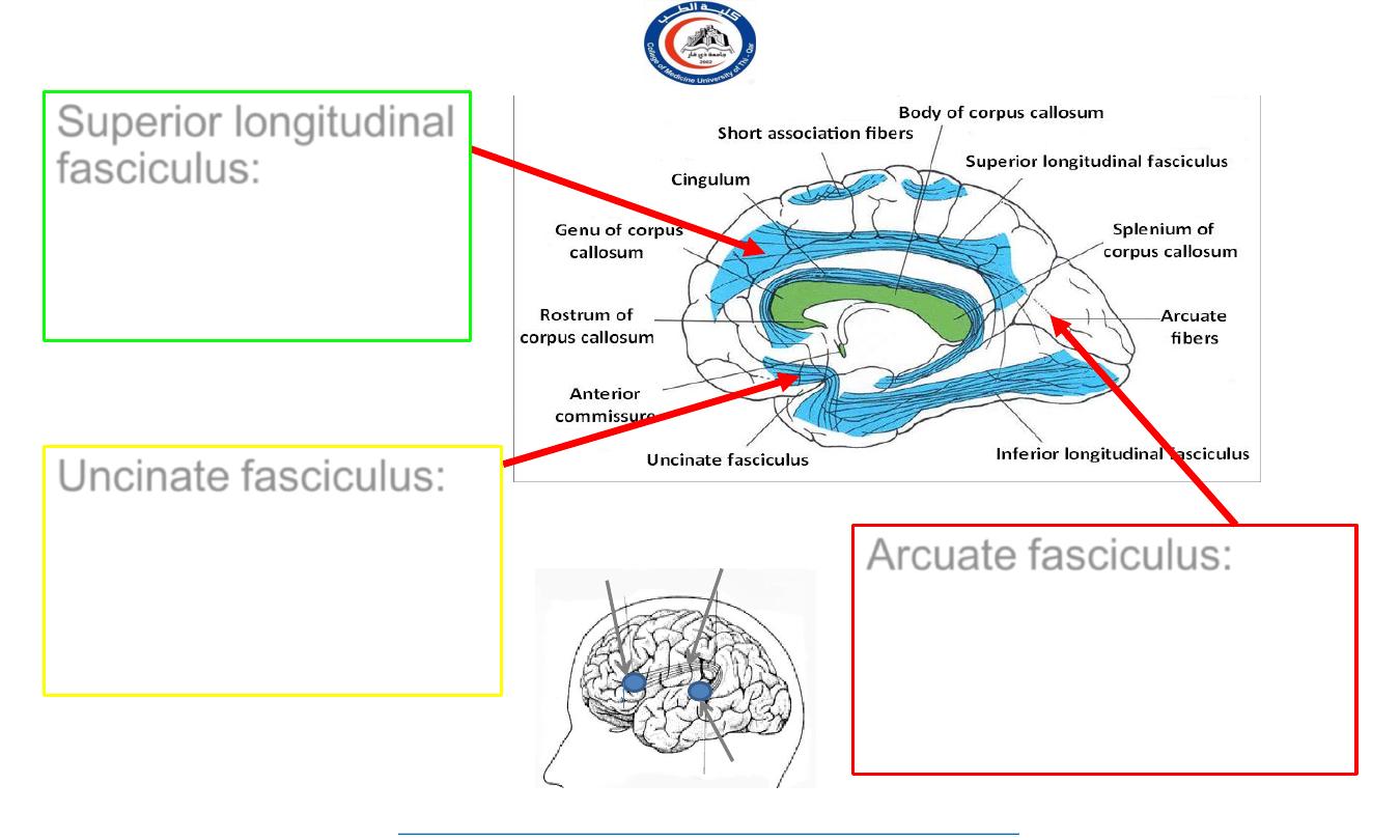
Superior longitudinal
fasciculus
:
connects
the frontal, parietal,
temporal and
occipital lobes
Uncinate fasciculus:
connects frontal to
temporal lobe,
contributing to the
regulation of behavior
Arcuate fasciculus:
connect gyri in frontal to
temporal lobes,
important for language
function
Wernicke’s Area
Broca’s
Area
Arcuate Fasciculus
7
University Of Thi-Qar
College Of medicine
Anatomy lecture . 2
nd
stage
Dr.Rafid Al-Temimi
Dr.Rafid Remthan AL-Temimi,Clinical Radiology,CAMB, 2020
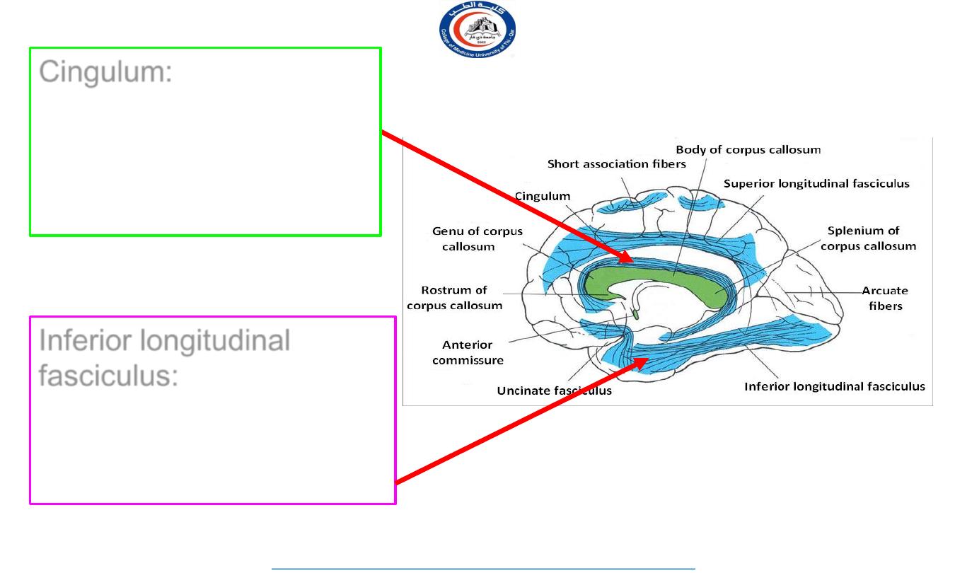
Cingulum:
connects
frontal & parietal lobes to
the para-hippocampal
gyrus and adjacent
temporal gyri
Inferior longitudinal
fasciculus:
connects
occipital to temporal pole
& contributes to visual
recognition
8
University Of Thi-Qar
College Of medicine
Anatomy lecture . 2
nd
stage
Dr.Rafid Al-Temimi
Dr.Rafid Remthan AL-Temimi,Clinical Radiology,CAMB, 2020
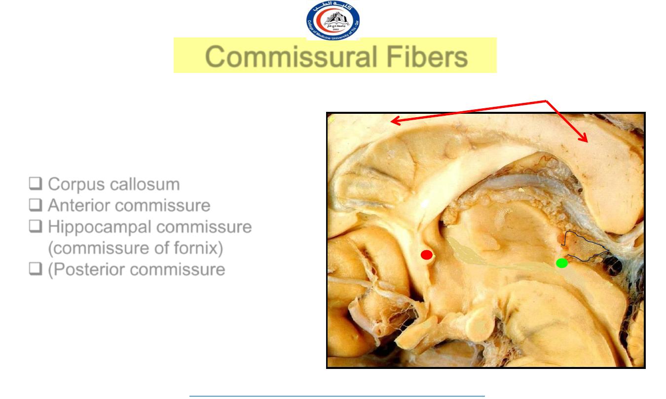
Commissural Fibers
Connect the corresponding regions
of the two hemispheres
Include:
Corpus callosum
Anterior commissure
Hippocampal commissure
(commissure of fornix)
(Posterior commissure
Corpus Callosum
F
P
9
University Of Thi-Qar
College Of medicine
Anatomy lecture . 2
nd
stage
Dr.Rafid Al-Temimi
Dr.Rafid Remthan AL-Temimi,Clinical Radiology,CAMB, 2020
Commissural Fibers
10
University Of Thi-Qar
College Of medicine
Anatomy lecture . 2
nd
stage
Dr.Rafid Al-Temimi
Dr.Rafid Remthan AL-Temimi,Clinical Radiology,CAMB, 2020
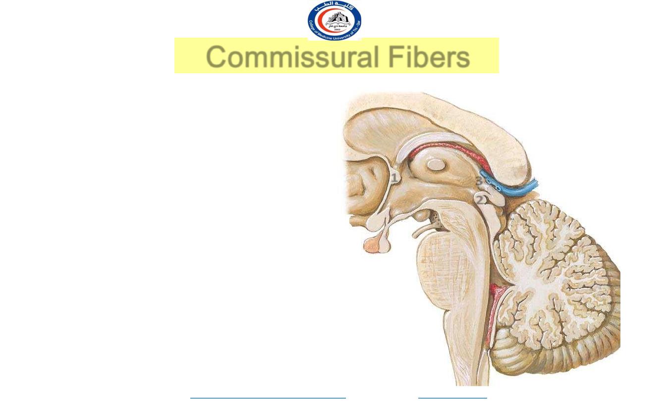
• 1- Anterior commissure :-
Connect both
temporal lobes
.
• 2- Posterior commissure :-
• Contain fibers from pretectal nuclei
concerned with light reflex
• 3- habenular commissures :-
• Connect habenular nuclei which are
connected to the amygdala & hippocampus
1
2
3
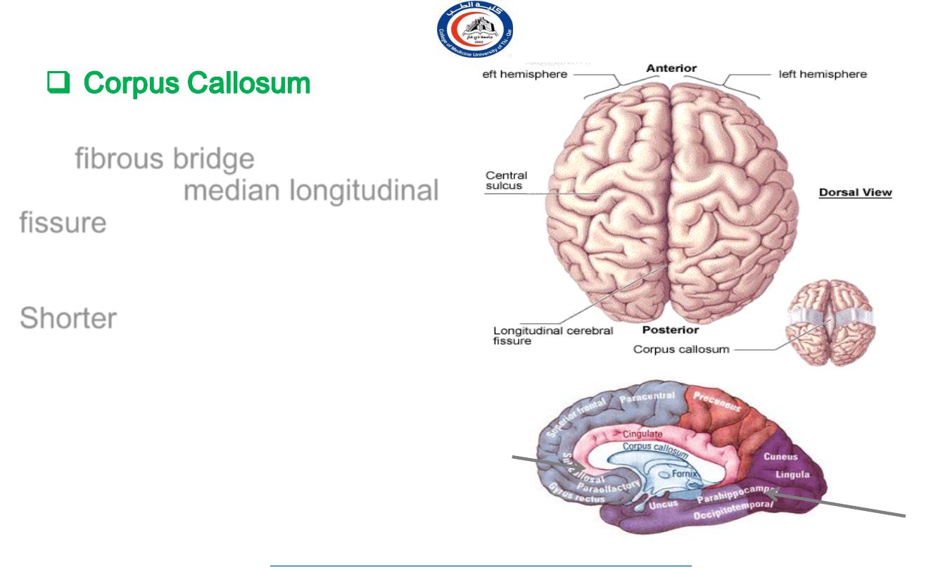
Is a
fibrous bridge
located in the
depth of the
median longitudinal
fissure
Connects the two cerebral
hemispheres together
Shorter
craniocaudally than is the
hemisphere
Cranial end is nearer to the frontal
pole of hemisphere as compared to
caudal end to the occipital pole
11
University Of Thi-Qar
College Of medicine
Anatomy lecture . 2
nd
stage
Dr.Rafid Al-Temimi
Dr.Rafid Remthan AL-Temimi,Clinical Radiology,CAMB, 2020
The fibers in the
corpus callosum
connect the corresponding regions
of the two hemispheres with each
other (
except the inferior part of
the temporal lobes
)
C
C
12
University Of Thi-Qar
College Of medicine
Anatomy lecture . 2
nd
stage
Dr.Rafid Al-Temimi
Dr.Rafid Remthan AL-Temimi,Clinical Radiology,CAMB, 2020
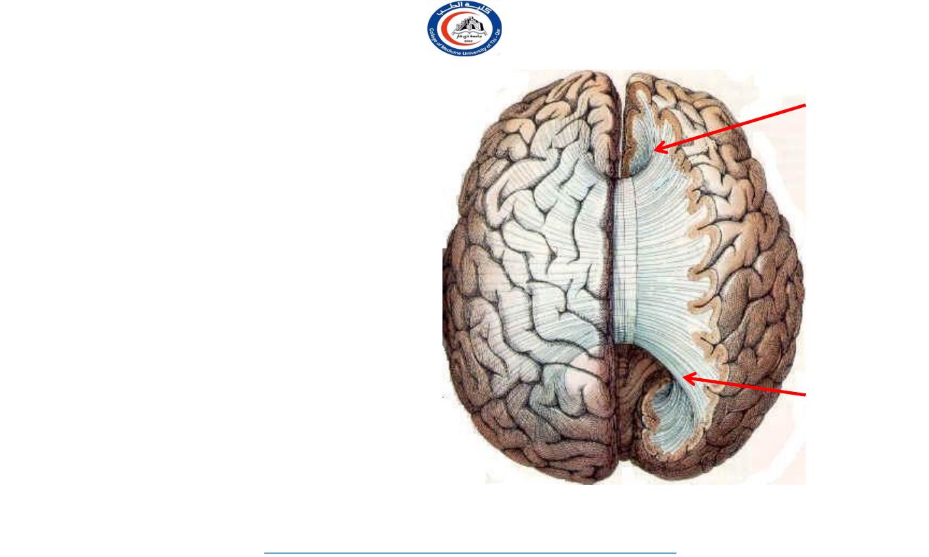
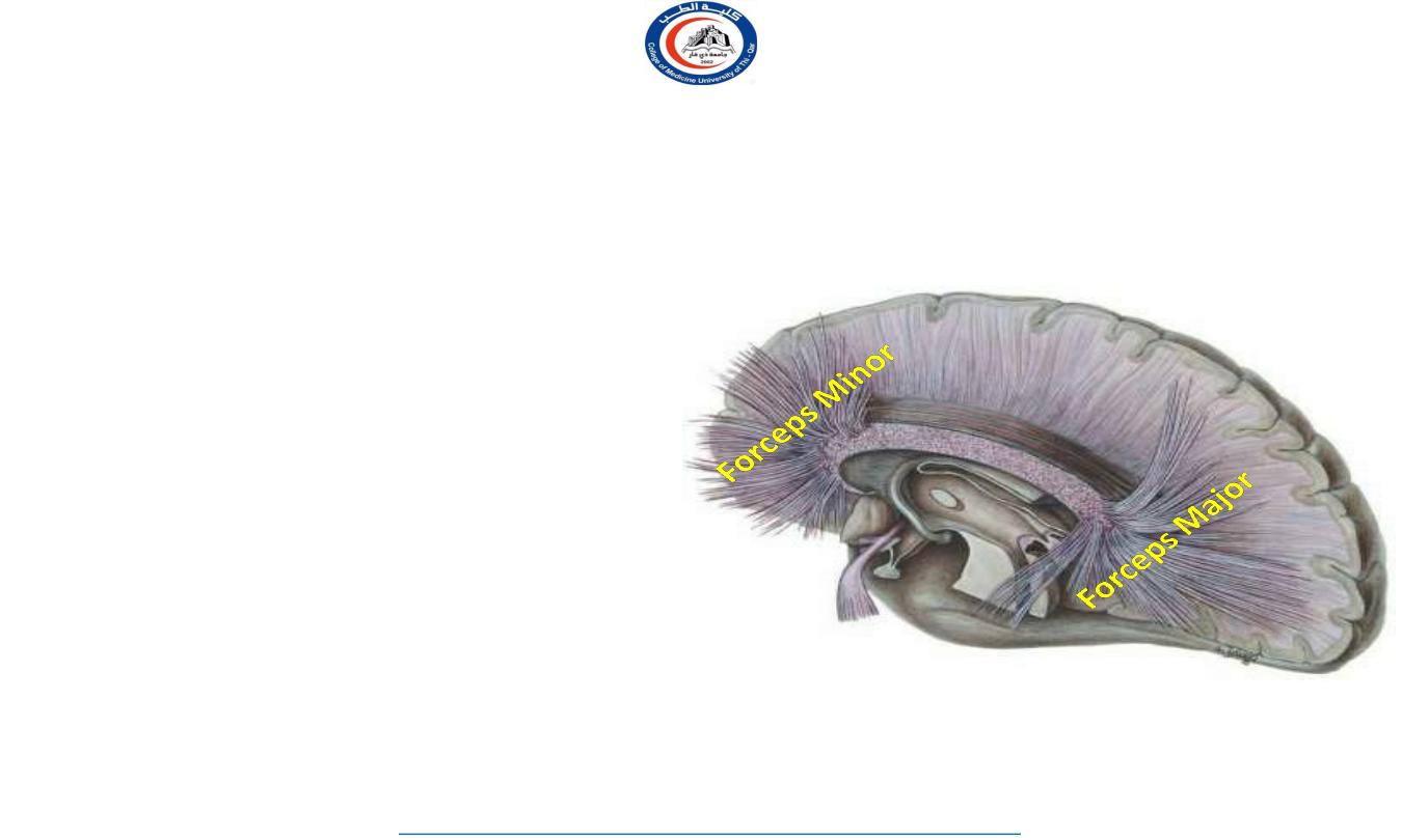
•
Fibers linking the two
frontal
poles
with each other, curve
forward
& form
u-shaped
anterior forceps (forceps
minor)
• Fibers linking the two
occipital
poles
with each other, curve
backward
& form
u-shaped
posterior forceps (forceps
major)
F
P
O
C
C
Anterior
forceps
Posterior
forceps
13
University Of Thi-Qar
College Of medicine
Anatomy lecture . 2
nd
stage
Dr.Rafid Al-Temimi
Dr.Rafid Remthan AL-Temimi,Clinical Radiology,CAMB, 2020
14
University Of Thi-Qar
College Of medicine
Anatomy lecture . 2
nd
stage
Dr.Rafid Al-Temimi
Dr.Rafid Remthan AL-Temimi,Clinical Radiology,CAMB, 2020
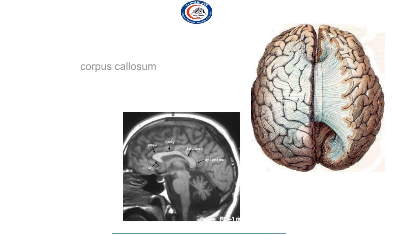
• 1- rostrum
• 2- genu
- Contains the forceps in
which connect areas the
frontal lobes
in the body extend
as radiation of the
• 3- body
- Fibers
laterally
corpus
- When descend in the lateral wall of the
inferior & posterior horns of lateral
ventricle are called tapetum.
• 4- splenum
- contains the forceps major which connect
areas in occipital lobes
2
1
3
4
Parts of corpus callosum
…
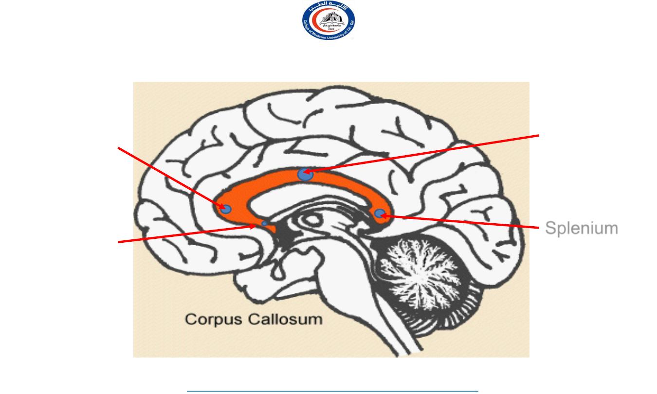
Parts of Corpus Callosum
Splenium
Body
Genu
Rostrum
15
University Of Thi-Qar
College Of medicine
Anatomy lecture . 2
nd
stage
Dr.Rafid Al-Temimi
Dr.Rafid Remthan AL-Temimi,Clinical Radiology,CAMB, 2020
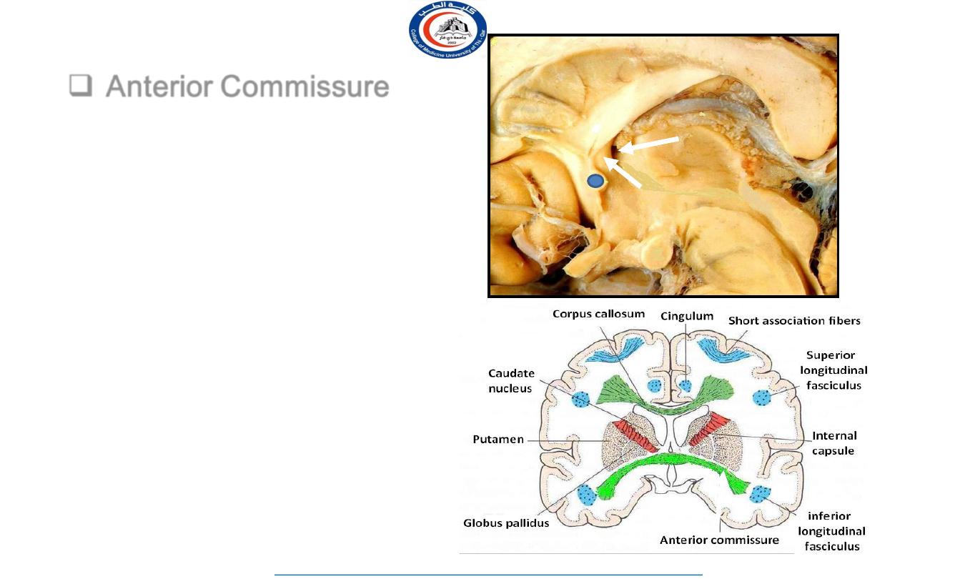
Anterior Commissure
Bundle of fibers runs
transversely in front of the
anterior columns of fornix
Connects the
inferior and
middle temporal gyri
&
the
olfactory regions
of the two
hemispheres
Anterior
column of
fornix
fornix
IVF
16
University Of Thi-Qar
College Of medicine
Anatomy lecture . 2
nd
stage
Dr.Rafid Al-Temimi
Dr.Rafid Remthan AL-Temimi,Clinical Radiology,CAMB, 2020
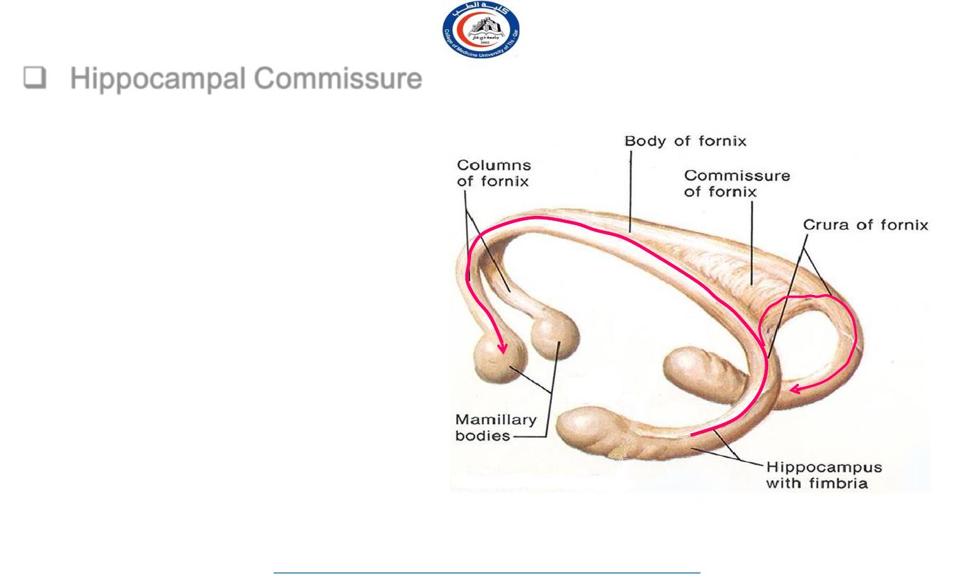
Hippocampal Commissure
Bundle of fibers runs transversely
between the crura of the fornix
Connect the
two hippocampi
with
each other
(note that hippocampo-mamillary
fibers do not cross)
17
University Of Thi-Qar
College Of medicine
Anatomy lecture . 2
nd
stage
Dr.Rafid Al-Temimi
Dr.Rafid Remthan AL-Temimi,Clinical Radiology,CAMB, 2020
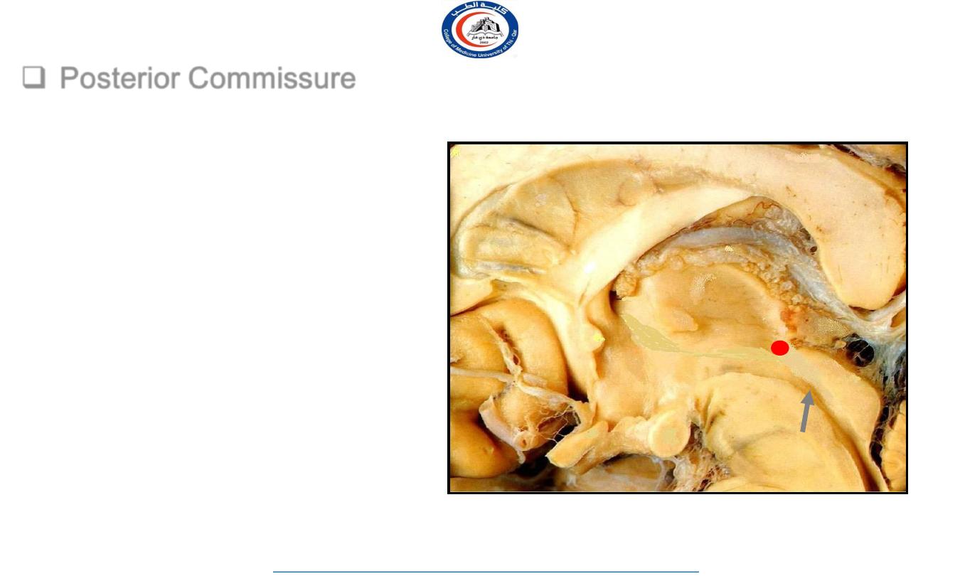
Posterior Commissure
Rounded band of white fibers
Crossing the midline on the
dorsal aspect of the upper end
of the cerebral aqueduct
(located
between superior colliculus & pineal
body)
Connects the
left and right
midbrain.
Plays important role in
the bilateral pupillary reflex
SC
P
IC
Cerebral
aqueduct
18
University Of Thi-Qar
College Of medicine
Anatomy lecture . 2
nd
stage
Dr.Rafid Al-Temimi
Dr.Rafid Remthan AL-Temimi,Clinical Radiology,CAMB, 2020
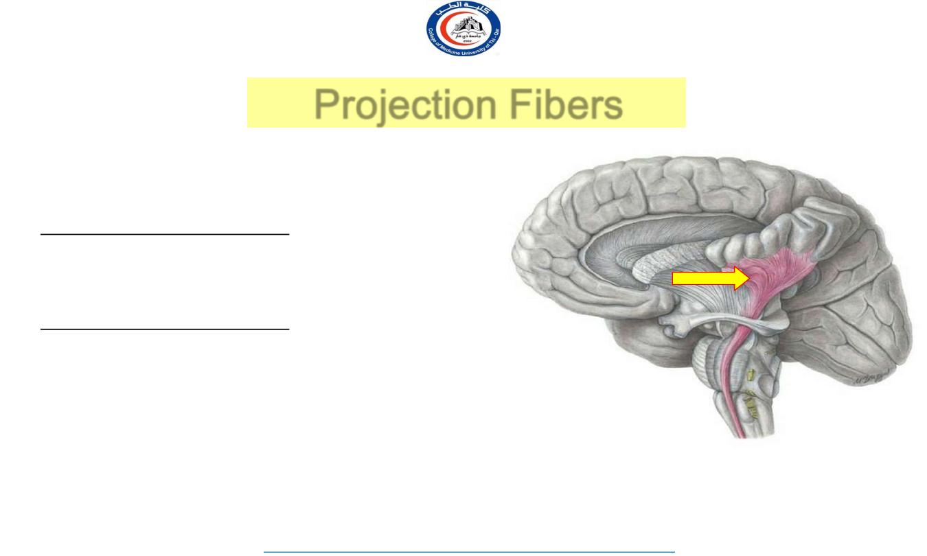
Projection Fibers
Fibers running vertically through the
hemispheres
Consist of:
Cortical afferent fibers
conveying impulses
to
the cerebral cortex:
(mainly thalamo-
cortical fibers)
Cortical efferent fibers
carrying impulses
away
from
the cortex to the lower centers:
(corticostriate, corticobulbar,
corticopontine, corticospinal, & descending
autonomic fibers)
19
University Of Thi-Qar
College Of medicine
Anatomy lecture . 2
nd
stage
Dr.Rafid Al-Temimi
Dr.Rafid Remthan AL-Temimi,Clinical Radiology,CAMB, 2020
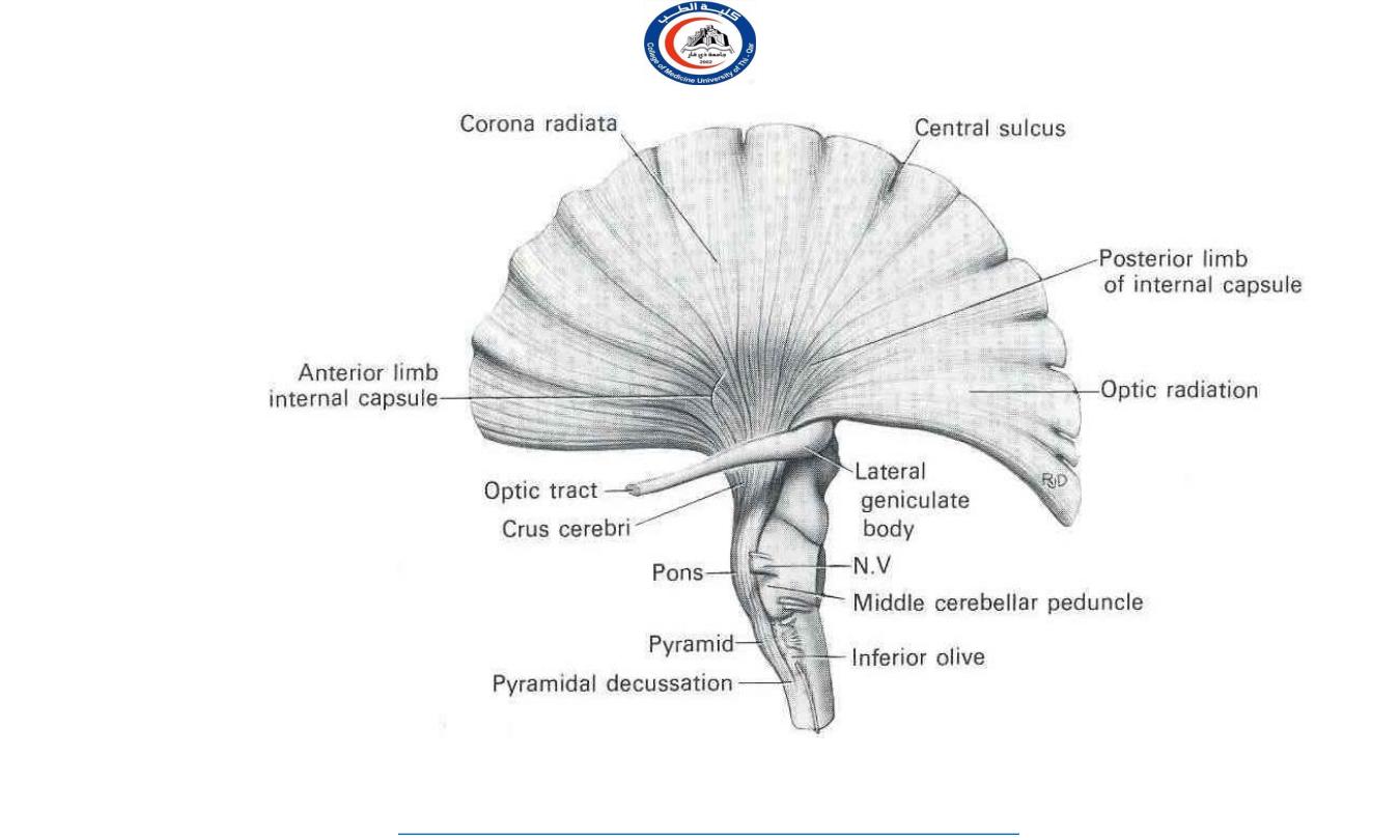
Deeper to the cortex, these fibers
are arranged radially as the
corona
radiata
Then the fibers converge to form a
sheath, called the
internal capsule
,
that passes between the thalamus
and the basal ganglia
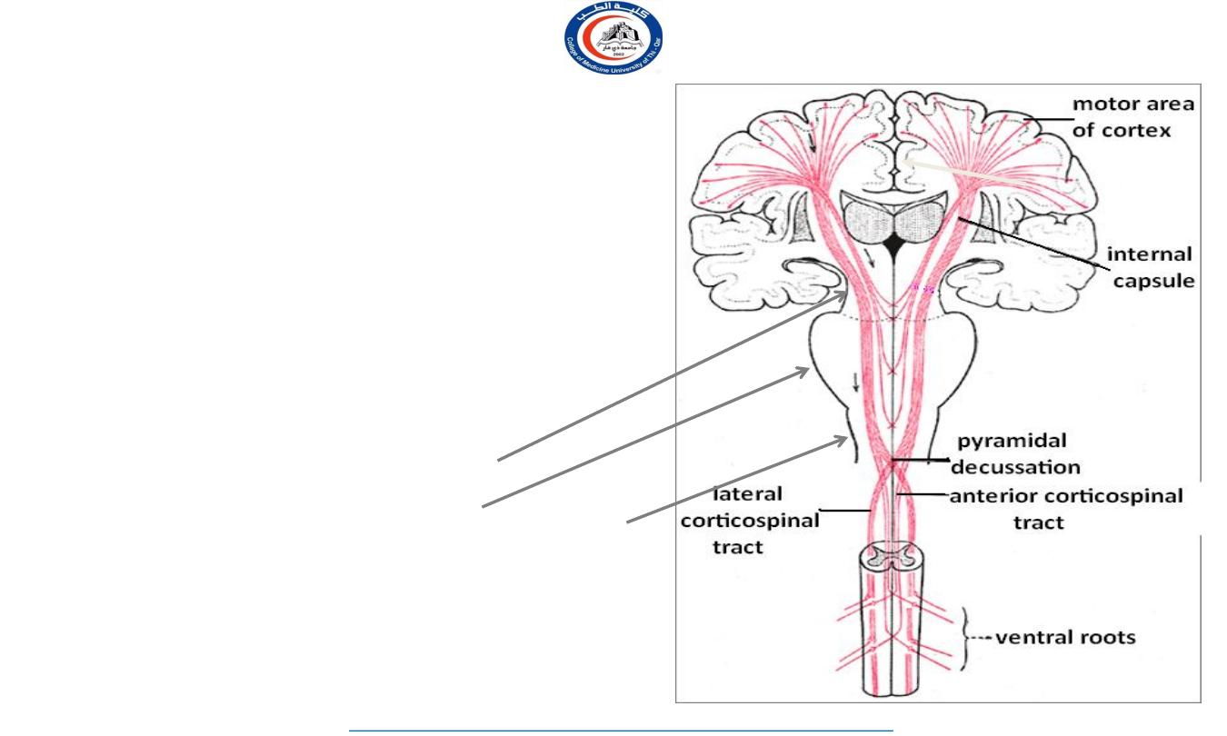
Continue in the:
Crus of the midbrain
Basilar part of pons
Pyramid of medulla oblongata
Continue in the spinal cord as the
corticospinal tracts
corona
radiata
20
University Of Thi-Qar
College Of medicine
Anatomy lecture . 2
nd
stage
Dr.Rafid Al-Temimi
Dr.Rafid Remthan AL-Temimi,Clinical Radiology,CAMB, 2020
21
University Of Thi-Qar
College Of medicine
Anatomy lecture . 2
nd
stage
Dr.Rafid Al-Temimi
Dr.Rafid Remthan AL-Temimi,Clinical Radiology,CAMB, 2020

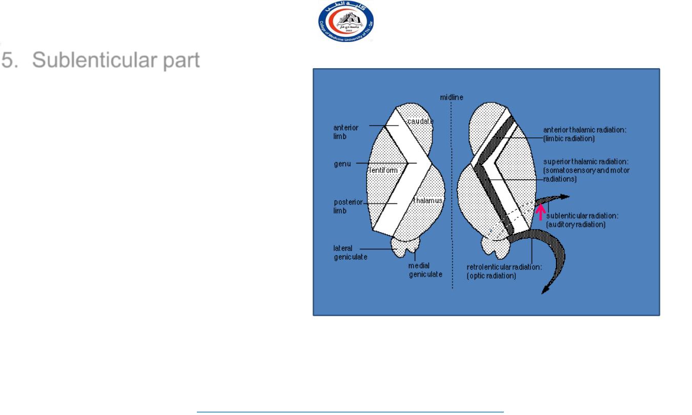
Internal Capsule
Bundle of
projection fibers
,
passes
through the interval between the
thalamus
and the
basal ganglia
BG
Th
22
University Of Thi-Qar
College Of medicine
Anatomy lecture . 2
nd
stage
Dr.Rafid Al-Temimi
Dr.Rafid Remthan AL-Temimi,Clinical Radiology,CAMB, 2020
AL
G
PL
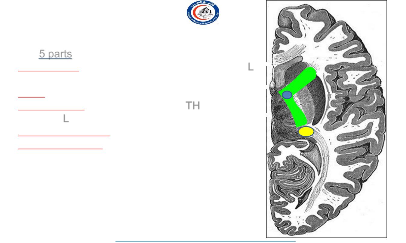
•
Has 5 parts:
1. Anterior limb
:
between caudate
(C)
& lentiform
(L)
nuclei
2. Genu
3. Posterior limb
:
between thalamus
(TH)
& lentiform
nucleus
(L)
4. Retrolenticular part
:
caudal to lentiform nucleus
5. Sublenticular part:
below lentiform nucleus
(canion)
C
Th
L
1
2
3
4
23
University Of Thi-Qar
College Of medicine
Anatomy lecture . 2
nd
stage
Dr.Rafid Al-Temimi
Dr.Rafid Remthan AL-Temimi,Clinical Radiology,CAMB, 2020
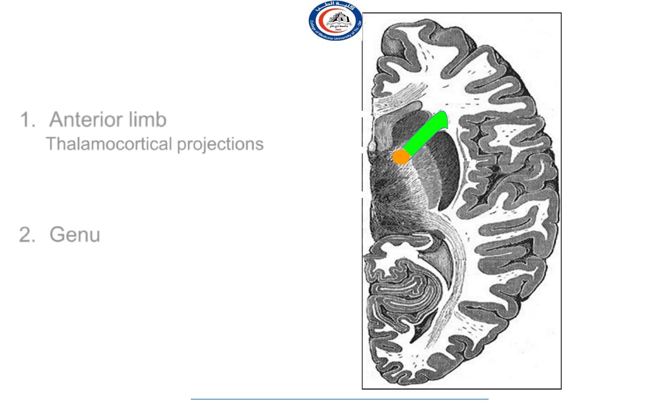
1. Anterior limb
contains:
Thalamocortical projections
that
connect mediodorsal nucleus of
thalamus with the prefrontal cortex
Frontopontine fibers
2. Genu
contains:
Corticobulbar fibers
which connect the
cortex with cranial nerve
motor nuclei
in
the brainstem
24
University Of Thi-Qar
College Of medicine
Anatomy lecture . 2
nd
stage
Dr.Rafid Al-Temimi
Dr.Rafid Remthan AL-Temimi,Clinical Radiology,CAMB, 2020
1.
.
2.
.
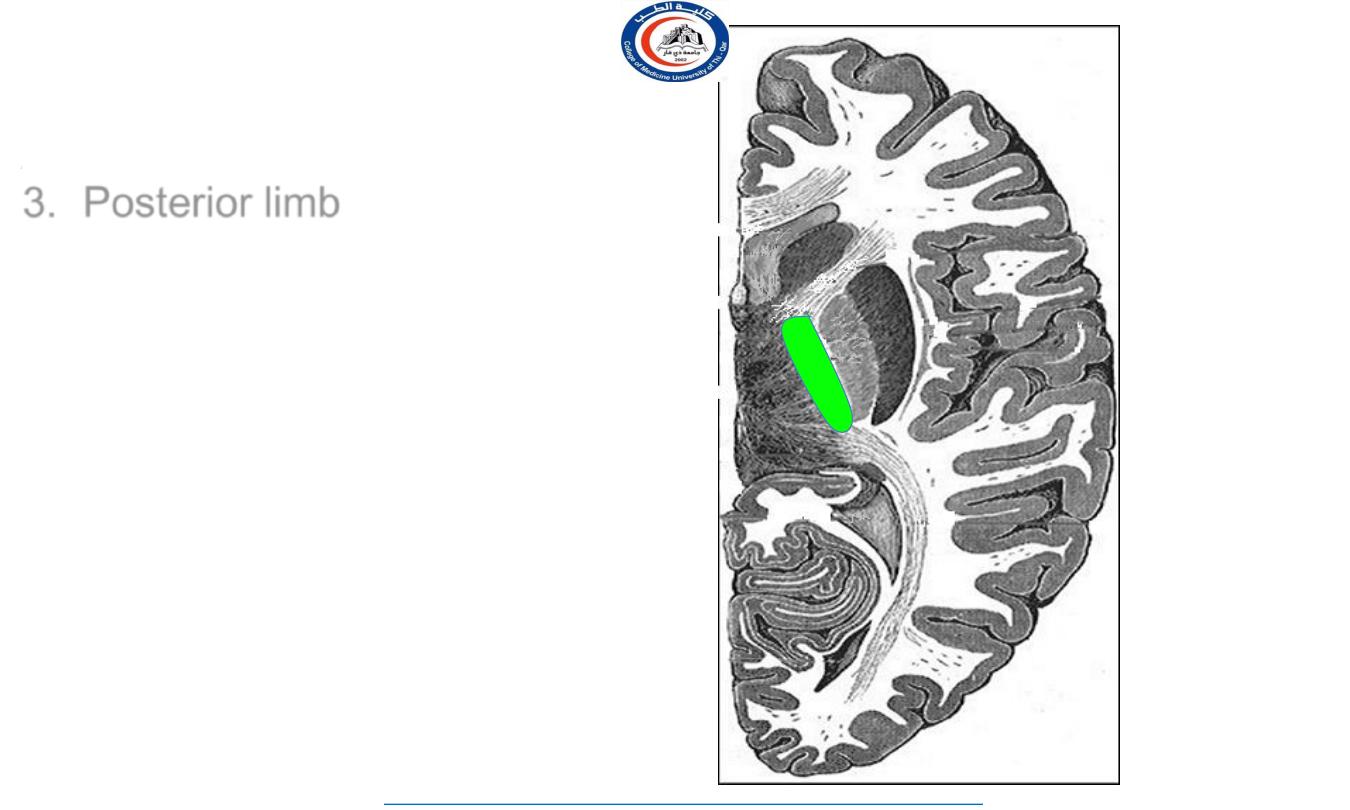
3. Posterior limb
contains
:
1.
Corticospinal
2.
Corticobulbar
3.
Thalamocortical projections
from:
VPN to the primary
somatosensory cortex
VAN & VLN to motor regions of
cortex
25
University Of Thi-Qar
College Of medicine
Anatomy lecture . 2
nd
stage
Dr.Rafid Al-Temimi
Dr.Rafid Remthan AL-Temimi,Clinical Radiology,CAMB, 2020
1.
.
2.
.
3.
.
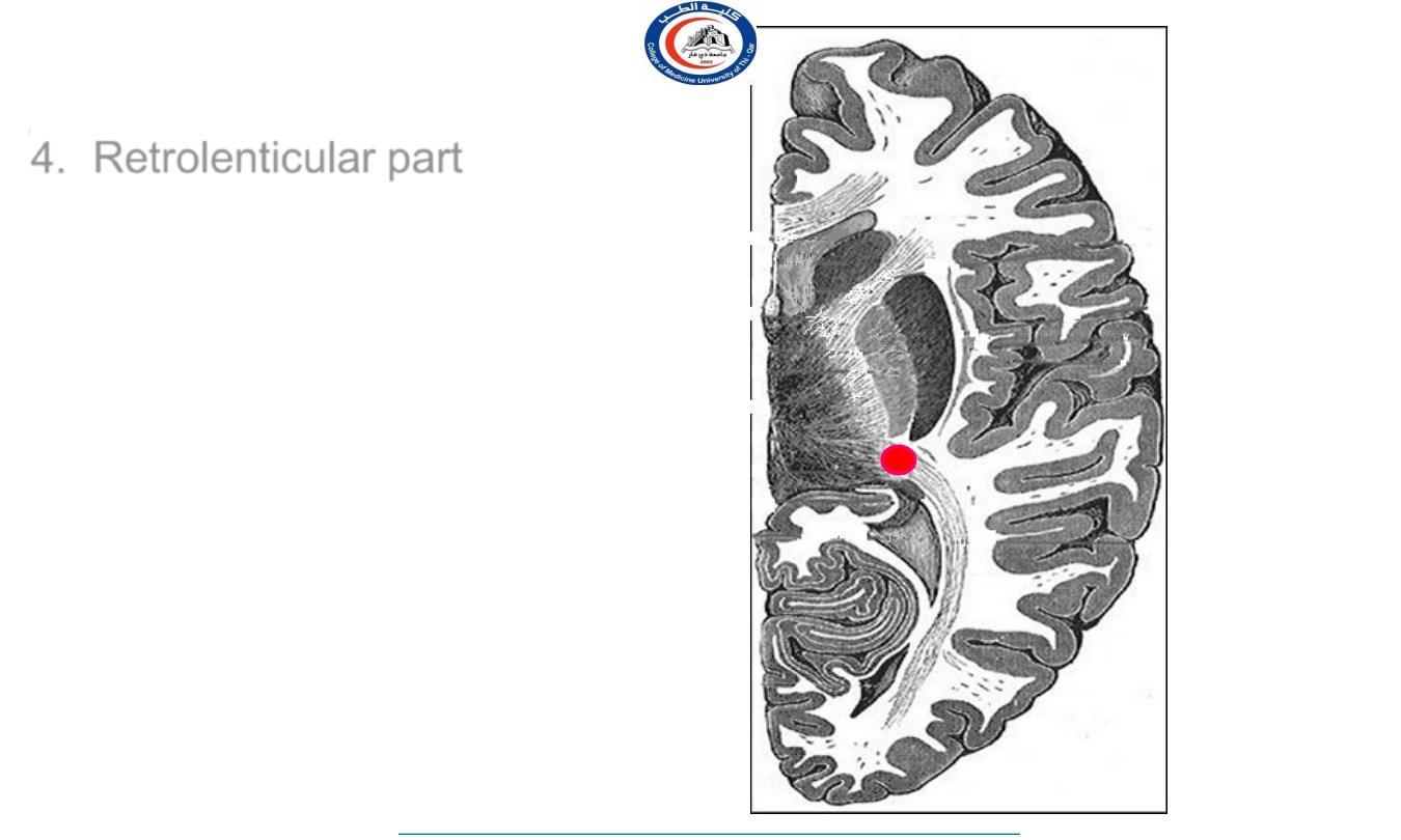
4. Retrolenticular part
contains
thalamocortical projections:
Geniculocalcarine fibers
(visual
radiation), from the lateral geniculate
nucleus of thalamus to the visual
cortex in the occipital lobe
& few
Geniculotemporal fibers
(auditory radiation) from the medial
geniculate nucleus of thalamus to the
auditory cortex in the temporal lobe
26
University Of Thi-Qar
College Of medicine
Anatomy lecture . 2
nd
stage
Dr.Rafid Al-Temimi
Dr.Rafid Remthan AL-Temimi,Clinical Radiology,CAMB, 2020
1.
.
2.
.
3.
.
4.
.
5. Sublenticular part
contains
thalamocortical projections
:
geniculo-temporal fibers
(auditory radiation) from the
medial geniculate nucleus of
thalamus to the auditory
cortex in the temporal lobe
27
University Of Thi-Qar
College Of medicine
Anatomy lecture . 2
nd
stage
Dr.Rafid Al-Temimi
Dr.Rafid Remthan AL-Temimi,Clinical Radiology,CAMB, 2020
28
University Of Thi-Qar
College Of medicine
Anatomy lecture . 2
nd
stage
Dr.Rafid Al-Temimi
Dr.Rafid Remthan AL-Temimi,Clinical Radiology,CAMB, 2020

29
University Of Thi-Qar
College Of medicine
Anatomy lecture . 2
nd
stage
Dr.Rafid Al-Temimi
Dr.Rafid Remthan AL-Temimi,Clinical Radiology,CAMB, 2020