Sublingual glands
AnatomyPaired gland lying in the anterior part of the floor of the mouth between the mucous membrane, the mylohoid muscle and the body of the mandible close the mental symphysis.
Head drains by numerous excretory ducts (ducts of Rivinus) directly into the oral cavity.
The tail drains into the submandibular duct or directly into the mouth.
Cysts
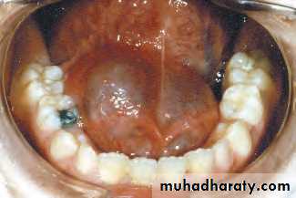
Ranula: mucous extravasation cyst.Characteristic frog belly.
Resolve spontaneously
May require surgery but with high morbidity.
Surgery include removal of the gland.
Incision and drainage… high recurrence.
Rana….. Frog in Latin
Sublingual glands
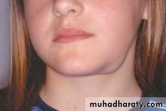
Plunging ranula
rare form of mucous retention cyst.
Mucus collects below the gland and perforates through mylohyoid muscle diaphragm to enter the neck.
Soft, fluctuant painless double shaped swelling in submandibular or submental region of the neck.
DX: US or MRI
Aspiration… thick yellow treacly fluid, distinguishing the cyst from lymphangioma.
Treatment by surgery. Cervical approach is contraindicated.
Removal of the gland and aspirate of the saliva.
The sac is formed of connective tissue not epithelium so melts away once the leak of saliva is resolved.
Sublingual glands
TumoursExtremely rare and are usually malignant 90%
Rubry painless swelling in the floor of the mouth.
Pain or lingual nerve paresthesia indicate a high grade tumour.
Dx punch biopsy.
Treatment: en block wide excision involving the overlying mucosa and adjacent periosteum with simultaneous neck dissection.
Immediate reconstruction of intraoral defect with radial artery forearm free flap or anterolateral thigh flap.
Minor salivary glands
Anatomy
800 minor salivary glands in the oral cavity.
Distributed in the lips, cheeks, palate, floor of the mouth ,retromolar area, upper aerodigestive.
Contributes to 10% of the total salivary volume.
Cysts
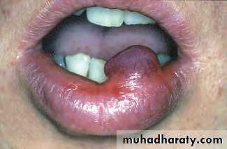
They are extravasation rather than retention variety.Lower lip and the floor of the mouth
Painless, usually but not always translucent.
Some resolve spontaneously and some require surgical excision.
Rare recurrence
Minor salivary glands
TumoursThey are malignant.
Commonly in the palate, upper lip and retromolar regions.
A well defined rubbery lump is a salivary gland tumour until proven otherwise.
Benign: painless, firm, slow growing
Benign tumour less than 1cm… excisional biopsy and the defect left to heal by secondary intention.
Larger than 1cm … punch biopsy if prove benign then formal excision.
Malignant neoplasms
Mostly low grade resembling benign lumps.
Firm, the overlying mucosa may be pink, blue or black.
High gade lesions usually become necrotic with ulceration as late presentation.
Low grade tumour in the palate… wide excision, the defect left to heal by secondary intention.
Lesions that perforate the palate require partial or total maxillectomy.
The defect managed by prosthetic obturation or immediate reconstruction.
Minor salivary glands
Necrotising sialometaplasia
This is a well established but rare entity.
Typically it occurs in the palate and mimics an aggressive cancer.
It presents as a deep punched out ulcer with an indurated margin.
It can not be distinguished from a neoplastic lesion except by biopsy.
The diagnosis is sugested by rapid onset in a young person.
The lesion resolves spontaniously with symptomatic treatment.
