
Physiology of excitable tissues lecture 5 Assi. Prof. Dr. Zahid M. kadhim
1
SYNAPSES
neurons generate electrical signals in the form of action potentials that
transmit messages from one area of the cell to another, and then from one
nerve cell to another through synapses. synapses are junctions where the
axon or some other portion of one cell (the presynaptic cell ) terminates on
the dendrites, soma, or axon of another neuron or, in some cases, a muscle
or gland cell (the postsynaptic cell). Synapses are dynamic structures,
increasing and decreasing in complexity and number with use, experience
and certainly age.
Two types of synapses are found in the nervous system: electrical
synapses and chemical synapses. Electrical synapses operate by allowing
electrical signals to be transmitted from one neuron to another through gap
junctions. Chemical synapses operate through the release of
neurotransmitters that activate signal transduction mechanisms in the target
cell.
Electrical synapses
Electrical synapses exist between neurons and either other neurons or glial
cells. At electrical synapses, the plasma membranes of adjacent cells are
linked together by gap junctions such that when an electrical signal is
generated in one cell, it is directly transferred to the adjacent cell by means
of ions flowing through the gap junctions. Second messenger molecules can
also move through these junctions.
Electrical synapses allow rapid communication between adjacent
neurons that synchronizes the electrical activity in these cells. This
communication is often bidirectional, although some gap junctions allow
current flow in only one direction.
The communication can be excitatory or inhibitory at the same
synapse, as either a depolarizing or a hyperpolarizing current can spread
through these junctions.
Electrical synapses have been identified in the retina of the eye and
certain areas of the cortex, areas of the brainstem that regulate breathing.

Physiology of excitable tissues lecture 5 Assi. Prof. Dr. Zahid M. kadhim
2
Chemical synapse
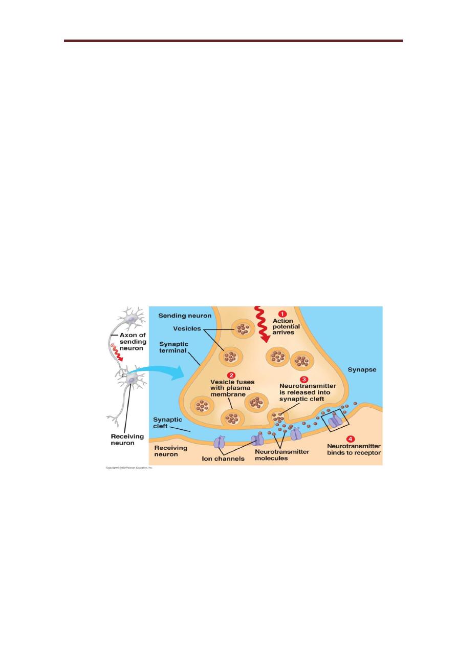
Almost all neurons transmit messages to other cells at chemical synapses. In
a chemical synapse, first neuron (the presynaptic neuron) secretes a
neurotransmitter into the extracellular fluid (synaptic cleft) in response to
an action potential arriving at its axon terminal. The neurotransmitter then
binds to receptors on the plasma membrane of a second cell (the
postsynaptic neuron), triggering in that cell an electrical signal that may or
may not initiate an action potential, depending on a number of
circumstances. A neuron can form synapses with other neurons or with
effector cells such as muscle or gland cell and is called a neuroeffector
junction.
Functional Anatomy of chemical synapse
Most often the presynaptic neuron’s axon terminal forms a synapse
with a dendrite of the postsynaptic neuron, in which case the synapses are
referred to as axodendritic synapses. However, synapses between the axon
terminal and soma of the postsynaptic cell, called axosomatic synapses, also
occur and have the same function as axodendritic synapses. The presynaptic
neuron’s axon terminal sometimes forms a synapse with the postsynaptic
neuron’s axon terminal, in which case the synapse is called an axoaxonic
synapse. Axoaxonic synapses have a special function in modulating
communication at axodendritic and axosomatic synapses. Dendrodendritic
synapses have also been identified.
In all cases, the axon terminal of the presynaptic neuron releases
neurotransmitters into the synaptic cleft. Once released into the synaptic
cleft, the neurotransmitters diffuse rapidly across the cleft and bind to
receptors on the postsynaptic neuron. The binding of the neurotransmitter
to the receptors produces a response in the postsynaptic neuron.
The axon terminal of the presynaptic neuron contains numerous small,
membrane-bound compartments called synaptic vesicles, which store
neurotransmitter molecules. Most neurotransmitters are synthesized in the
cytosol of the axon terminal, where the enzymes for their synthesis are
located. After synthesis, neurotransmitters are actively transported into
synaptic vesicles, where they are stored until their eventual release by
Physiology of excitable tissues lecture 5 Assi. Prof. Dr. Zahid M. kadhim
3
exocytosis. while the vesicles and the proteins contained in their walls are
synthesized in the neuronal cell body and transported along the axon to the
endings by fast axoplasmic transport.
Calcium ions play important role in synaptic transmission. Cytosolic
calcium triggers the release of neurotransmitter by exocytosis. Calcium
channels in the plasma membrane of presynaptic neuron open when the
axon terminal is depolarized. Calcium will flow down its electrochemical
gradient into the axon terminal, thereby increasing its concentration in the
cytosol of axon terminal. Calcium then causes the membranes of synaptic
vesicles to fuse with vesicle attachment sites on the inner surface of the
axon terminal membrane and undergo exocytosis, which releases the
neurotransmitters into the synaptic cleft.
The amount of neurotransmitter released depends on the
concentration of calcium in the cytosol of the axon terminal, which depends
on the frequency of action potentials in the presynaptic neuron.
Chemical synapse
Following a single action potential, neurotransmitter release stops
within a few milliseconds because the voltage-gated calcium channels close
soon after opening, and because calcium ions are actively transported out of
the axon terminal on a continual basis, bringing the cytosolic calcium
concentration back to its resting level. If a second action potential arrives
before neurotransmitter is cleared from the synaptic cleft, however, then

Physiology of excitable tissues lecture 5 Assi. Prof. Dr. Zahid M. kadhim
4
the cytosolic calcium levels increase causing more neurotransmitter to be
released from the presynaptic cell, thereby increasing the amount of
neurotransmitter in the synaptic cleft. When a series of action potentials
arrives at an axon terminal in a short time, cytosolic calcium levels increase
even more, thereby releasing even more neurotransmitter. As a
consequence, the concentration of neurotransmitter in the synaptic cleft
increases as the frequency of action potentials increases.
The binding of a neurotransmitter molecule to a receptor is a brief and
reversible process. If neurotransmitter molecules were to remain indefinitely
in the synaptic cleft following their release, they would bind to receptors
over and over again, inducing a continual response in the postsynaptic
neuron. Continual binding of neurotransmitter to receptor does not occur
because a number of processes quickly clear the neurotransmitter from the
cleft, thereby terminating the signal.
Some neurotransmitter molecules are degraded by enzymes, which
may be located on the postsynaptic neuron’s plasma membrane, on the
presynaptic neuron’s plasma membrane, on the plasma membranes of
nearby glial cells, in the interstitial fluid of the synaptic cleft, or even in the
cytoplasm of the presynaptic neuron or glial cells. Other neurotransmitter
molecules can be actively transported back into the presynaptic neuron that
released them, a process known as reuptake. Once inside the neuron, these
neurotransmitter molecules are usually degraded and the breakdown
products recycled to form new neurotransmitter molecules. Still other
neurotransmitter molecules in the synaptic cleft simply diffuse out of the
cleft.
As a result, neurotransmitter is usually present in the synaptic cleft for
only a few milliseconds after its release from the presynaptic neuron.
Once an impulse reaches the presynaptic terminals, a response can be
obtained in the postsynaptic neuron after a a time lag called Synaptic delay
.The delay is due to the time it takes for the synaptic mediator to be released
from presynaptic neuron and diffuse through the synaptic cleft and act on
the postsynaptic neuronal receptors.
Signal Transduction Mechanisms at Chemical Synapses

Physiology of excitable tissues lecture 5 Assi. Prof. Dr. Zahid M. kadhim
5
Neurotransmitter can induce either a fast or slow response in a postsynaptic
neuron.
The fast response occurs whenever a neurotransmitter binds to a
channel-linked receptor, also called an ionotropic receptor. All channel-
linked receptors are ligand-gated channels. The binding of the
neurotransmitter opens the ion channel, allowing one or more specific ions
to permeate the plasma membrane and change the electrical properties of
the postsynaptic neuron.
Slow responses, by contrast, are mediated through G protein–linked
receptors called metabotropic receptors. In the nervous system, G proteins
can trigger either the opening or the closing of ion channels, depending on
the specific synapse.
These G protein–regulated ion channels respond to the
binding of neurotransmitter more slowly than the channels that mediate the
fast response, with durations ranging from milliseconds to hours, depending
on the synapse, but with the same ultimate effect.
Excitatory Synapses
An excitatory synapse is one that brings the membrane potential of the
postsynaptic neuron closer to the threshold for generating an action
potential; that is, excitatory synapses depolarize the postsynaptic neuron.
Sometimes, a single stimulus applied to a neuron does not lead to the
formation of a propagated action potential in the postsynaptic neuron.
Instead, the stimulation produces a transient partial depolarization that
brings membrane potential closer to threshold without firing. During this
time, the excitability of the neuron to other stimuli is increased, and another
stimulus of low intensity that does not cause action potential under normal
circumstances capable of causing depolarization and consequently this
potential is called an excitatory postsynaptic potential (EPSP).
Inhibitory synapse
An inhibitory synapse is a synapse that takes the membrane potential of the
postsynaptic neuron away from the action potential threshold by
hyperpolarizing the neuron or, alternatively, stabilizes the membrane
potential at the resting value by opening channels for either potassium ions

Physiology of excitable tissues lecture 5 Assi. Prof. Dr. Zahid M. kadhim
6
or chloride ions, so a potential stronger than normal is needed to get action
potential and is called an inhibitory postsynaptic potential (IPSP).
Temporal summation
when two subthreshold potentials (EPSP) arrive at a neuron at the same
time or the second potential arrive before the first one decay, the two
potentials summate and can induce an action potential of that neuron.
Spatial summation
when two subthreshold stimuli (EPSP) arrive at two different sites of the
neuron, they can summate and induce an action potential.
Neural Integration
Communication in the nervous system is generally not a process in which
one presynaptic cell communicates to one postsynaptic cell. Instead, the
axon of one neuron often has several collaterals that communicate to
several other neurons, an arrangement called divergence. Likewise, a given
neuron typically receives communication from many neurons (hundreds or
thousands), an arrangement called convergence.