
Physiology of excitable tissues lect. 1 Asst. Prof. Dr. Zahid M. Kadhim
1
Nervous system
The human central nervous system (CNS) contains about (100 billion)
neurons. It also contains 10–50 times this number of glial cells. The CNS
is a complex organ; it has been calculated that 40% of the human genes
participate, at least to a degree, in its formation. The neurons are the
basic building blocks of the nervous system.
Cellular elements of central nervous system
Glial cells (figure 1-1)
The word glia is Greek for glue. It accounts for 90% of the cells in the
nervous system. Their main functions include providing structural
integrity to the nervous system and chemical and anatomical support
that permits neurons to carry out their functions. The role of glial cells
in neural communication, however, may go beyond simple support. For
example, the higher up the organism is on the evolutionary scale, the
more glial cells in the brain. Thus humans have more glial cells than any
other animal. Recent studies suggest that glial cells may also play
important roles in intercellular communication.
Types of glial cells
There are two major types of glial cells in the vertebrate nervous
system: microglia and macroglia.
Microglia, the brain immune cells (figure 1-2)
Microglia is scavenger cells that resemble tissue macrophages and
remove debris resulting from injury, infection, and disease (eg, multiple
sclerosis, AIDS-related dementia, Parkinson disease, and Alzheimer
disease). Microglia arises from macrophages outside the nervous

Physiology of excitable tissues lect. 1 Asst. Prof. Dr. Zahid M. Kadhim
2
system and is physiologically and embryologically unrelated to other
neural cell types.
Macroglia
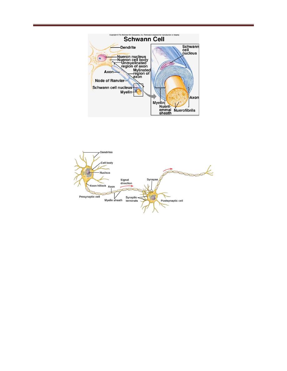
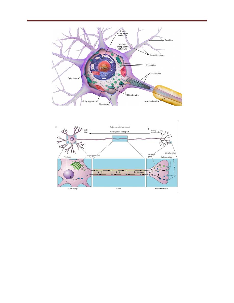
Consist of 3 types of cells:- oligodendrocytes figure 1-4, Schwann cells
figure 1-5, and astrocytes. Oligodendrocytes and Schwann cells are
involved in myelin formation around axons in the CNS and peripheral
nervous system, respectively.
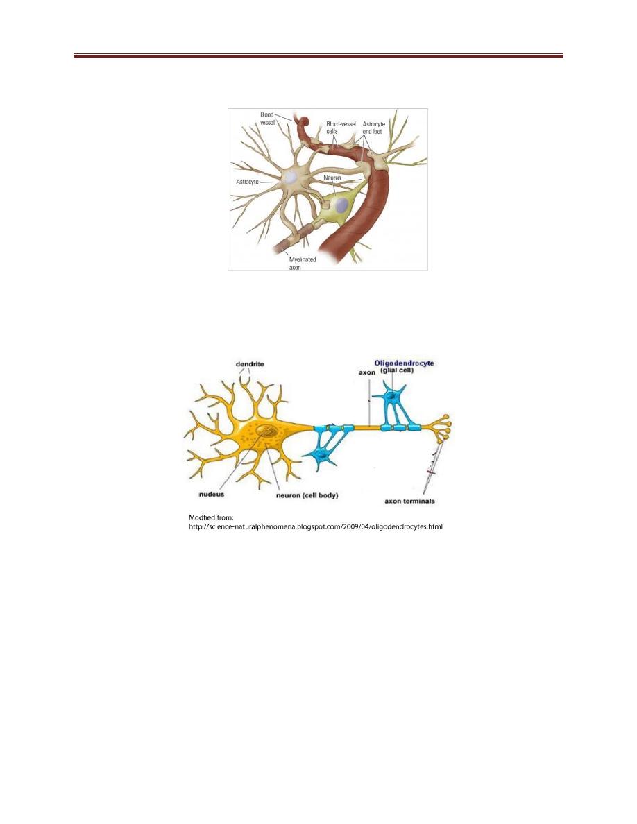
Astrocytes (figure 1-3), which are found throughout the brain and it's
the most abundant of other glial cells, send processes (end foot) to
blood vessels, where they induce capillaries to form the tight junctions
making up the blood–brain barrier.
Neurons figure 1-6
The typical neuron consists of 3 parts, cell body (soma), receptor part
(dendrite) & affector part (axon).
The cell body (soma) figure 1-7, contains the cell nucleus, endoplasmic
reticulum, Golgi apparatus, and most of the free ribosomes.
Mitochondria are located in the cell body, but also throughout the
neuron. The cell body carries out most of the functions such as protein
synthesis and cellular metabolism. Although mature neurons retain
their nuclei, they lose the ability to undergo cell division.
Clinical correlation: absence of blood brain barrier makes brain susceptible to
injury by chemicals and toxins present in the blood and this is seen in newborn
babies with physiological jaundice (elevation of bilirubin) in whom blood brain
barrier is not yet developed resulting in permanent damage to the brain

Physiology of excitable tissues lect. 1 Asst. Prof. Dr. Zahid M. Kadhim
3
Dendrites, several processes that extend outward from the cell body
and branch extensively where it receive action potential from
neighboring neuron through specialized junctions called synapses.
Neuron also has a long fibrous axon that originates from a somewhat
thickened area of the cell body, the axon hillock. The axon divides into
pre-synaptic terminals.
The axoplasmic flow (figure 1-8) or transport occurs along
microtubules that run along the length of the axon and it’s of two
types:- orthograde transport moves proteins, secretory vesicles… etc
from the cell body toward the axon terminals; while retrograde
transport which is in the opposite direction (from the nerve ending to
the cell body). Synaptic vesicles recycle in the membrane, but some
used vesicles are carried back to the cell body and deposited in
lysosomes. Some materials taken up at the ending by endocytosis,
including nerve growth factor (NGF) and some viruses are also
transported back to the cell body.
The axon functions in the rapid transmission of information over
relatively long distances in the form of electrical signals called action
potentials, which are brief, large changes in membrane potential during
which the inside of the cell becomes positively charged relative to the
outside.
The axon hillock—the site where the axon originates from the cell
body—is specialized in most neurons for the initiation of action
potentials. Once initiated, action potentials are transmitted to the axon
terminal. The axon terminal is specialized to release neurotransmitter
on arrival of an action potential. The released neurotransmitter

Physiology of excitable tissues lect. 1 Asst. Prof. Dr. Zahid M. Kadhim
4
molecules carry a signal to a postsynaptic cell, usually to a dendrite or
the cell body of another neuron or to the cells of an effector organ.
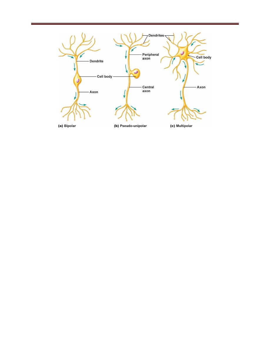
Structural Classification of Neurons figure 1-8
Neurons can be classified structurally according to the number of
processes (axons and dendrites) that project from the cell body into:-
Bipolar neurons are generally sensory neurons with two projections, an
axon, and a dendrite, coming off the cell body. Typical bipolar neurons
function in the senses of olfaction (smell) and vision.
Most sensory neurons, however, are a subclass of bipolar neurons
called pseudo-uni-polar neurons. This name arises because the axon
and dendrite projections appear as a single process that extends in two
directions from the cell body.
Multipolar neurons, the most common neurons, have multiple
projections from the cell body; one projection is an axon, all the others
are dendrites.
Functional Classification of Neurons
Three functional classes of neurons exist: efferent neurons, afferent
neurons, and interneurons.
Efferent neurons transmit information from the central nervous system
to effector organs & include the motor neurons that extend to skeletal
muscle and the neurons of the autonomic nervous system
The function of afferent neurons is to transmit information from either
sensory receptors (which detect information pertaining to the outside
environment) or visceral receptors (which detect information

Physiology of excitable tissues lect. 1 Asst. Prof. Dr. Zahid M. Kadhim
5
pertaining to conditions in the interior of the body) to the central
nervous system for further processing.
The third functional class of neurons is interneurons, which account for
99% of all neurons in the body. They are located entirely in the central
nervous system. Interneurons perform all the functions of the central
nervous system, including processing sensory
information from afferent
neurons, creating and sending out commands to effector organs
through efferent neurons, and carrying out complex functions of the
brain such as thought, memory, and emotions.
Structural Organization of Neurons in the Nervous System
Neurons are arranged within the nervous system in an orderly fashion,
with those having similar functions tending to be grouped together. In
addition, neurons are aligned in such a way that cell bodies and
dendrites of adjacent cells tend to be grouped together, and axons of
adjacent cells tend to be grouped together. In the central nervous
system, cell bodies of neurons are often grouped into nuclei, and the
axons travel together in bundles called pathways, tracts, or
commissures. In the peripheral nervous system, cell bodies of neurons
are clustered together in ganglia, and the axons travel together in
bundles called nerves.

Physiology of excitable tissues lect. 1 Asst. Prof. Dr. Zahid M. Kadhim
6
Figure 1-1 glial cells
Figure 1-2 microglia the brain immune cells
Physiology of excitable tissues lect. 1 Asst. Prof. Dr. Zahid M. Kadhim
7
Figure 1-3 astrocyte
Figure 1-4 oligodendrocyte
figure 1-5 Schwann cells
Physiology of excitable tissues lect. 1 Asst. Prof. Dr. Zahid M. Kadhim
8
Figure 1-6 neuron
Figure 1-7 neuron’s cell body (soma)
Physiology of excitable tissues lect. 1 Asst. Prof. Dr. Zahid M. Kadhim
9
Figure 1-8 axoplasmic flow of neurons
Figure 1-9 structural classification of neurons
Physiology of excitable tissues lect. 1 Asst. Prof. Dr. Zahid M. Kadhim
10
Figure 1-10 resting membrane potential