
11
College of Medicine/Babylon University
Medical Physics Module
Lecture 2:
Physics of The Skeleton
Objectives
: after the end of this lecture, the student must know:
1- The different function of bones in the body
2- How can we measure bone strength
3- Calcium homeostasis and the role of calcium in bone formation and
strength
Bone has at least six functions in the body:
1- support
2- Locomotion
3- Protection
4- Storage of chemicals
5- Nourishment
6- -Sound transmission ( in the middle ear)
1- The body’s muscles attached to the bones through tendons and ligaments,
and the system of bones plus muscles support the body.
2- Bone joints permit movement of one bone with respect to another. The
destruction of joints in arthritis can seriously limit locomotion.
3- The skull protect the brain and several of most important sensory organs
(eye and ears). The ribs form protective cage for heart and lungs.
4- The bones acts as chemical bank for storing elements for future use by the
body.
For example: constant level of calcium is needed in the blood to maintain normal
body functions, if the calcium level falls too low, it activate parathyroid glands to
release more parathormone hormone into the blood, and this causes bones to
release the needed calcium to the blood and restore it to normal value.

12
College of Medicine/Babylon University
Medical Physics Module
5- The teeth are specialized bones can cut food and serve nourishment.
6- The smallest bones of the body are ossicles in the middle ear. These three
bones acts as levers, and provide matching system for converting sound
vibration in air to sound vibration in fluid in cochlea. They are the only
bones that attain full adult size before birth.
What Is Bone Made Of
There are large percentage of calcium (Ca
+
) in the body Ca
+
has much heavier
nucleus than most other elements of the body, it absorbs x-ray much better than
surrounding soft tissue. This is the reason that x-rays show bones so well.
Using X-rays scattering indicates that bone mineral crystals are rod shaped of
diameter 20–70 Aº and length 50–100 Aº.
How Strong Are Your Bones
The strain ( ∆ L / L )
F / A α ∆ L / L
F / A = Y ∆ L / L
Y = F L / A ∆ L Y = young modulus
When the force increase the bone breaks at stress of about 120 N / mm
2
∆ L = L F / A Y ( elongation )
College of Medicine/Babylon University
Medical Physics Module
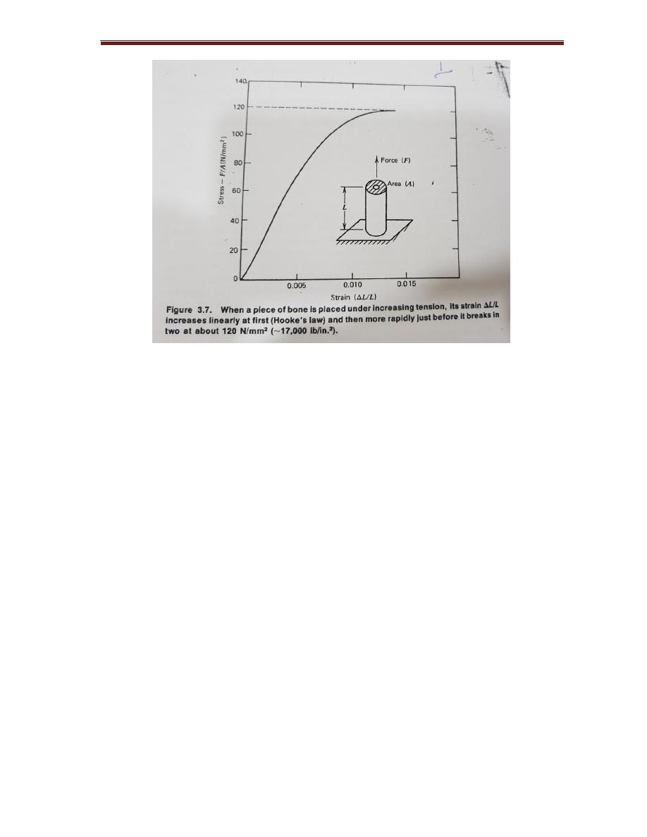
Figure 2-1:
when a piece of bone is placed under increasing tension, it’s strain
increase linearly at first (Hook’s law) and then more rapidly just before it breaks.
The viscosity of synovial fluid decreases under large sheer stress found in the
joint
The good lubricating properties of synovial fluid are thought to be due to the
presence hyaluronic acid and mucopolysaccharide of molecular weight 500000
which deform under load.
Example
: assume a leg has 1.2 m shaft of bone with cross-sectional area of 3 cm
2
(3x10
-4
m
2
) what is the amount of shortening , when all of the body weight of 700
N is supported on his leg?
∆ L = ( 1.2 m ) ( 7 x 10
2
N ) / 3x 10
-4
m
2
( 1.8 x 10
10
N/m
2
= 1.5x 10
-4
m = 0. 15 mm
In running the force on the hip bone when the heel strikes the ground may be
four times the body’s weight.
In normal walking the force on the hip are about twice the body’s weight.

14
College of Medicine/Babylon University
Medical Physics Module
same force applied over a long period of time. This property called
viscoelasticity.
The local electrical fields may also play a rule.
When bone is bent it generates an electrical charges on its surfaces called
piezoelectricity,
may act as physical stimulus for bone growth and repair.
Experiments with animal bone fractures have shown that bone heals faster if an
electrical potential is applied across the broken bone.
The synovial membrane encase the joints and retains the lubricating synovial
fluid. The surfaces of joint are articular cartilages. They are smooth somewhat
rubbery material attached to the solid bone.
The lubricating properties of fluid depends on its viscosity, the viscosity of
synovial fluid decreases under large shear stress found in the joint.
Measurement Of Bone Minerals in The Body
There are many physical techniques for studying bones in the body. A few years
ago, osteoporosis (decrease bone density) was difficult to detect, until the
patient got broken hip or crushed vertebra, where it’s too late to use preventive
therapy.
The strength of bone depends of the mass of bone mineral present. The available
techniques for studying bones are:
1- X-ray image to measure bone mineral, it’s an old technique with several
problems like:
A- x-ray beam has many different energies, and the absorption of x-ray by
calcium varies rapidly with energy.

15
College of Medicine/Babylon University
Medical Physics Module
B- There are scattered radiation when it reaches the film.
C- The film is a poor detector for making quantitative measurements.
2- Photon absorbiometric technique by (JRC ) author ,1960
The three problems of x-ray use are eliminated here by using:
A- mono energetic x–ray or gamma ray
source.
B- A narrow beam to minimize scatter.
C- A scintillation detector that detects all photons.
The determination of bone mineral mass by using
MB α log ( I˳/ I )
Where I˳ is the initial intensity, MB bone mineral
mass
MB = K log ( I˳ / I )
This instrument is a modern clinical bone scanner that uses photon absorption
technique.
Since bone is living tissue, it undergoes changes throughout life. A continuous
process destroying old bone and building new bone, called bone remodeling is
slow work.
Bone remodeling is performed by specialized bone cells, the osteoclast that
destroy the bone, and osteoblast build it, we have the equivalent of a new
skeleton about every 7 years. Each day osteoclast destroy bone containing
about 0.5 gm of calcium (the bone have about 1000 gm of calcium, and
osteoblast build new bone using about the same amount of calcium).
While the body is young and growing the osteoblast do more than osteoclasts,
but after the body is 35 to 40 years old the activity of osteoclasts is greater than

16
College of Medicine/Babylon University
Medical Physics Module
of the osteoblasts, resulting in a gradual decrease in bone mass that continues
until death.
This decrease apparently faster in woman than in men and leads to serious
problems in older women, this condition called osteoporosis results in
spontaneous fractures, especially in the spine and hips.
One osteoclast can destroy bone 100 times faster than one osteoblast can build
new bone, as in other aspects of life.
Small group discussion:
Qx/ Explain why, there is no attraction between two bodies?
Qx/ To calculate force at the fifth lumbar vertebra (L5) with body tipped forward at
60º to the vertical and with weight of 225N in the hands can approach ?
Qx/ Explain, there is friction inside body parts or organs?
Qx/ A 30 years old male presented with shivering (shaking of body) and found that his
temperature 39C°, explain the mechanism of elevating body temperature and how
can we reduced the body temperature.
Qx/ Explain how piezoelectricity of bone fasten healing of fractures
Qx/ Why x-ray shows bone so well
Qx/ 55 years old female experience pain in her joints mainly the knee, from physical
point of view explain the causes of pain.
Qx/ What are the medical effects of orbiting satellite in space journey on the bones?