The Larynx
• Anatomy & PhysiologyLarynx
Lies in front of the laryngo- pharynx from the level of the third to the sixth cervical vertebrae.Consists of a framework of cartilages, connected by ligaments and membranes, lined by a mucous membrane and moved by muscles.
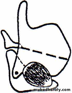
Infantile larynx
Absolutely and relatively smaller.Funnel-shaped.
Much softer therefore collapse more easily in forced inspiration.
Lies high up under the tongue.
The plane of its inlet is less oblique.
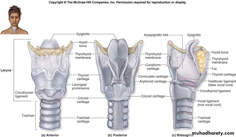
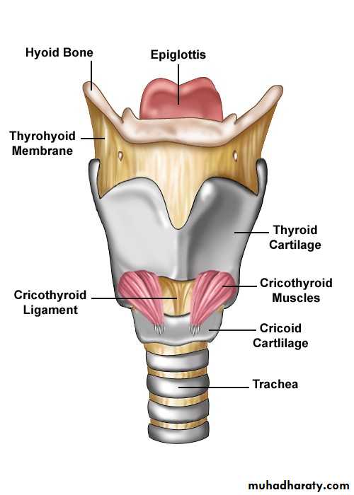
Laryngeal cartilages
Unpaired crtilages Thyroid cartilage
The largest, each half consists of:
1. Ala (lamina)
The two alae meet in the midline, forming an angle of about 90ْ in men, about 120ْ in women.
2. Superior cornu.
3. Inferior cornu.
Cricoid cartilage
Thicker and stronger , a signet ring, narrow in front, broad behind.
Cartilage of epiglottis
Rises up behind the tongue. It is a thin leaf-like sheet of elastic fibrocartilage.
Paired cartilages
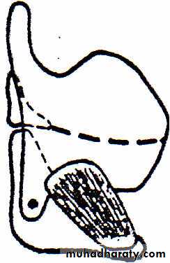
1. Arytenoid cartilages
Largest. pyramidal in shape.
Posterior surface is triangular and concave. It extends laterally into a
muscular process.
Anterolateral surface is convex. It extends forward into a vocal process.
Medial surface is narrow, smooth
Inferior surface or base, is concave. It articulates with the cricoid cartilage.
Apex curves backwards to articulate with the corniculate cartilage
2. Corniculate cartilages
3. Cuneiform cartilages
Laryngeal ligaments and membranes
1. Intrinsic 2. Extrinsic
Thyrohyoid membrane.Cricotracheal membrane.
Hyo-epiglottic ligament.
Cricothyroid membrane
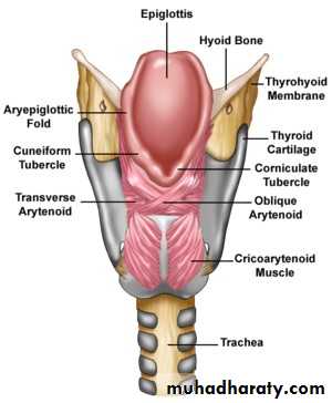
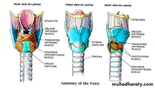
Laryngeal muscles
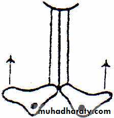
1. Intrinsic A. Abductors of the vocal cords
Posterior crico-arytenoid muscle. Opens the glottis.
Origin - from the depression on the posterior surface of the cricoid lamina.
Direction - upwards and outwards.
Insertion - into the
back of the muscular
process of the
arytenoid cartilage.

B.Adductors of the vocal
Lateral crico-arytenoid muscle:Origin - from the upper border of the
arch of the cricoid cartilage.
Direction - upwards and backwards.
Insertion into the front of the
muscular process of the arytenoid
cartilage.
Transverse portion of interarytenoid muscle.
External portion of thyro-arytenoid muscle.
C.Tensors of the vocal cords
There are two on each side:1.Cricothyroid muscle.
2.Internal portion of thyro-arytenoid (vocalis) muscle .
D.Opener of the laryngeal inlet
Thyro-epiglottic muscle is a part of the thyro-arytenoid musclesE.Closers of the laryngeal inlet
Oblique portion of interarytenoid muscleAryepiglottic muscle
2. Extrinsic
'Strap' muscles of the neck1. Sternothyroid muscle
2. Thyrohyoid muscle3.Sternohyoid m
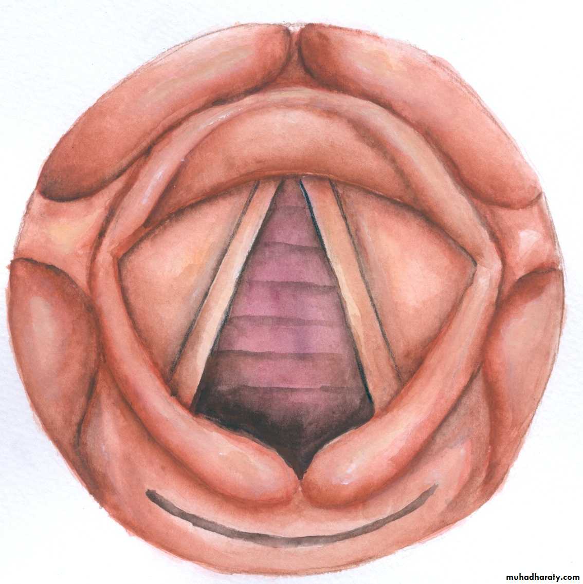
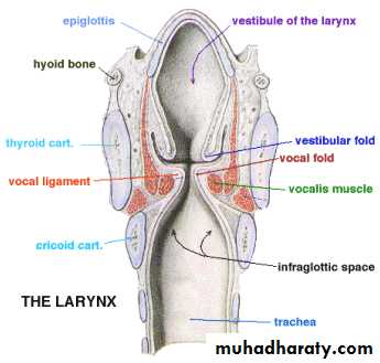
Cavity of the Larynx
Extends from the inlet into the laryngopharynx, to the lower border of the cricoid cartilage.It is divided into three parts by two folds of mucous membrane:
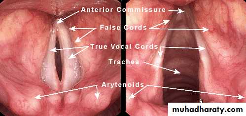
1. False vocal cords
2. True vocal cords
The mucosal folds divide the cavity into the following parts:
a. Vestibule
Lies between the inlet and the edges of the false cords
b. Ventricle of larynx
A recess between the false and true vocal cords.
c. Subglottic space
Lies between the true vocal cords and the lower border of the cricoid cartilage .
Mucous membrane of the larynx
All is Ciliated columnar epithelium except that covers vocal cords & upper part of vestibule of larynx is Stratified squamous epithelium
Blood supply of the larynx
The larynx is supplied by:
1. Laryngeal branches of superior thyroid artery.
2. Laryngeal branches of inferior thyroid artery.
3. Cricothyroid branches of superior thyroid artery.
Anastomose freely with one another. Veins accompany the arteries.
Nerve supply of the larynx
The larynx is supplied by branches of the vagus nerve.Superior laryngeal nerve has two,laryngeal branches:
1. Internal branch. Entirely sensory supplies the cavity of the larynx the above the vocal cords.
2. External branch motor supplies the cricothyroid muscle
Recurrenr (inferior) laryngeal nerve divides into:
1. An anterolateral (motor) branch which supplies all the intrinsic muscles of the larynx except the cricothyroid muscle
2. Posteromedial (sensory) branch which supplies the cavity of the larynx
below the level of the vocal cords.
Lymphatic drainage of the larynx
The edges of the vocal cords divide the lymphatic system of the larynx into two parts:1. Supraglottic above the vocal cords. The vessels drain into:
Pre-epiglottic nodes.
Upper deep cervical nodes
2. Subglottic below the vocal cords. The vessels drain to:
Prelaryngeal and pretracheal nodes.
Lower deep cervical nodes
The vocal cords themselves have practically no lymphatic vessels.
Applied physiology of the larynx
FUNCTIONS OF THE LARYNX
1.Protection of the lower air passages1. Closure of the laryngeal inlet
The aryepiglottic folds move towards one another and close the inlet2. Closure of the glottis
3. Cessation of respiration
4. Cough reflex …'watchdog of the lungs'
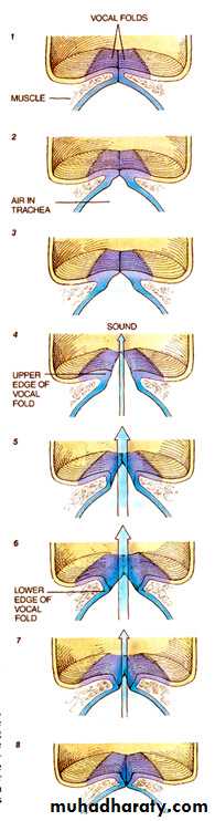
2.Phonation
Voice is produced by vibration or the vocal cords 1. Pitch
The vibrations of the cords cut the air column into puffs and the frequency of the puffs determines the pitch produced. The-larynx is therefore a wind instrument. The average individual human voice can produce a frequency range of two octaves in singing.
2. 'Volume'
The intensity of sound produced by the larynx depends on the air pressure generated in the lungs by contraction of the abdominal and thoracic muscles.
3.Respiration
The larynx plays a part in the mechanism of respiration by reflex adjustments of the glottic aperture.4.Fixation of the chest
When the larynx is closed the thoracic cage becomes fixed permitting climbing or digging. Since the ribs cannot rise freely , a fixed support is given to the pectoral muscles.