THE PAROTID GLAND- tumour- like lesions
Sialasenosis (sialoisis)Non inflammatory swelling
Associated with conditions eg DM, alcoholism, pregnancy, bulimia and idiopathic.
Prolong malnutrition produces sialosis by process of hypertrophy to compensate for swings in acid balance.Drug induced ; commonly sympathomimetic.
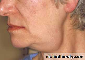
Age; 40-70, soft and often symmetrical swelling, hamster-like appearanceIn DM and drug induce silaosis, may which may associated with neuropathy which interferes with salivary gland function and subsequent acinar cell atrophy.
Rx; no effective treatment ; treat the underlying cause.

THE PAROTID GLAND- degenerative conditions
Sjogren’s syndrome
autoimmune condition causing progressive destruction of the salivary and lacrimal gland s.
Primary Sjogren’s syndrome occurs without an associated connective tissue diseases, the symptoms are more sever than the secondary one and it has higher lymphomatous transformation.
Incidence 0.5%-2%, F:M 10:1, enlarged salivary gland (parotid more than submadiblular), pain, xerostomia.
Pathology: progressive lymphocytic infiltration, acinar cell destruction and proliferation of duct epithelium in all salivary and lacrimal gland tissue.
Dx; depends on history.
Mx; symptomatic, ophthalmological assessment and artificial tear for keratoconjunctivitis sica , artificial salivary substitute for dry mouth.Cx; non Hodgkin’s B-cell lymphpma 4.3%
THE PAROTID GLAND- degenerative conditionsXerostomia
salivary flow decreases with age
Causes.
Chronic anxiety states and depression.
Dehydration.
Anticholinergic drugs especially antidepressants
Salivary gland disorders eg Sjogren’s syndrome
Radiotherapy to the head an neck.
Sialorrhea
Increase salivary flow
Caused by certain drugs and oral infection.
Mx
antisialogogues.
Intraparanchymal botulinum toxin injection.
Uncotrollable drooling managr by surgery:
Blilateral submandibular duct repositioning and simultaneous sublingual gland excision.
Bilateral submanidibular gland excision.
Transposition of parotid ducts and simultaneous submandibular gland excision
* Restiing salivary flow arise from the submandibular gland
THE SUBMANDIBULAR GLANDS-anatomy
Paired glands lie below the mandible on either side.
There is a larger superficial lobe and a smaller deep lobe.
Important anatomical relationship
Lingual nerve
Hypoglossal nerve
Anterior facial vein
Facial artery
Marginal mandibular branch of the facial nerve
It drains by Wharton’s duct and opens in the anterior floor of the mouth at the sublingual papilla.
Ectopic / aberrant salivary gland tissue
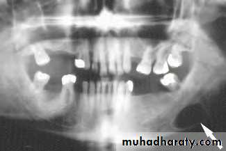
Stafne bone cyst, the most common ectopic salivary tissue.
Asymptomatic clearly demarcated radiolucency of the angle of the mandible.
No treatment
THE SUBMANDIBULAR GLANDS-inflammatory disorders
Acute submandibular sialadenitisviral. Paramyxovirus (mumps)
Bacterial. More common than viral sialadenitis and occurs secondary to stone obstruction.
Chronic submandibular sialadenitis.
THE SUBMANDIBULAR GLANDS-obstruction and trauma
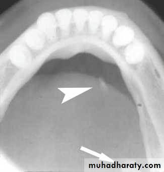
sialolithiasis; the most common cause of obstruction 80%, because the submandibular secretions usually viscus.80% radio-opaque and can identified by x-ray
The stones composed mainly of phosphate and oxalate salts.
Stricture is the 2nd most common cause of obstruction.
Floor of mouth pathology or external pressure accounts 5-10%
C/F: painful swelling, precipitated by eating , the swelling occurs rapidly and resolve spontaneously over 1-2 hrs.(meal time syndrome)
The most common sites of impaction are the of the gland and near the punctum.
Examination: enlarged firm tender swelling. Pus may be visible from sublingual papilla or expressed by bimanual palpation.

THE SUBMANDIBULAR GLANDS-obstruction and trauma
Mxsmall less than 4 mm .. Retrieved by Dormia basket (min invasive procedure performed under local anaesthesia either endoscopically –sialendoscopy- or under US control.
Larger … extrscorporeal or intracorporial lithotripsy. Then retrieved as above.
Stone in submandibular duct in the floor of the mouth anterior to the point crossing the lingual nerve (second molar region)…. The stone removed under local anaesthetics.
Stone at the hilum of the gland…. Stone retrieval via intraoral approach under GA
Stone retrieval success rate 95%
Failure of stone retraction….. Submandiular gland excision
Indications of submandibular gland excision
Sialadentis when min invasive methods have failed.
Salivary tumours
Complications of submandibular gland excision
Haematoma
Wound infection
Marginal mandibular nerve injury
Lingual nerve injury
Hypoglossal nerve injury
Transection of the nerve to the mylohoid muscle producing submental skin anaesthesia.
THE SUBMANDIBULAR GLANDS-tumours
It presents as a slow-growing, painless swellingon examination,it is difficult to differentiate from submandibular lymphadenopathy.
This can be resolved by US examination.
Most salivary neoplasms, even malignant tumours, are often slow-growing, painless swellings. The difficulty is to always distinguish between benign and malignant lesions prior to excision.Pain is not a reliable indication of malignancy
rapid growth, facial nerve palsy, lymph node enlargement and skin tethering are signs of a high-grade malignant lesion.The most common malignant tumour is an adenocystic carcinoma (40%),
THE SUBMANDIBULAR GLANDS-tumours
InvestigationUS with FNAC/True-Cut biopsy , the investigation of choice ( with carful history 95% of malignancy can be identified)
CT&MRI scanning for preoperative planning.
Open surgical biopsy is contraindicated , it may seed the tumour into surrounding tissue making it impossible to eradicate microscopic deposit.
Management
Benign: surgical excision
Malignant depend on stage of the disease. Larger and more aggressive the lesion the more radical surgery required.