Immunity
It is protection against infections.Immune system is the collection of cells and molecules that are responsible for 1- defending us against the countless pathogenic microbes in our environment; 2- prevent the proliferation of cancer cell; and 3- mediated the healing of damaged tissue.
Defense against microbes consists of two types of reactions:-
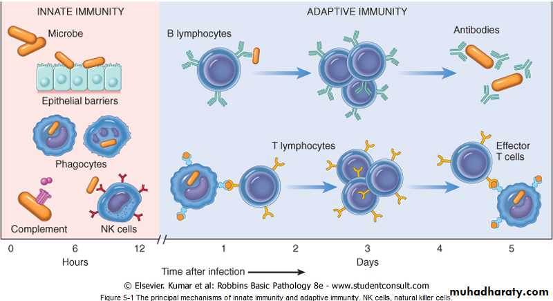
1- Innate immunity (also called natural, or native, immunity) is mediated by cells and proteins that are always present and poised to fight against microbes and are called into action immediately in response to infection. The major components of innate immunity are:-
A- epithelial barriers of the skin, gastrointestinal tract, and respiratory tract, which prevent microbe entry .
B- phagocytic leukocytes (neutrophils and macrophages).
C- a specialized cell type called the natural killer (NK) cell.
D- several circulating plasma proteins, the most important of which are the proteins of the complement system .
1
2- Adaptive immunity (also called acquired, or specific, immunity). Adaptive immunity is normally silent and responds (or "adapts") to the presence of infectious microbes by becoming active, expanding, and generating potent mechanisms for neutralizing and eliminating the microbes. The components of the adaptive immune system are lymphocytes and their products. By convention , the terms "immune system" and "immune response" refer to adaptive immunity.
2
3
Types of adaptive immunity
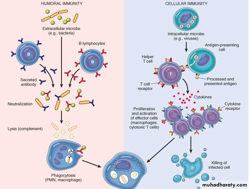
1-Humoral immunity, mediated by soluble antibody proteins that are produced by B lymphocytes (also called B cells).2-Cell-mediated (or cellular) immunity, mediated by T lymphocytes (also called T cells) . Antibodies provide protection against extracellular microbes in the blood, mucosal secretions, and tissues. T lymphocytes are important in defense against intracellular microbes. They work by either directly killing infected cells (accomplished by cytotoxic T lymphocytes) or by activating phagocytes to kill ingested microbes, via the production of soluble protein mediators called cytokines (made by helper T cells).
4
When the immune system is inappropriately triggered or not properly controlled, the same mechanisms that are involved in host defense cause tissue injury and disease.
The reaction of the cells of innate and adaptive immunity may be manifested as inflammation which is a beneficial process, but it is also the basis of many human diseases .
5
6
CELLS AND TISSUES OF THE IMMUNE SYSTEM
1- lymphocytes, which are the mediators of adaptive immunity.2- specialized antigen-presenting cells (APCs), which capture and display microbial and other antigens to the lymphocytes.
3- various effector cells, that eliminating the antigens (typically, microbes).
7
Lymphocytes
Lymphocytes are present in the circulation and in various lymphoid organs. Lymphocytes develop from precursors in the generative lymphoid organs as two types :-
T lymphocytes are so called because they mature in the thymus,
B lymphocytes mature in the bone marrow.
Each T or B lymphocyte expresses receptors for a single antigen, and the total population of lymphocytes (numbering about 1012 in humans) is capable of recognizing tens or hundreds of millions of antigens.
T Lymphocytes
Thymus-derived, or T, lymphocytes are the effector cells of cellular immunity and provide important stimuli for antibody responses to protein antigens. T cells constitute 60% to 70% of the lymphocytes in peripheral blood and are the major lymphocyte population in splenic periarteriolar sheaths and lymph node interfollicular zones. T cells do not detect free or circulating antigens. Instead, the vast majority (>95%) of T cells recognize only peptide fragments of protein antigens that are displayed on other cells bound to proteins of the major histocompatibility complex (MHC; or in humans, human leukocyte antigen [HLA] complex). The MHC was discovered on the basis of studies of graft rejection or acceptance (tissue, or "histo," compatibility).
the normal function of MHC molecules is to display peptides for recognition by T lymphocytes. thus perform their function of killing infected cells or activating phagocytes or B lymphocytes that have ingested protein antigens.
8
B Lymphocytes
Bone marrow-derived, or B, lymphocytes comprise 10% to 20% of the circulating peripheral lymphocyte population. They are also present in bone marrow and in the follicles of peripheral lymphoid tissues (lymph nodes, spleen, tonsils, and other mucosal tissues). B cells are synthesize antibodies, also called immunoglobulins (Ig) as IgG; IgM; IgA; IgE; IgD . After stimulation, B cells differentiate into plasma cells, which secrete large amounts of antibodies, the mediators of humoral immunity .9
Natural Killer Cells
Natural killer (NK) cells are lymphocytes that arise from the common lymphoid progenitor that gives rise to T and B lymphocytes. NK cells are cells of innate immunity . they do not have specificities as diverse as do T cells or B cells. NK cells use a limited set of activating receptors to recognize molecules expressed on stressed or infected cells or cells with DNA damage, and then kill these cells .NK cells express inhibitory receptors that recognize self class I MHC molecules, which are expressed on all healthy cells; engagement of these inhibitory receptors typically overrides the activating receptors and thus prevents activation of the NK cells. Infections (especially viral infections) and stress are associated with loss of expression of class I MHC molecules. So the NK cells are released from their inhibition and destroy the unhealthy host cells.
10
Antigen-Presenting Cells
These cells are specialized to capture microbial antigens and display these to lymphocytes. these cells are Dendritic cells (DCs) and macrophages.1-Dendritic Cells
Cells with fine dendritic cytoplasmic processes occur as two functionally distinct types.
A- Interdigitating DCs, are non phagocytic cells that express high levels of class II MHC and T-cell co stimulatory molecules.
Immature DCs reside in epithelia, where they are located to capture entering microbes; an example is the Langerhans cell of the epidermis.
Mature DCs are present in the T-cell zones of lymphoid tissues, where they present antigens to T cells circulating through these tissues. DCs are also present in the interstitium of many non lymphoid organs, such as the heart and lungs, where they can capture the antigens of microbes that have invaded the tissues.
B- follicular dendritic cells (FDCs). They are located in the germinal centers of lymphoid follicles in the spleen and lymph nodes. These cells bear receptors for the Fc tails of IgG and for complement proteins, and hence efficiently trap antigen bound to antibodies and complement.
2-Macrophages ingest microbes and other particulate antigens and display them for recognition by T lymphocytes. These T cells in turn activate the macrophages to kill the microbes, the central reaction of cell-mediated immunity.
11
Effector Cells
Many different types of leukocytes perform the adaptive immune response, which is to eliminate infections. These include :-
1- NK cells are frontline effector cells because of their ability to rapidly react against "stressed" cells.
2- Antibody-secreting plasma cells are effector cells of humoral immunity.
3-T lymphocytes, both CD4+ helper T cells and CD8+ CTLs, are effector cells of cell-mediated immunity. These lymphocytes often function in host defense together with other cells.
4- Macrophages, bind microbes that are coated with antibodies or complement, and phagocytose and destroy these microbes, thus serving as effector cells of humoral immunity. Macrophages also respond to signals from helper T cells and improve their ability to destroy phagocytosed microbes, thus serving as effector cells of cellular immunity.
T lymphocytes secrete cytokines that recruit and activate other leukocytes, such as neutrophils and eosinophils, and all these cell types function in defense against various pathogens.
Lymphoid Tissues
The lymphoid tissues of the body are divided into
1- Generative (primary) organs, where lymphocytes express antigen receptors and mature. these incude thymus and bone marrow .
2- peripheral (secondary) lymphoid organs, where adaptive immune responses develop. the peripheral organs are the lymph nodes, spleen, and mucosal and cutaneous lymphoid tissues
12
HYPERSENSITIVITY DISEASES
Under normal conditions , the immune response prevents disease , but occasionally ,the inapproperiate activation of the immune system can lead to debilitating or life-threatening illness like :-1- Allergic or hypersensitivity reactions.
2- Transplantation immunopathology.
3-Autoimmune disorders.
4- Immunodeficiency states.
13
1- Allergic or hypersensitivity reactions
Hypersensitivity it is define as an exaggerated immune response to a foreign agent resulting in injury to the host .its caused by immune response to environmental antigens called allergens that produce inflammation and cause tissue injury.
Allergens Any foreign substances capable of inducing an immune response .many different chemicals of natural and synthetic origin are known as allergens.
Complex and natural organic chemicals especially proteins are more likely to cause an immediate hypersensitivity response whereas simple organic compound ,inorganic chemicals ,and metals more commonly cause delayed hypersensitivity reactions . exposure to allergen can be through inhalation ,ingestion ,injection ,or skin contact .
Hypersensitivity disorders are four types :-
Type I: IgE – mediated disorders (immediate (Type I) Hypersensitivity ).
Type II: Antibody –mediated (ctotoxic)disorders.
Type III: Immune –Complex disorder .
Type IV: Cell-mediated hypersensitivity reactions.
14
Causes of Hypersensitivity Diseases
1-Autoimmunity. Normally, the immune system does not react against an individual's own antigens. This phenomenon is called self-tolerance. Sometimes, self-tolerance fails, resulting in reactions against one's own cells and tissues that are called autoimmunity. The diseases caused by autoimmunity are referred to as autoimmune diseases.2- Reactions against microbes. There are many types of reactions against microbial antigens that may cause disease. In some cases, the reaction appears to be excessive or the microbial antigen is persistent. If antibodies are produced against such antigens, the antibodies may bind to the microbial antigens to produce immune complexes, which deposit in tissues and trigger inflammation; as in poststreptococcal glomerulonephritis .T-cell responses against persistent microbes may give rise to severe inflammation, as in tuberculosis and other infections . In viral hepatitis, the virus that infects liver cells is not cytopathic, but it is recognized as foreign by the immune system. Cytotoxic T cells try to eliminate infected cells, and this normal immune response damages liver cells.
3- Reactions against environmental antigens. Most healthy individuals do not react strongly against common environmental substances (e.g., pollens, animal danders, or dust mites), but almost 20% of the population is "allergic" to these substances.
15
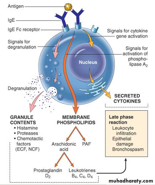
IgE – mediated disorders (immediate (Type I) Hypersensitivity )
this type of hypersensitivity is a tissue reaction that occurs rapidly (within minutes) after the interaction of antigen with IgE antibody . These disorders triggered by binding of an allergen with specific IgE found on the surface of mast cells and basophiles .Mast cell (tissue cells ) and basophiles (blood cells) are derived from blood precursor cells .mast cells are distributed throughout connective tissue , near surfaces that exposed to environmental antigens especially in areas beneath the skin and mucous membranes of respiratory ,gastrointestinal ,and geneto urinary tracts ,and adjacent to blood and lymph vessels .
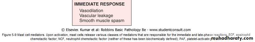
Mast cells and basophile have granules that contain potent mediator of allergic reactions . during the sensitization , the allergen specific IgE antibodies attach to receptors on these mast cells triggers a series of events that lead to degranulation of the sensitized mast cells , causing release of their allergy producing mediators which include :-
- Histamine a potent vasodilator that increase the permeability of capillaries and venules , smooth muscle contraction, and increased secretion of mucous .
- Acetylcholine produce bronchial smooth muscle contraction and dilation of small blood vessels .
-Prostaglandin and leukotrienes is the most abundant mediator generated by mast cells. It produce responses similar to histamine and acetylcholine .
- proteases which may damage tissues and also generate kinins and cleave complement components to produce additional chemotactic and inflammatory mediator .
- Cytokines. Activation of mast cells results in the synthesis and secretion of several cytokines that are important for the late-phase reaction. These include TNF and chemokines, which recruit and activate leukocytes.
16
17
Type I hypersensitivity reactions may present as a systemic disorder (anaphylaxis) or localized reaction (atopy).
Systemic anaphylactic reactions
Result from parenteral administration ( injection ) of protein antigens (e.g., in bee or wasp venom) or drugs (e.g., penicillin) or radiographic contrast dyes .
May also result from ingested allergens ( seafood ,nuts , and legumes).
Signs and symptoms
anaphylaxis has rappid onset (within minutes ), there will be :-
1- itching
2-urticaria .
3- skin erythema appear
4- respiratory difficulty caused by pulmonary bronchoconstriction and hypersecretion of mucus.
5- Laryngeal edema may exacerbate matters by causing upper airway obstruction.
6- the musculature of the entire gastrointestinal tract may be affected, with resultant vomiting, abdominal cramps, and diarrhea.
7- , there may be systemic vasodilation with fall in blood pressure (anaphylactic shock), and the patient may progress to circulatory collapse and death within minutes.
18
Local reactions (atopic)
occur when the antigen is confined to a particular site, such as skin (contact, causing urticaria), gastrointestinal tract (ingestion, causing diarrhea), or lung (inhalation, causing bronchoconstriction). The common forms of skin and food allergies, hay fever, and certain forms of asthma are examples of localized allergic reactions.
The term atopy is used to imply familial predisposition to such localized reactions.
Type I hypersensitivity reactions plays an important protective role in parasitic infections. IgE antibodies are produced in response to many helminthic infections, and their physiologic function is to target helminths for destruction by eosinophils and mast cells.
19
Type II, Antibody –mediated (ctotoxic) disorders
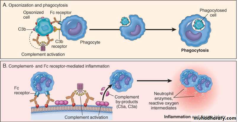
These disorders are caused by antibodies directed interaction with target antigens on the surface of cells or other tissue components. The antigens may be normal molecules intrinsic to cell membranes or extracellular matrix, or they may be adsorbed exogenous antigens (e.g., a drug metabolite). Antibody-mediated abnormalities are the underlying cause of many human diseases ex. mismatched blood transfusion ,hemolytic disease in newborn ,and Autoimmune hemolytic anemia.Mechanisms of Antibody-Mediated Diseases
Antibodies cause disease by targeting cells for phagocytosis , by activating the complement system, and by interfering with normal cellular functions .
1- Opsonization and phagocytosis. When circulating cells, such as erythrocytes or platelets, are coated (opsonized) with antibodies (IgG) , the cells become targets for phagocytosis by neutrophils and macrophages . These phagocytes express receptors for the Fc tails of IgG antibodies and for breakdown products of the C3 complement protein, and use these receptors to bind and ingest opsonized particles. Opsonized cells are usually eliminated in the spleen, and this is why splenectomy is of some benefit in autoimmune thrombocytopenia and hemolytic anemia ..
2-Inflammation. Antibodies bound to cellular or tissue antigens activate the complement system by the "classical" pathway . Products of complement activation recruit neutrophils and monocytes, triggering inflammation in tissues, opsonize cells for phagocytosis, and lyse cells, especially erythrocytes.
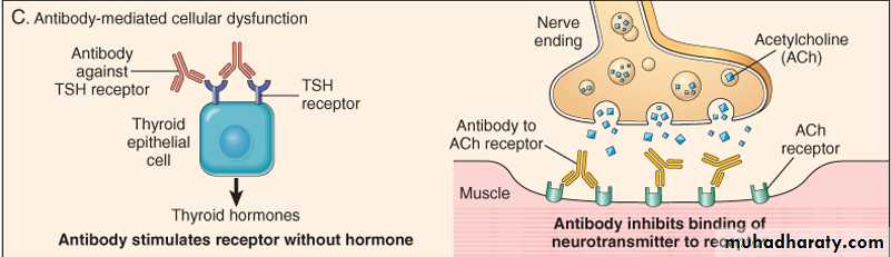
3-Antibody-mediated cellular dysfunction. In some cases, antibodies directed against cell surface receptors impair or dysregulate cellular function without causing cell injury or inflammation .
20
Effector mechanisms of antibody-mediated injury. A, Opsonization of cells by antibodies and complement components, and ingestion of opsonized cells by phagocytes. B, Inflammation induced by antibody binding to Fc receptors of leukocytes and by complement breakdown products. C, Antireceptor antibodies disturb the normal function of receptors
21
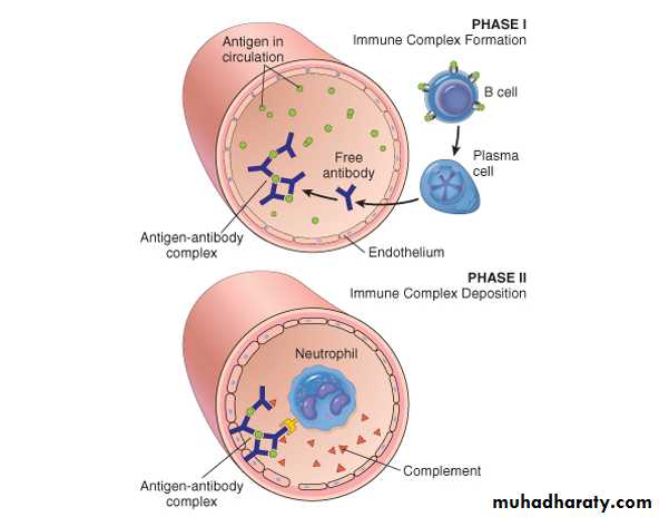
Type III: Immune –Complex disorder
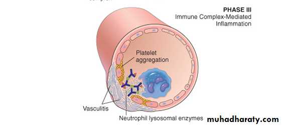
They are mediated by formation of Antigen-antibody complexes that activate complement which will generate chemotactic vasoactive mediators that cause tissue damage by a variety of mechanism , including alterations in blood flow ,increase vascular permeability ,and the destructive action of the inflammatory cells.The reaction occur when the antigen combines with antibody in the circulation or at extravascular sites where antigen may have been deposited.
Immunecomplexes formed in the circulation produce damage when the come in contact with vessel lining or are deposit in tissue ,as renal glomerulus , skin venules , lung ,and joint synovium .
There are two types of antigens that cause immunecomplex mediated injury:-
1-exogenous antigen such as viral and bacterial proteins.
2- endogenous antigen such as self antigen associated with autoimmune disorders or nucleoproteins .
Type III reactions are responsible for
1-the acute glomerulonephritis after streptococcal infection .
2- the manefestaion of autoimune disorders such as Systemic lupus erythematosus.
3- acute serum sickness its type of a systemic immunecoplex disease triggered by the deposition of insoluble antigen –antibody (IgM , IgG) complex in blood vessel , joint , heart, and kidney tissues .the deposited complex activate complement ,increase vascular permeability ,and recruit phagocytic cells all of which can promote tissue damage and edema. this syndrome consisting rash , lymphadenopathy , arthralgias ,and neurologic disorders that appear in 7 of more after injection of horse antisera for prevention the tetanus .
22
Immune complex disease. The sequential phases in the induction of systemic immune complex-mediated diseases (type III hypersensitivity).
23
Type IV: Cell-mediated hypersensitivity reactions.
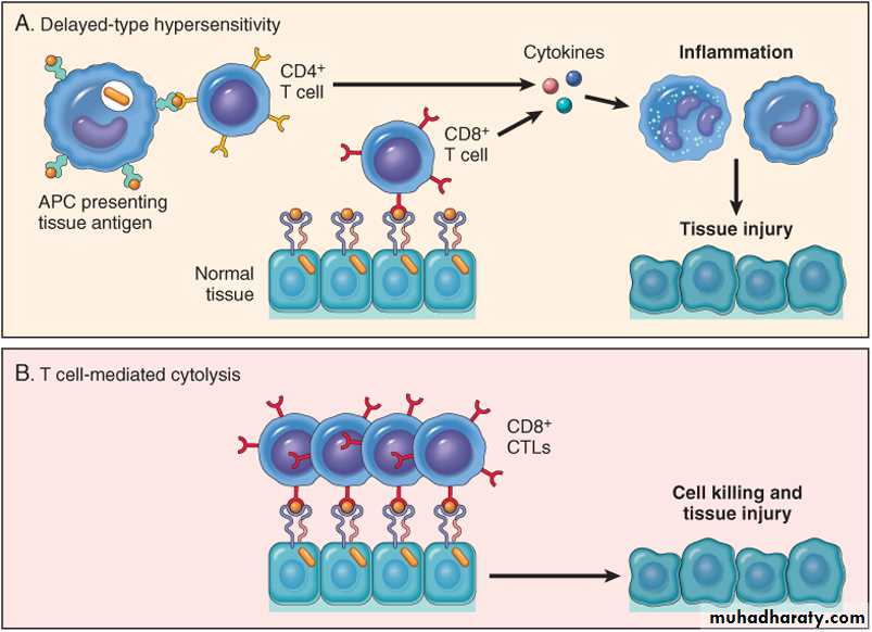
Occur 24-72 hrs after exposure of sensitized individual to the offending antigen .they mediated by T helper lymphocytes that are directly cytotoxic or secret inflammatory mediators like cyokines that cause tissue changes. Cyokines will attract T or B lymphocytes as well as monocytes , neutrophils , eosinophils ,and basophiles .some of the cytokines promote activation of macrophages that function as phagocytic and antigen presenting cells .Two types of T-cell reactions are capable of causing tissue injury and disease: (1) delayed-type hypersensitivity (DTH), initiated by CD4+ T cells, and (2) direct cell cytotoxicity, mediated by CD8+ T cells.
- The best known example of delayed-type hypersensitivity is the reaction to the tuberculin test(tuberculin is a protein extract of the tubercle bacillus), in which inactivated or purified protein derivative is injected under the skin . In previously sensitized person to the tubercle bacillus ,redness and induration of the area develop within 8-12 hrs ,reaching to a peak in 24-72hrs . A positive tuberculin test indicate that the person had sufficient exposure to Mycbacterium tuberculosis to incite hypersensitivity reaction .
Certain type of antigens include cell mediated immunity .
Prolonged DTH reactions against persistent microbes may result in a special morphologic pattern of reaction called granulomatous inflammation. The initial perivascular CD4+ T-cell replaced by macrophages over a period of 2 to 3 weeks; these accumulated macrophages typically exhibit morphologic evidence of activation, that is, they become large, flat, and eosinophilic (denoted as epithelioid cells). The epithelioid cells occasionally fuse under form multinucleated giant cells. A microscopic aggregate of epithelioid cells, typically surrounded by a collar of lymphocytes, is called a granuloma.
-Direct T cell cytotoxicity -mediated hypersensitivity, CD8+ CTLs kill antigen-bearing target cells. CTLs play a critical role in resistance to virus infections and some tumors. CTLs play an important role in the rejection of solid-organ transplants and may contribute to many immunologic diseases, such as type 1 diabetes (in which insulin-producing β cells in pancreatic islets are destroyed by an autoimmune T-cell reaction).
24
Mechanisms of T-cell-mediated (type IV) hypersensitivity reactions. A, In delayed-type hypersensitivity reactions, CD4+ T cells respond to tissue antigens by secreting cytokines that stimulate inflammation and activate phagocytes, leading to tissue injury. B, In some diseases, CD8+ CTLs directly kill tissue cells. APC, antigen-presenting cell.
25
26
Typical manifestation
mechanism
Initiation time
Descriptive name
Type
1-systemic anaphylaxis .
2- localized anaphylaxis .
3 –asthma.
4- food allergy
5-eczema
AG cross linkage of IgE bound to mast cells and basophils with release vasoactive mediators.
2-30min
IgG mediated hypersensitivity
Type I
1-blood transfusion reaction
2- autoimmune hemolytic anemia
Ab directed to the cell surface antigens mediate cell destruction via complement
5-8h
Antibody mediated cytotoxic hypersensitivity
Type II
1-arthus reactions
2- serum sickness
3-glomerulonephritis
4- systemic lupus-
AG-Ab complex deposited in various tissues induce complement activation and inflammatory response
2-8h
Immunecomplex mediated type hypersensitivity
Type III
1-Contact
Dermatitis
2-tubercular lesion
3-graftregection
Sensitized T cells release cytokines which activate macrophages or T ceiis which mediate direct cellular damage
24-72 h
Cell mediated hypersensitivity
Type VI